Friday, June 20, 2014

Ceremonial PTSD Therapies Favored by Native American Veterans


This is interesting, and it confirms a growing trend I have seen in aboriginal peoples seeking to return to the spiritual traditions that once nourished their people. One of the best books I have seen on the subject is Healing the Soul Wound: Counseling with American Indians and Other Native Peoples (2006) by Eduardo Duran.


Ceremonial PTSD therapies favored by Native American veterans

Medical News Today | Friday 20 June 2014

Native American veterans battling Post Traumatic Stress Disorder find relief and healing through an alternative treatment called the Sweat Lodge ceremony offered at the Spokane Veterans Administration Hospital.

In the Arizona desert, wounded warriors from the Hopi Nation can join in a ceremony called Wiping Away the Tears. The traditional cleansing ritual helps dispel a chronic "ghost sickness" that can haunt survivors of battle.

These and other traditional healing therapies are the treatment of choice for many Native American veterans, - half of whom say usual PTSD treatments don't work - according to a recent survey conducted at Washington State University. The findings will be presented at the American Psychological Association conference in Washington D.C. this August.

The study is available online at https://www.surveymonkey.com/s/nativeveterans.

Led by Greg Urquhart and Matthew Hale, both Native veterans and graduate students in the WSU College of Education, the ongoing study examines the attitudes, perceptions, and beliefs of Native American veterans concerning PTSD and its various treatment options. Their goal is to give Native veterans a voice in shaping the types of therapies available in future programs.

"Across the board, Native vets don't feel represented. Their voices have been silenced and ignored for so long that they were happy to provide feedback on our survey," said Hale.

Historically, Native Americans have served in the military at higher rates than all other U.S. populations. Veterans are traditionally honored as warriors and esteemed in the tribal community. A 2012 report by the Department of Veterans Affairs showed that the percentage of Native veterans under age 65 outnumbers similar percentages for veterans of all other racial groups combined.

The WSU survey provides a first-hand look at the veterans' needs, but more importantly, reveals the unique preferences they have as Native American veterans, said Phyllis Erdman, executive associate dean for academic affairs at the college and mentor for the study.

Cultural worldview

Urquhart said many Native veterans are reluctant to seek treatment for PTSD because typical western therapy options don't represent the Native cultural worldview.

"The traditional Native view of health and spirituality is intertwined," he explained. "Spirit, mind, and body are all one - you can't parcel one out from the other - so spirituality is a huge component of healing and one not often included in western medicine, although there have been a few studies on the positive effects of prayer."

For many years, the U.S. government banned Native religious ceremonies, which subsequently limited their use in PTSD programs, said Urquhart. Seeking to remedy the situation, many Veterans Administration hospitals now offer traditional Native practices including talking circles, vision quests, songs, drumming, stories, sweat lodge ceremonies, gourd dances and more. Elders or traditional medicine men are also on staff to help patients process their physical and emotional trauma.

"PTSD is a big issue and it's not going away anytime soon," said Hale who identifies as Cherokee and was a mental health technician in the Air Force.

Urquhart, who is also Cherokee and developed mild symptoms of PTSD after a tour as a cavalry scout in Iraq, said there have been very few studies on Native veterans and PTSD. He and Hale designed their survey to be broader and more inclusive than any previous assessments. It is the first to address the use of equine therapy as a possible adjunct to both western treatments and Native ceremonial approaches.

Standard treatments disappointing

So far, 253 veterans from all five branches of the military have completed the survey, which includes 40 questions, most of them yes or no answers. It also includes an open-ended section where participants can add comments. The views reflect a diverse Native population ranging from those living on reservations to others who live in cities.

The majority of survey takers felt that "most people who suffer from PTSD do not receive adequate treatment," said Urquhart. For Native veterans who did seek standard treatment, the results were often disappointing. Sixty percent of survey respondents who had attempted PTSD therapy reported "no improvement" or "very unsatisfied."

Individual counseling reportedly had no impact on their PTSD or made the symptoms worse for 49 percent of participants. On the other hand, spiritual or religious guidance was seen as successful or highly successful by 72 percent of Native respondents. Animal assisted therapy - equine, canine, or other animals - was also highly endorsed.

"The unique thing about equine therapy is that it's not a traditional western, sit-down-with-a-therapist type program. It's therapeutic but doesn't have the stigma of many therapies previously imposed on Native Americans," said Urquhart.

Strongly supportive of such efforts, Erdman is expanding the long-running WSU Palouse Area Therapeutic Horsemanship (PATH) program to include a section open to all veterans called PATH to Success: A Warrior's Journey.

Giving veterans a voice

Urquhart, Hale, and teammate, Nasreen Shah say their research is gaining wide support in Native communities throughout the nation.

The team plans to distribute the survey results to all U.S. tribes, tribal governments, Native urban groups, and veteran warrior societies. They also hope the departments of Veterans Affairs and Indian Health Services will take notice and continue to incorporate more traditional healing methods into their programs.

As one Iroquois Navy veteran commented on the survey, "Traditional/spiritual healing can be very effective together with in depth education and background in modern treatment methods."

A Nahua Army veteran agreed, writing, "Healing ceremonies are absolutely essential, as is story telling in front of supportive audiences. We need rituals to welcome back the warriors."

Effects of Early Life Adverse Experiences on the Brain: Implications from Maternal Separation Models in Rodents

Adverse Childhood Experiences (ACE) Study Pyramid

In the past, I have posted quite a few time about how adverse childhood experiences (ACE) can have a profound impact on physical and mental health (see here, here, here, here, here, and here, for example). This new study looks at how maternal separation in a mouse model impacts brain development.

It' easy to dismiss research like this because it involves mice, not humans. First of all, we could never do this research with humans - it's not ethical. More importantly, however, nearly all mammals hve similar parent/offspring bonding drives and needs, so for the first few months of life, we can model these behaviors in rodents with relative assurance that they will at least partially translate to humans.

In this particular mini-review, the researchers are seeking a molecular explanation of how early life stress impacts brain development and dysfunction (hint: stress hormones).

Full Citation: 
Nishi, M., Horii-Hayashi, N. and Sasagawa, T. (2014, Jun 17). Effects of early life adverse experiences on the brain: implications from maternal separation models in rodents. Frontiers in Neuroscience: Neuroendocrine Science; 8:166. doi: 10.3389/fnins.2014.00166

Effects of early life adverse experiences on the brain: implications from maternal separation models in rodents

Mayumi Nishi, Noriko Horii-Hayashi and Takayo Sasagawa
  • Department of Anatomy and Cell Biology, Nara Medical University, Kashihara, Japan
During postnatal development, adverse early life experiences affect the formation of neuronal networks and exert long-lasting effects on neural function. Many studies have shown that daily repeated maternal separation (MS), an animal model of early life stress, can regulate the hypothalamic-pituitary-adrenal axis (HPA axis) and affect subsequent brain function and behavior during adulthood. However, the molecular basis of the long-lasting effects of early life stress on brain function has not been fully elucidated. In this mini review, we present various cases of MS in rodents and illustrate the alterations in HPA axis activity by focusing on corticosterone (CORT). We then show a characterization of the brain regions affected by various patterns of MS, including repeated MS and single time MS at various stages before weaning, by investigating c-Fos expression. These CORT and c-Fos studies suggest that repeated early life stress may affect neuronal function in region- and temporal-specific manners, indicating a critical period for habituation to early life stress. Next, we introduce how early life stress can impact behavior, namely by inducing depression, anxiety or eating disorders, and alterations in gene expression in adult mice subjected to MS.

Introduction


As our contemporary society changes rapidly, changes in family structure can have a large influence on the mother–child relationship, as well as on other social environmental factors. In adult patients with various neuropsychiatric disorders, childhood abuse including sexual and/or physical abuse and neglect, is one of the most serious causes (Bremne and Vermetten, 2001; Heim and Nemeroff, 2001; Teicher et al., 2006). Adverse experiences occurring during critical periods of development, such as perinatal life, harmfully influence behavior, and physiological functions, including growth, metabolism, reproduction, and immune responses. Stressful environments in early life may induce permanent rather than transient consequences in animals. Previous studies have indicated that early unfavorable events augment the risk of behavioral disorders in adulthood, including neuropsychiatric disorders, such as depression (Kendler et al., 2002) and psychosis (Morgan et al., 2007). In rodent and primate models, adverse environments during the neonatal periods seem to play a critical role in developing the brain systems important to regulate behavior and stress responsiveness. In particular, the responsiveness of the hypothalamic-pituitary-adrenal (HPA) axis can be deteriorated by interrupting usual mother-pup interactions, which may induce persistent changes in the neurobiology, physiology, and emotional behavior in adult animals (Ellenbroek et al., 1998; Lyons et al., 1998; Pryce et al., 2005; Enthoven et al., 2008; Nishi et al., 2013).

In this mini review, we will focus on the response of corticosterone (CORT), an end product of the HPA axis in rodents, and c-Fos expression for examining the activated brain regions induced by maternal separation (MS), a model of rodent early life stress. Furthermore, we will also present alterations of behavioral aspects and alterations in gene expression.

Early MS


The inventive studies of Levine and colleagues, and consequently of Meaney, Plotsky, and their collaborators have demonstrated that changes in rodents' early postnatal experiences can induce profound long-lasting effects on emotionality and stress response (Levine, 1967; Meaney, 2001; Plotsky et al., 2005), which have spurred the employment of the rodent MS for investigating early life stress. This early life stress model is based on the evidence that unfavorable events in early life cause the vulnerability for developing various kinds of diseases in later life. In this type of study, MS should be carefully discussed in comparison to the appropriate control group, which may or may not be undisturbed from mother.

The procedure of MS showed a variety of the duration (e.g., 60 min–24 h) and the number of days (e.g., 1–14 days, 15–21 days) for the separation experiences among laboratories (Biagini et al., 1998; Caldji et al., 2000; Barreau et al., 2004; Arborelius and Eklund, 2007; Carrera et al., 2009; Tjong et al., 2010). In MS paradigm, many experiments, but certainly not all, have demonstrated that separation of pups from their mothers during the early postnatal period permanently increased anxiety-like behaviors in adulthood (Francis et al., 1999; Huot et al., 2001, 2004; Menard et al., 2004). As to the HPA axis activity, the response to stress is relatively low during early postnatal life (Walker et al., 1991; Levine, 2005), while MS could lead to life-long hyperactivity of the HPA axis (Holmes et al., 2005; Lippmann et al., 2007; Aisa et al., 2008; Marais et al., 2008). In contrast, short-term disturbance (e.g., 15 min), which has been called “handling,” appeared to reduce anxiety-like behaviors, decrease HPA axis tone and reduce the response to stress in adulthood (Levine, 2005; Plotsky et al., 2005). The process of handling may imitate natural mice rearing, whereby the mother leaves her pups for short periods of time to collect foods. Thus, the short-term MS, handling, might be considered a more natural event.

The effect of MS also varies depending upon whether pups are separated in a group of littermates during MS or isolated singly. Miyazaki and colleagues recently reported that rat pups isolated singly from the mother during PND7 to PND11 presented disturbance of cortical function, whereas pups separated but gathered from PND7 to PND11 showed no cortical disruption (Miyazaki et al., 2012).

Characterization of Maternally Separated Animals


Serum Level of CORT

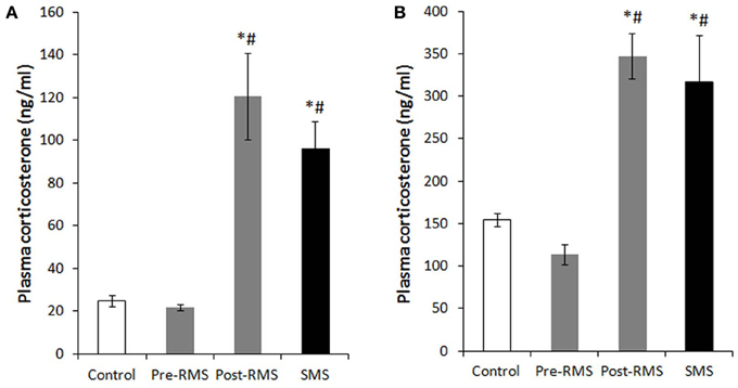
In rodents, there is an unique period during which the HPA axis shows a rapid regression known as the stress hyporesponsive period (SHRP) (Levine, 2001). This period extends from PND4 to PND14 in rats and from PND2 to PND12 in mice. During the course of SHRP, ACTH in increased and baseline plasma glucocorticoid levels are lower than normal (Rosenfeld et al., 1991). Because, during ontogeny, the maintenance of low and stable levels of CORT is necessary for normal growth and development of the central nervous system (CNS), the SHRP is hypothesized to be neuroprotective against stress-induced excessive stimulation of glucocorticoid receptors (GRs) (Sapolsky and Meaney, 1986; Sapolsky, 1996). In rodents, the presence of the mother appears to suppress HPA axis activity, which primarily preserves the SHRP. Indeed, even during the SHRP, MS is a compelling inducer of a stress response. Meaney and his colleagues suggest that the quality of the mother-pup interactions, such as increased maternal licking, grooming, and arched-back nursing, is an important aspect for the preservation of this dampened HPA axis activity (Francis et al., 1999). The disturbance of SHRP induced by MS could cause an excessive exposure of the brain to high concentrations of glucocorticoids and activation of GRs, which may subsequently regulate brain and behavior in later life. Enhanced secretion of stress-induced CORT was observed in pups separated from their mothers for 1 h on PND2 to PND9 (McCormick et al., 1998). Nevertheless, a recent study indicated that repeated MS for 8 h daily from PND3 to PND5 rapidly desensitized the HPA axis activity of neonatal mice (Enthoven et al., 2008). We also reported that repeated MS for 3 h daily from PND1 to PND14 did not elevate a baseline level of CORT on PND14, whereas a single-time MS for 3 h at PND14 raised a baseline CORT level (Figure 1) (Horii-Hayashi et al., 2013). In contrast to the effects of MS on neonatal animals, repeated MS for 3 h daily from PND1 to PND14 significantly raises a CORT level in adulthood, as reported by many studies (Ryu et al., 2008; Jahng et al., 2010; Horii-Hayashi et al., 2013).
FIGURE 1  
http://www.frontiersin.org/files/Articles/87697/fnins-08-00166-HTML/image_m/fnins-08-00166-g001.jpg

Figure 1. Plasma CORT levels of repeated maternal separation (RMS) and single-time maternal separation (SMS) mice on PND14 and PND21 (Horii-Hayashi et al., 2013). The graphs show plasma CORT concentrations of PND14 (A) and PND21 (B) (n = 5–9 for each group). Blood samples were collected before (pre-RMS) and after (post-RMS) the final separation from RMS mice and after the separation from SMS mice. *P < 0.05 vs. control, #P < 0.05 vs. Pre-MS.

Activated Brain Regions Analyzed by c-Fos Expression

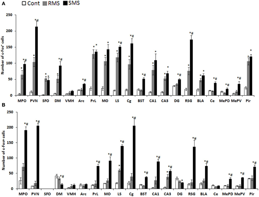
The expression of the immediate early gene product c-Fos is a reliable molecular marker to investigate neuronal activation. The examination of c-Fos expression has revealed that many brain regions are activated by MS, which differs depending on age and the type of stress. We recently analyzed the c-Fos expression induced by repeated MS and single-time MS during different developmental stages and time periods. Mice were exposed to 3 h repeated MS daily from PND1 to PND14 or from PND14 to PND21, or to single-time MS at PND14 or PND21 (Horii-Hayashi et al., 2013). We clarified that MS activated many brain regions and that c-Fos expression patterns changed developmentally (Figure 2). Single-time MS at both ages activated many regions of the hypothalamus and limbic forebrain, while the pattern of c-Fos expression in the repeated MS groups were significantly different on PND14 and PND21. In repeated MS of PND14 mice, the c-Fos expression levels in many regions were markedly increased compared with age-matched controls, excepting the VMH, Arc, BST, DG, Ce, MePV, and MePD. By contrast, in repeated MS on PND21 mice, c-Fos expression was reduced to control levels in all observed brain regions except for the LS and CA3. These findings suggest that repetition of a homotypic stimulus suppresses c-Fos expression by PND21, but that such suppression is barely observed on PND14. Moreover, in animals exposed to repeated homotypic stress during the postnatal period, increase in adrenal CORT secretion does not always associate with increased c-Fos expression in the PVN. Such developmental differences in c-Fos expression detected in the repeated MS groups may be associated with a developmental critical period for stress responses involving the HPA axis, during which animals are more susceptible to MS and other environments. In rodents, the critical period is the first two postnatal weeks. Thus, in early life, a repeated stress will be unlikely to suppress c-Fos expression. In turn, inappropriately activated c-Fos target genes may drastically alter how neurons function in critical neural circuits. Indeed, the suppression of increased c-Fos expression in repeated MS of PND14 mice was observed in specific regions (BST, Ce, MePD, and MePV) that form anatomical neural connections. These regions are referred to as an extended amygdala, which are closely associated with anxiety, fear, and psychiatric disorders (Davis et al., 2010). Therefore, even at PND14, repeated homotypic stress may reduce neural activity in the circuit of the extended amygdala. Moreover, in the SFO, where neurons are influenced by osmolality, calcium, and sodium concentrations in the systemic circulation (Smith and Ferguson, 2010), c-Fos expression was increased in both repeated and single-time MS mice, as compared to controls, on PND14. However, there were no changes in any of the groups on PND21. This difference may reflect the increased resistance of physical growth to the hyperosmolality induced by deprivation of lactation.
FIGURE 2  
http://www.frontiersin.org/files/Articles/87697/fnins-08-00166-HTML/image_m/fnins-08-00166-g002.jpg

Figure 2. c-Fos expression in the hypothalamus and limbic forebrain after MS (Horii-Hayashi et al., 2013). The graphs show the numbers of c-Fos-positive cells on PND14 (A) and PND21 (B) in non-separated control (white bar), RMS (gray bar), and SMS (black bar) mice (n = 4–5 for each group). In both RMS and SMS, the sampling point is just after MS procedure. *P < 0.05 vs. control; #P < 0.05 vs. RMS. MPO, medial preoptic area; PVN, paraventricular nucleus; SFO, subfornical organ; DM, dorsomedial hypothalamic nucleus; VMH, ventromedial hypothalamic nucleus; PrL, prelimbic cortex; MO, medial orbital cortex; LS, lateral septum; Cg, cingulate cortex; BST, bed nucleus of stria terminalis; CA1, hippocampal area CA1; CA3, hippocampal area CA3; DG, dentate gyrus; RSG, retrosplenial granular cortex; La, lateral amygdaloid nucleus; BLA, anterior part of the basolateral amygdaloid nucleus; Ce, central amygdaloid nucleus; MePD, posterodorsal part of the medial amygdaloid nucleus; MePV, posteroventral part of the medial amygdaloid nucleus; Pir, piriform cortex.

Behavioral Changes Induced by MS in Rodents

Early life adverse experiences including MS is one of the greatest contributing factors for mental health problems across life stages (Levine, 2005), relating not only to risk for mental health disorders but also to transdiagnostic features common in many psychological disorders (Glaser et al., 2006). I will introduce some of the behavioral aspects observed in animal model of MS.

Depression- and anxiety-like behaviors

Numerous studies have demonstrated a strong relationship between traumatic events during early life and development of behavioral abnormalities later in life. Early life adversity, such as that induced by MS, child physical, sexual, and emotional abuse, and general neglect has been linked to serious psychiatric impairment in adulthood (MacMillan et al., 2001). Particularly, a stressful life event such as early parental loss is associated with unipolar and bipolar depression, as well as anxiety disorders, beyond familial or genetic factors (Kendler et al., 1992; Agid et al., 1999; Furukawa et al., 1999; Heim and Nemeroff, 2001). Many human studies have reported that major depression and anxiety disorders are frequent in adults with a history of childhood abuse (Stein et al., 1996; Felitti et al., 1998). There have been numerous reports of the behavioral changes induced by MS in animal studies. Neonatal MS induces permanent alterations in the characteristics of the HPA response to stress in the offspring later in life (Ladd et al., 1996; Vazquez et al., 2000). Many studies of repeated MS during the first 2 weeks of neonatal life showed depression- and anxiety-like behaviors in adulthood (Newport et al., 2002; Daniels et al., 2004; Lee et al., 2007; Ryu et al., 2009). In these studies, ambulation and rearing decreased, immobility during a forced swim test increased, and time spent in the closed arms of an elevated plus maze increased.

Fear response

Until recently, no one had investigated how early experiences affected fear retention and extinction development, although these forms of emotional learning could be critically involved in the pathogenesis and treatment of mental health problems. Recent several studies showed that the timing of the maturation of fear learning is not set in static, but can be dynamically regulated by early experiences. Although the exact mechanisms are still unknown, when rats are reared under stressful conditions then they exhibit adult-like fear retention and extinction behaviors at an earlier stage of development (Callaghan et al., 2013). Chocyk et al. reported that MS decreased freezing time in both contextual and auditory fear conditioning in adolescent and adult rats (Chocyk et al., 2014). These results suggest that early life stress may permanently affect fear learning and memory.

Food intake and response to food deprivation

Previous studies showed that repeated MS during the first 2 weeks after birth may not permanently affect food intake and body weight gain of the offspring as long as the pups are reared in a group (Iwasaki et al., 2000; Kalinichev et al., 2002; Ryu et al., 2008). In contrast, post-weaning social isolation promotes food intake and weight gain of adolescent MS pups, with impacts on anxiety-like behaviors (Ryu et al., 2008). Anhedonia to palatable food, one of the major symptoms of depression, was reported in adolescent MS pups with disruption of the mesolimbic dopaminergic activity in response to stress (Noh et al., 2008). Another study showed that sustained hyperphagia observed in the MS pups subjected to a fasting/re-feeding cycle repeated during adolescent period of MS pups induced a binge-like eating disorder, in which increased activity of the HPA axis responding to such metabolic challenges appeared to play a role, at least partly, in mediation with the hypothalamic neuro peptide Y (NPY) (Jahng, 2011).

Gene Expression

Many animal studies, including MS, have improved our knowledge of gene-environment interactions and elucidated the pathways that program an animal in response to its early life experiences (Meaney and Szyf, 2005). Epigenetic mechanisms involving DNA methylation, post-translational modification of histone proteins and non-coding RNAs (most notably micro-RNA) are major candidates for regulating gene expression and integrating intrinsic and environmental signals in the genome (Jaenisch and Bird, 2003). Murgatroyd and colleagues showed that in the parvocellular subdivision of the paraventricular nucleus of the hypothalamus, MS in mice persistently upregulates Avp gene expression associated with reduced DNA methylation of a region in the Avp enhancer. This early life stress-responsive region serves as a binding site for the methyl-CpG binding protein 2, which in turn is regulated through neuronal activity. They also found that the ability of methyl-CpG binding protein 2 to control transcription of the Avp gene and induce DNA methylation occurred by recruiting components of the epigenetic machinery (Murgatroyd et al., 2009; Murgatroyd and Nephew, 2013). Other groups investigated DNA methylation levels at a specific sequence motif upstream of the GR gene (Nr3c1) in the hippocampus of offspring, and found that subjecting pups to a single 24 h MS increases methylation levels (Kember et al., 2012). The epigenetic alterations of these genes suggest that the HPA axis could be dysregulated by MS. Importantly, however, the DNA methylation differences were also often strain specific (Kember et al., 2012). Taken together, these findings demonstrate the importance of investigating environmental effects on a range of genetic backgrounds, emphasizing the need for the further examination of environmental, genetic, and epigenetic interactions.

Conclusions


Adverse environments and experiences during the neonatal period can dramatically affect the development of the HPA axis that underlies adaptive behavioral responses. MS experiments, as a model of early life stress, demonstrate that CORT levels and c-Fos expression change depending upon the different experimental conditions of MS, e.g., age at testing and frequency of repetition. Furthermore, separation conditions (isolation with or without a littermate) could also influence the results of the MS experiments. MS can induce various behavioral changes manifested in later life, which could be caused, at least in part, by alterations in gene expression, particularly through epigenetic mechanisms.

Conflict of Interest Statement

The authors declare that the research was conducted in the absence of any commercial or financial relationships that could be construed as a potential conflict of interest.

Acknowledgments

This work was supported by Grants-in-Aid for Scientific Research (23390040 to Mayumi Nishi and AstraZeneca Research Grant 2009). We thank Dr. Julian G. Mercer, a chief editor of J Neuroendocrinology, for permitting the reuse of our own figures published in J Neuroendocrinology.

The Limits of Logic: Simon Blackburn, Beatrix Campbell, and Iain McGilchrist


In this thought-provoking conversation (debate?) from iai.tv [Institute of Art and Ideas], Shahidha Bari asks Cambridge philosopher and author of Think: A Compelling Introduction to Philosophy, Simon Blackburn, psychiatrist and author of The Master and His Emissary: The Divided Brain and the Making of the Western World, Iain McGilchrist [also author of The Divided Brain and the Search for Meaning, a $0.99 Kindle only essay companion to the book], and radical journalist Beatrix Campbell, whether we should embrace the irrational.

I'm not sure I agree with the premise, which would be that we are or we want to be rational beings. Mostly, we are irrational (emotional) beings who create elaborate rationalizations for our irrational beliefs and/or behaviors.

If that is the premise, I am not sure that we should so much embrace the irrational as we should seek to bring into the light of rational thought the non-rational/irrational beliefs that inform, influence, and motivate our behaviors.

Still, these are always interesting discussions.

The Limits of Logic

Simon Blackburn, Beatrix Campbell, Iain McGilchrist. Hosted by Shahidha Bari.


Logicians don't rule the world or get the most done. Could it be that a consistent world view is neither desirable nor achievable? If we abandon the straightjacket of rationality might this lead to a more powerful and exciting future, or is it a heresy that leads to madness?

The Panel: Shahidha Bari asks Cambridge philosopher and author of Think, Simon Blackburn, psychiatrist and author of The Master and His Emissary, Iain McGilchrist, and radical journalist Beatrix Campbell, whether we should embrace the irrational.
In association with:

Thursday, June 19, 2014

Jeremy O'Brien: "Quantum Technologies"


Jeremy O'Brien spoke recently at Google on Quantum Technologies, a topic he has written on extensively [see his 2009 paper, with Furusawa and Vu ckovi c, Photonic Quantum Technologies]. This is interesting stuff - and likely to be the future of computing technology.

Jeremy O'Brien: "Quantum Technologies"

June 17, 2014


Jeremy O'Brien visited Google LA to deliver a talk: "Quantum Technologies." This talk took place on April 1, 2014.

Abstract:

The impact of quantum technology will be profound and far-reaching: secure communication networks for consumers, corporations and government; precision sensors for biomedical technology and environmental monitoring; quantum simulators for the design of new materials, pharmaceuticals and clean energy devices; and ultra-powerful quantum computers for addressing otherwise impossibly large datasets for machine learning-artificial intelligence applications. However, engineering quantum systems and controlling them is an immense technological challenge: they are inherently fragile; and information extracted from a quantum system necessarily disturbs the system itself. Despite these challenges a small number of quantum technologies are now commercially available. Delivering the full promise of these technologies will require a concerted quantum engineering effort jointly between academia and industry. We will describe our progress in the Centre for Quantum Photonics to delivering this promise using an integrated quantum photonics platform---generating, manipulating and interacting single particles of light (photons) in waveguide circuits on silicon chips.

Bio:

Jeremy O'Brien is professor of physics and electrical engineering and director of the Centre for Quantum Photonics (CQP). He received his Ph.D. in physics from the University of New South Wales in 2002 for experimental work on correlated and confined electrons in organic conductors, superconductors and semiconductor nanostructures, as well as progress towards the fabrication of a phosphorus in silicon quantum computer. As a research fellow at the University of Queensland (2001-2006) he worked on quantum optics and quantum information science with single photons. CQP's efforts are focused on the fundamental and applied quantum mechanics at the heart of quantum information science and technology, ranging from prototypes for scalable quantum computing to generalised quantum measurements, quantum control, and quantum metrology.

"Journal of Religion and Health" Publishes Paper Blaming Schizophrenia on Demons


WTF?!

I shouldn't be surprised - or maybe I am surprised that a "real" journal published this crap.

But, then, I've had religious fundamentalists tell people with dissociative identity disorder that the parts are demons and if they simply reject Satan and embrace Jesus everything will be fine.

This is from Real Clear Science.

Published Paper Blames Schizophrenia on Demons


Posted by Ross Pomeroy June 17, 2014


IS SCHIZOPHRENIA CAUSED by demons? A Turkish researcher seems to think so, and his article on the topic was just published in the Journal of Religion and Health, a scientific journal owned by Springer, a German-based publishing company.

The first two-thirds of M. Kemal Irmak's paper, "Schizophrenia or Possession?", read normally enough. You learn about the devastating symptoms of schizophrenia, current treatment approaches, and the nature of the delusions and hallucinations that schizophrenics experience. And then you arrive at this little doozy:
"One approach to this hallucination problem is to consider the possibility of a demonic world."
The abrupt transition from established science to outlandish woo is positively comical. And once the quackery starts, it doesn't stop. You're first treated to a background on all things demonic (boldness added to emphasize the absurdity):
In our region, demons are believed to be intelligent and unseen creatures that occupy a parallel world to that of mankind. In many aspects of their world, they are very similar to us. They marry, have children, and die. The life span, however, is far greater than ours (Ashour 1989). Through their powers of flying and invisibility, they are the chief component in occult activities. The ability to possess and take over the minds and bodies of humans is also a power which the demons have utilized greatly over the centuries (Littlewood 2004; Gadit and Callanan 2006; Ally and Laher 2008). Most scholars accept that demons can possess people and can take up physical space within a human’s body (Asch 1985). They possess people for many reasons. Sometimes it is because they have been hurt accidentally, but possession may also occur because of love (Ashour 1989; Philips 1997). When the demon enters the human body, they settle in the control center of the body–brain.
Once the groundwork for demons is laid, Irmak expounds on the link between schizophrenia and possession:
There exist similarities between the clinical symptoms of schizophrenia and demonic possession. Common symptoms in schizophrenia and demonic possession such as hallucinations and delusions may be a result of the fact that demons in the vicinity of the brain may form the symptoms of schizophrenia... The hallucination in schizophrenia may therefore be an illusion—a false interpretation of a real sensory image formed by demons... On the other hand, auditory hallucinations expressed as voices arguing with one another and talking to the patient in the third person may be a result of the presence of more than one demon in the body.
Irmak then describes a personal anecdote about a local faith healer in Turkey who employs "good" demons to expel evil ones. Based on this experience, Irmak concludes:
Above considerations have led to the suggestion that it is time for medical professions to consider the possibility of demonic possession in the etiology of schizophrenia, especially in the cases with hallucinations and delusions. Therefore, it would be useful for medical professions to work together with faith healers to define better treatment pathways for schizophrenia.
THE EDITOR OF the Journal of Religion and Health is Dr. Curtis Hart, a lecturer in public health at Weill Cornell Medical College. RealClearScience reached out to him for comment.
"The article was published in hopes that it would provoke discussion," he said. "The Journal does not agree that demons are a real entity."
There are currently no plans to retract the paper, but two rebuttals are already slated for a future issue, Curtis added. The journal's publisher, Springer, recently made headlines by withdrawing 16 gibberish papers spotted by an independent computer scientist. The nonsense papers were created with a computer program, SciGen.

RCS gave Irmak a chance to defend his paper.
"There is no scientific evidence to support the existence of demons," he admitted. "This is like the argument of creation or evolution. It is a matter of belief and I think the existence of demons cannot be proved by scientific methods."
For the record, there is an overwhelming amount of scientific evidence in support of evolution, which is not at all a matter of belief.

Irmak also insisted that readers of his paper watch the Academy Award-winning film A Beautiful Mind, which chronicles the life of genius mathematician John Nash, who suffers from schizophrenia.

"I think the creatures who disturb John Nash are demons," he said.

SCHIZOPHRENIA IS A debilitating brain disorder characterized by hallucinations, confused thinking, and abnormal social behavior. Sufferers often struggle to recognize what is real and what isn't. Its causes are still up for debate, but most experts believe that it's tied to an imbalance in brain chemistry and is -- to a degree -- genetically inherited.

One thing for certain, however, is that schizophrenia is not caused by demonic possession. Any "scientific" journal that posits otherwise lends credibility to every witch doctor or religious fanatic who has attempted to exorcise demons from patients with serious mental illness. The world needs less magic and more evidence-based medicine.

(Image: Shutterstock)

Wednesday, June 18, 2014

Tom Ireland - What Does Mindfulness Meditation Do to Your Brain?


Via Scientific American, an article from Tom Ireland on the ways mindfulness meditation changes the brain, not least of which is shrinking the amygdala (fear and stress center) and thickening the prefrontal cortex (executive function).

What Does Mindfulness Meditation Do to Your Brain?

By Tom Ireland | June 12, 2014

The views expressed are those of the author and are not necessarily those of Scientific American.

As you read this, wiggle your toes. Feel the way they push against your shoes, and the weight of your feet on the floor. Really think about what your feet feel like right now – their heaviness.

If you’ve never heard of mindfulness meditation, congratulations, you’ve just done a few moments of it. More people than ever are doing some form of this stress-busting meditation, and researchers are discovering it has some quite extraordinary effects on the brains of those who do it regularly.

Originally an ancient Buddhist meditation technique, in recent years mindfulness has evolved into a range of secular therapies and courses, most of them focused on being aware of the present moment and simply noticing feelings and thoughts as they come and go.


Credit: Sebastien Wiertz via Flickr

It’s been accepted as a useful therapy for anxiety and depression for around a decade, and mindfulness websites like GetSomeHeadSpace.com are attracting millions of subscribers. It’s being explored by schools, pro sports teams and military units to enhance performance, and is showing promise as a way of helping sufferers of chronic pain, addiction and tinnitus, too. There is even some evidence that mindfulness can help with the symptoms of certain physical conditions, such as irritable bowel syndrome, cancer, and HIV.

Yet until recently little was known about how a few hours of quiet reflection each week could lead to such an intriguing range of mental and physical effects. Now, as the popularity of mindfulness grows, brain imaging techniques are revealing that this ancient practice can profoundly change the way different regions of the brain communicate with each other – and therefore how we think – permanently.


Mindfulness practice and expertise is associated with a decreased volume of grey matter in the amygdala (red), a key stress-responding region. (Image courtesy of Adrienne Taren)

No fear

MRI scans show that after an eight-week course of mindfulness practice, the brain’s “fight or flight” center, the amygdala, appears to shrink. This primal region of the brain, associated with fear and emotion, is involved in the initiation of the body’s response to stress.

As the amygdala shrinks, the pre-frontal cortex – associated with higher order brain functions such as awareness, concentration and decision-making – becomes thicker.

The “functional connectivity” between these regions – i.e. how often they are activated together – also changes. The connection between the amygdala and the rest of the brain gets weaker, while the connections between areas associated with attention and concentration get stronger.

The scale of these changes correlate with the number of hours of meditation practice a person has done, says Adrienne Taren, a researcher studying mindfulness at the University of Pittsburgh.

“The picture we have is that mindfulness practice increases one’s ability to recruit higher order, pre-frontal cortex regions in order to down-regulate lower-order brain activity,” she says.

In other words, our more primal responses to stress seem to be superseded by more thoughtful ones.

Lots of activities can boost the size of various parts of the pre-frontal cortex – video games, for example – but it’s the disconnection of our mind from its “stress center” that seems to give rise to a range of physical as well as mental health benefits, says Taren.

“I’m definitely not saying mindfulness can cure HIV or prevent heart disease. But we do see a reduction in biomarkers of stress and inflammation. Markers like C-reactive proteins, interleukin 6 and cortisol – all of which are associated with disease.”

Feel the pain

Things get even more interesting when researchers study mindfulness experts experiencing pain. Advanced meditators report feeling significantly less pain than non-meditators. Yet scans of their brains show slightly more activity in areas associated with pain than the non-meditators.

“It doesn’t fit any of the classic models of pain relief, including drugs, where we see less activity in these areas,” says Joshua Grant, a postdoc at the Max Plank Institute for Human Cognitive and Brain Sciences in Leipzig, Germany. The expert mindfulness meditators also showed “massive” reductions in activity in regions involved in appraising stimuli, emotion and memory, says Grant.

Again, two regions that are normally functionally connected, the anterior cingulate cortex (associated with the unpleasantness of pain) and parts of the prefrontal cortex, appear to become “uncoupled” in meditators.

“It seems Zen practitioners were able to remove or lessen the aversiveness of the stimulation – and thus the stressing nature of it – by altering the connectivity between two brain regions which are normally communicating with one another,” says Grant. “They certainly don’t seem to have blocked the experience. Rather, it seems they refrained from engaging in thought processes that make it painful.”


Credit: Balint Földesi via Flickr

Feeling Zen

It’s worth noting that although this study tested expert meditators, they were not in a meditative state – the pain-lessening effect is not something you have to work yourself up into a trance to achieve; instead, it seems to be a permanent change in their perception.

“We asked them specifically not to meditate,” says Grant. “There is just a huge difference in their brains. There is no question expert meditators’ baseline states are different.”

Other studies on expert meditators – that is, subjects with at least 40,000 hours of mindfulness practice under their belt – discovered that their resting brain looks similar, when scanned, to the way a normal person’s does when he or she is meditating.

At this level of expertise, the pre-frontal cortex is no longer bigger than expected. In fact, its size and activity start to decrease again, says Taren. “It’s as if that way of thinking has becomes the default, it is automatic – it doesn’t require any concentration.”

There’s still much to discover, especially in terms of what is happening when the brain comprehends the present moment, and what other effects mindfulness might have on people. Research on the technique is still in its infancy, and the imprecision of brain imaging means researchers have to make assumptions about what different regions of the brain are doing.

Both Grant and Taren, and others, are in the middle of large, unprecedented studies that aim to isolate the effects of mindfulness from other methods of stress-relief, and track exactly how the brain changes over a long period of meditation practice.

“I’m really excited about the effects of mindfulness,” says Taren. “It’s been great to see it move away from being a spiritual thing towards proper science and clinical evidence, as stress is a huge problem and has a huge impact on many people’s health. Being able to take time out and focus our mind is increasingly important.”

Perhaps it is the new age, quasi-spiritual connotations of meditation that have so far prevented mindfulness from being hailed as an antidote to our increasingly frantic world. Research is helping overcome this perception, and ten minutes of mindfulness could soon become an accepted, stress-busting part of our daily health regimen, just like going to the gym or brushing our teeth.


About the Author: Tom Ireland is managing editor at the Society of Biology and a freelance journalist covering mostly health, education and science. Follow on Twitter @Tom_J_Ireland.

The views expressed are those of the author and are not necessarily those of Scientific American.

Neuron Tells Stem Cells to Grow New Neurons


Interesting new research out of Duke University on how specific neurons tell neuronal stem cells where to go to repair damage. Below this press release is the abstract and citation from Nature Neuroscience (where the article is, of course, embargoed).

Neuron Tells Stem Cells to Grow New Neurons

Researchers identify first piece of new brain-repair circuit

June 2, 2014 | By Karl Leif Bates


In this artist's representation of the adult subependymal neurogenic niche (viewed from underneath the ependyma), electrical signals generated by the ChAT+ neuron give rise to newborn migrating neuroblasts, seen moving over the underside of ependymal cells. Illustration by O’Reilly Science Art.

Durham, NC - Duke researchers have found a new type of neuron in the adult brain that is capable of telling stem cells to make more new neurons. Though the experiments are in their early stages, the finding opens the tantalizing possibility that the brain may be able to repair itself from within.

Neuroscientists have suspected for some time that the brain has some capacity to direct the manufacturing of new neurons, but it was difficult to determine where these instructions are coming from, explains Chay Kuo, M.D. Ph.D., an assistant professor of cell biology, neurobiology and pediatrics.

In a study with mice, his team found a previously unknown population of neurons within the subventricular zone (SVZ) neurogenic niche of the adult brain, adjacent to the striatum. These neurons expressed the choline acetyltransferase (ChAT) enzyme, which is required to make the neurotransmitter acetylcholine. With optogenetic tools that allowed the team to tune the firing frequency of these ChAT+ neurons up and down with laser light, they were able to see clear changes in neural stem cell proliferation in the brain.

The findings appeared as an advance online publication June 1 in the journal Nature Neuroscience.

The mature ChAT+ neuron population is just one part of an undescribed neural circuit that apparently talks to stem cells and tells them to increase new neuron production, Kuo said. Researchers don't know all the parts of the circuit yet, nor the code it's using, but by controlling ChAT+ neurons' signals Kuo and his Duke colleagues have established that these neurons are necessary and sufficient to control the production of new neurons from the SVZ niche.

"We have been working to determine how neurogenesis is sustained in the adult brain. It is very unexpected and exciting to uncover this hidden gateway, a neural circuit that can directly instruct the stem cells to make more immature neurons," said Kuo, who is also the George W. Brumley, Jr. M.D. assistant professor of developmental biology and a member of the Duke Institute for Brain Sciences. "It has been this fascinating treasure hunt that appeared to dead-end on multiple occasions!"

Kuo said this project was initiated more than five years ago when lead author Patricia Paez-Gonzalez, a postdoctoral fellow, came across neuronal processes contacting neural stem cells while studying how the SVZ niche was assembled.

The young neurons produced by these signals were destined for the olfactory bulb in rodents, as the mouse has a large amount of its brain devoted to process the sense of smell and needs these new neurons to support learning. But in humans, with a much less impressive olfactory bulb, Kuo said it's possible new neurons are produced for other brain regions. One such region may be the striatum, which mediates motor and cognitive controls between the cortex and the complex basal ganglia.

"The brain gives up prime real estate around the lateral ventricles for the SVZ niche housing these stem cells," Kuo said. "Is it some kind of factory taking orders?" Postdoctoral fellow Brent Asrican made a key observation that orders from the novel ChAT+ neurons were heard clearly by SVZ stem cells.

Studies of stroke injury in rodents have noted SVZ cells apparently migrating into the neighboring striatum. And just last month in the journal Cell, a Swedish team observed newly made control neurons called interneurons in the human striatum for the first time. They reported that interestingly in Huntington's disease patients, this area seems to lack the newborn interneurons.

"This is a very important and relevant cell population that is controlling those stem cells," said Sally Temple, director of the Neural Stem Cell Institute of Rensselaer, NY, who was not involved in this research. "It's really interesting to see how innervations are coming into play now in the subventricular zone."

Kuo's team found this system by following cholinergic signaling, but other groups are arriving in the same niche by following dopaminergic and serotonergic signals, Temple said. "It's a really hot area because it's a beautiful stem cell niche to study. It's this gorgeous niche where you can observe cell-to-cell interactions."

These emerging threads have Kuo hopeful researchers will eventually be able to find the way to "engage certain circuits of the brain to lead to a hardware upgrade. Wouldn't it be nice if you could upgrade the brain hardware to keep up with the new software?" He said perhaps there will be a way to combine behavioral therapy and stem cell treatments after a brain injury to rebuild some of the damage.

The questions ahead are both upstream from the new ChAT+ neurons and downstream, Kuo says. Upstream, what brain signals tell ChAT+ neurons to start asking the stem cells for more young neurons? Downstream, what's the logic governing the response of the stem cells to different frequencies of ChAT+ electrical activity?

There's also the big issue of somehow being able to introduce new components into an existing neuronal circuit, a practice that parts of the brain might normally resist. "I think that some neural circuits welcome new members, and some don't," Kuo said.

In addition to Paez-Gonzalez, Asrican, and Kuo, Erica Rodriguez, a graduate student in the neurobiology training program, is also an author. This research was supported by the National Institutes of Health, David & Lucile Packard Foundation, and George Brumley Jr. Endowment.

More Information
Contact: Karl Leif Bates
Phone: (919) 681-8054
Email: karl.bates@duke.edu © 2014 Office of News & Communications
615 Chapel Drive, Box 90563, Durham, NC 27708-0563 | (919) 684-2823
* * * * *

Full Citation:
Paez-Gonzalez, P, Asrican, B, Rodriguez, E, and Kuo, CT, (2014, June 1). Identification of distinct ChAT+ neurons and activity-dependent control of postnatal SVZ neurogenesis. Nature Neuroscience; doi:10.1038/nn.3734 - ePub ahead of print

Identification of distinct ChAT+ neurons and activity-dependent control of postnatal SVZ neurogenesis

Patricia Paez-Gonzalez, Brent Asrican, Erica Rodriguez & Chay T Kuo

Abstract

Postnatal and adult subventricular zone (SVZ) neurogenesis is believed to be primarily controlled by neural stem cell (NSC)-intrinsic mechanisms, interacting with extracellular and niche-driven cues. Although behavioral experiments and disease states have suggested possibilities for higher level inputs, it is unknown whether neural activity patterns from discrete circuits can directly regulate SVZ neurogenesis. We identified a previously unknown population of choline acetyltransferase (ChAT)+ neurons residing in the rodent SVZ neurogenic niche. These neurons showed morphological and functional differences from neighboring striatal counterparts and released acetylcholine locally in an activity-dependent fashion. Optogenetic inhibition and stimulation of subependymal ChAT+ neurons in vivo indicated that they were necessary and sufficient to control neurogenic proliferation. Furthermore, whole-cell recordings and biochemical experiments revealed direct SVZ NSC responses to local acetylcholine release, synergizing with fibroblast growth factor receptor activation to increase neuroblast production. These results reveal an unknown gateway connecting SVZ neurogenesis to neuronal activity-dependent control and suggest possibilities for modulating neuroregenerative capacities in health and disease.

Claude Steele - Stereotype Threat: How It Affects Us and What We Can Do About It


Claude Steele is the author of Whistling Vivaldi: How Stereotypes Affect Us and What We Can Do (Issues of Our Time) (2010), a book that escaped my attention but seems to offer some important insights into how stereotypes narrow our experience of the world.
The acclaimed social psychologist offers an insider’s look at his research and groundbreaking findings on stereotypes and identity.

Claude M. Steele, who has been called “one of the few great social psychologists,” offers a vivid first-person account of the research that supports his groundbreaking conclusions on stereotypes and identity. He sheds new light on American social phenomena from racial and gender gaps in test scores to the belief in the superior athletic prowess of black men, and lays out a plan for mitigating these “stereotype threats” and reshaping American identities.
Below the talk given at UC Berkeley is a longer review of the book from the Harvard Education Review.

Claude Steele - Stereotype Threat: How It Affects Us and What We Can Do About It

Published on June 17, 2014


Claude Steele, internationally renowned social scientist and Executive Vice Chancellor and Provost, will discuss his theory of stereotype threat, which has been the focus of much of his research and writing throughout his academic career. The theory examines how people from different groups, being threatened by different stereotypes, can have quite different experiences in the same situation. It has also been used to understand group differences in performance ranging from the intellectual to the athletic. Steele's recent book, "Whistling Vivaldi: And Other Clues to How Stereotypes Affect Us and what we Can Do," published in 2010, was based on this research and lays out a plan to mitigate the negative effects of "stereotype threat."
* * * * *

 From the Harvard Education Review:

Whistling Vivaldi: And Other Clues to How Stereotypes Affect Us

Claude M. Steele
New York: W. W. Norton, 2010. 

242 pp. $25.95. 
 
Why are students of color not graduating from college at the same rate as white students? Why might white students be reluctant to take courses with a substantial number of students of color in them? What can educators do to address these problems?
In his new book, Whistling Vivaldi: And Other Clues to How Stereotypes Affect Us, social psychologist Claude Steele helps us find answers to these questions based on findings from social psychology experiments. Steele’s book sets forth an argument for understanding how contextual factors—not individual characteristics or personal beliefs motivated by prejudice or malice—help explain so-termed “racial achievement gaps” in education and ongoing societal racial and ethnic segregation.

In an accessible, page-turning account written for a general audience, Steele explains how identity contingencies—the conditions that a given social identity forces us to face and overcome in a particular setting—affect our everyday behavior and perpetuate broader societal problems. Expanding on his prior work, he focuses on a specific type of identity contingency: stereotype threat, or the fear of what people could think about us solely because of our race, gender, age, etc. An African American male walking down the street at night, for example, faces the threat of being seen as potentially violent. Steele recounts how, to deflect this stereotype threat, African American New York Times writer Brent Staples whistled Vivaldi while walking the streets of Hyde Park at night to signal to white people that he was educated and nonviolent. Another example of stereotype threat would be a white student in a class that is predominantly nonwhite facing the threat of being perceived as racist. Steele explains how such threats follow us like a “cloud.”

Steele summarizes research findings that show how the concerns students face as a result of these stereotype threats affect a wide range of educational outcomes. He explains how the threat of a stereotype and the extra efforts required of students who try to dispel it interfere with academic performance. The additional stress and anxiety, which can operate without awareness, can lead to underperformance in the classroom or on standardized tests relative to ability. Stereotype threat can also undermine feelings of belonging, competence, and aspiration. Importantly, Steele explains how contextual cues, such as being in an environment where there are few students or faculty of color, or where the curriculum marginalizes the experiences of students of color, are enough to trigger a stereotype threat that undermines performance.

Steele offers practices educators can use to help counteract these messages. For instance, self-affirmation exercises in the classroom, particularly for students of color, can be enough to counter negative messages that trigger stereotype threat. Some other practices include emphasizing incremental views of intelligence (i.e., intelligence as an expandable as opposed to fixed characteristic) and facilitating faculty-to-student or student-to-student mentoring and cross-racial interactions. Steele’s insights are so helpful that I was disappointed when he relegated some other important discussions to footnotes, such as when he outlines how findings about the effects of stereotype threat call into question past research that suggests families, not schools, are responsible for the achievement gap.

In the later chapters of the book, Steele focuses on how identity threats influence interracial interactions more broadly. He explains how our actions, conscious or not, contribute to persistent racial segregation as, understandably, each of us may retreat to the safety of a more homogeneous environment that does not trigger the risk of a stereotype threat. But Steele’s outlook is hopeful: the factors that contribute to our living segregated lives also have the potential to help us bridge our differences. We are all affected by identity threats, and awareness of this commonality should help us empathize with the experience of others.

Overall, Steele provides strong evidence demonstrating how situational cues affect student performance, and educators can benefit from the practical implications of his research in their efforts to remedy racial inequities in education. While the importance of addressing structural factors should not be overlooked, simple institutional practices can counter the otherwise powerful cues that trigger stereotype threat for students of color. The findings presented in this book unearth the powerful and prevalent ways in which group identity affects us all and demonstrate the need to acknowledge this fact: we need to be “identity conscious” if we are going to improve race relations across
our society.

l.m.g.

Tuesday, June 17, 2014

Documentary - Stuck in Traffick (Child Sex Trafficking from Survivors' Perspective)

I don't like "trigger warnings" in general, but this feels like an exception. This documentary is likely to be triggering to anyone who has been through childhood sexual abuse. Please be aware of your own feelings and avoid this if necessary.

This documentary is based in Phoenix, AZ, a couple of hours north of Tucson. In Arizona, child sex trafficking in the fastest growing criminal activity, as it is in the U.S. and the rest of the world (it is the 2nd largest criminal activity in the world now).

Definition of ST.jpg

Part of the problem in AZ is that the drug cartels, who straddle the U.S.-Mexico border, have realized that they can only sell drugs once, but they can sell a child over and over and over again.

According to Sold No More, a Tucson-based anti-trafficking group:

Trafficking FAQs

  • Human trafficking is now the 2nd largest and fastest growing illegal trafficking activity in the world.  (United Nations Office on Drugs and Crime, 2008)
  • The annual market value of human trafficking is $32+ billion. (Ibid.)  80% of victims  are women and 50% are children.  (Trafficking in Persons Report 2007, U.S. Department of State)
  • Globally, one million children are forced to work in the sex industry every year.   (Child Exploitation and Obscenity Section, 2007)
  • Among the millions trafficked each year, hundreds of thousands are teenage girls, some as young as five years of age. (Ibid)
  • In the U.S., handlers, “pimps,” can make $100,000 a year, per child. 
  • Child pornography is a multi-billion dollar industry and among the fastest growing criminal segments on the Internet.  Child pornography fuels the child sex trade.   (National Center for Missing and Exploited Children)
  • 450,000 children run away from home each year in the U.S..  A third of those lured into sex slavery are taken within 48 hours of leaving home. (Child Exploitation and Obscenity Section, 2007)
  • The average age a child is first commercially sexually exploited in the U.S. is 13 years of age.  (Ibid)
  • Fewer than 1,000 minor sex trafficking victims in the U.S. have been assisted by  federal, state, and local law enforcement agencies since 2001.  (Ibid)
  • 10% of trafficked children were kidnapped.  (Karla Dial, “Reaching Into the Dark,” Citizen Magazine, March 2010, 5)
Here is a striking graphic of the situation:

Infograph Web.jpg

This is tough to watch - but awareness breeds action. We need to put an end to this form of slavery.

Stuck in Traffick


Prostitute. Hooker. Floozy. Call girl. Lady of the Night. Streetwalker.

Society stamps a negative label on the women of this dangerous criminal industry. However, a fine line must be drawn between those women doing it by choice and the victims who have no choice.

Sex trafficking is the fastest growing criminal industry in the world. There are more than 100,000 minors forced into the sex trade every year in the U.S., with an average age of only 13. They are first recruited, forced, coerced, or abducted by a pimp or his workers. Then they are beaten, raped, brainwashed, and treated as commodities. Some of these victimized women can bring in $15,000 a week. The pimp usually receives every penny.

There are more slaves today than at any other time in our history. Slavery in the form of sex trafficking is among us in a hidden and quiet fashion. It’s in our neighborhoods and in our malls. It’s in our suburbs, it's leaking into our schools, and it's all over the Internet. Education and awareness are the best tools we have at stopping this heinous crime.

Stuck in Traffick is a documentary that brings light to the underground world of sex trafficking and what can be done to help these victims.