Showing posts with label cortisol. Show all posts

Friday, June 20, 2014

Effects of Early Life Adverse Experiences on the Brain: Implications from Maternal Separation Models in Rodents

Adverse Childhood Experiences (ACE) Study Pyramid

In the past, I have posted quite a few time about how adverse childhood experiences (ACE) can have a profound impact on physical and mental health (see here, here, here, here, here, and here, for example). This new study looks at how maternal separation in a mouse model impacts brain development.

It' easy to dismiss research like this because it involves mice, not humans. First of all, we could never do this research with humans - it's not ethical. More importantly, however, nearly all mammals hve similar parent/offspring bonding drives and needs, so for the first few months of life, we can model these behaviors in rodents with relative assurance that they will at least partially translate to humans.

In this particular mini-review, the researchers are seeking a molecular explanation of how early life stress impacts brain development and dysfunction (hint: stress hormones).

Full Citation: 
Nishi, M., Horii-Hayashi, N. and Sasagawa, T. (2014, Jun 17). Effects of early life adverse experiences on the brain: implications from maternal separation models in rodents. Frontiers in Neuroscience: Neuroendocrine Science; 8:166. doi: 10.3389/fnins.2014.00166

Effects of early life adverse experiences on the brain: implications from maternal separation models in rodents

Mayumi Nishi, Noriko Horii-Hayashi and Takayo Sasagawa
  • Department of Anatomy and Cell Biology, Nara Medical University, Kashihara, Japan
During postnatal development, adverse early life experiences affect the formation of neuronal networks and exert long-lasting effects on neural function. Many studies have shown that daily repeated maternal separation (MS), an animal model of early life stress, can regulate the hypothalamic-pituitary-adrenal axis (HPA axis) and affect subsequent brain function and behavior during adulthood. However, the molecular basis of the long-lasting effects of early life stress on brain function has not been fully elucidated. In this mini review, we present various cases of MS in rodents and illustrate the alterations in HPA axis activity by focusing on corticosterone (CORT). We then show a characterization of the brain regions affected by various patterns of MS, including repeated MS and single time MS at various stages before weaning, by investigating c-Fos expression. These CORT and c-Fos studies suggest that repeated early life stress may affect neuronal function in region- and temporal-specific manners, indicating a critical period for habituation to early life stress. Next, we introduce how early life stress can impact behavior, namely by inducing depression, anxiety or eating disorders, and alterations in gene expression in adult mice subjected to MS.

Introduction


As our contemporary society changes rapidly, changes in family structure can have a large influence on the mother–child relationship, as well as on other social environmental factors. In adult patients with various neuropsychiatric disorders, childhood abuse including sexual and/or physical abuse and neglect, is one of the most serious causes (Bremne and Vermetten, 2001; Heim and Nemeroff, 2001; Teicher et al., 2006). Adverse experiences occurring during critical periods of development, such as perinatal life, harmfully influence behavior, and physiological functions, including growth, metabolism, reproduction, and immune responses. Stressful environments in early life may induce permanent rather than transient consequences in animals. Previous studies have indicated that early unfavorable events augment the risk of behavioral disorders in adulthood, including neuropsychiatric disorders, such as depression (Kendler et al., 2002) and psychosis (Morgan et al., 2007). In rodent and primate models, adverse environments during the neonatal periods seem to play a critical role in developing the brain systems important to regulate behavior and stress responsiveness. In particular, the responsiveness of the hypothalamic-pituitary-adrenal (HPA) axis can be deteriorated by interrupting usual mother-pup interactions, which may induce persistent changes in the neurobiology, physiology, and emotional behavior in adult animals (Ellenbroek et al., 1998; Lyons et al., 1998; Pryce et al., 2005; Enthoven et al., 2008; Nishi et al., 2013).

In this mini review, we will focus on the response of corticosterone (CORT), an end product of the HPA axis in rodents, and c-Fos expression for examining the activated brain regions induced by maternal separation (MS), a model of rodent early life stress. Furthermore, we will also present alterations of behavioral aspects and alterations in gene expression.

Early MS


The inventive studies of Levine and colleagues, and consequently of Meaney, Plotsky, and their collaborators have demonstrated that changes in rodents' early postnatal experiences can induce profound long-lasting effects on emotionality and stress response (Levine, 1967; Meaney, 2001; Plotsky et al., 2005), which have spurred the employment of the rodent MS for investigating early life stress. This early life stress model is based on the evidence that unfavorable events in early life cause the vulnerability for developing various kinds of diseases in later life. In this type of study, MS should be carefully discussed in comparison to the appropriate control group, which may or may not be undisturbed from mother.

The procedure of MS showed a variety of the duration (e.g., 60 min–24 h) and the number of days (e.g., 1–14 days, 15–21 days) for the separation experiences among laboratories (Biagini et al., 1998; Caldji et al., 2000; Barreau et al., 2004; Arborelius and Eklund, 2007; Carrera et al., 2009; Tjong et al., 2010). In MS paradigm, many experiments, but certainly not all, have demonstrated that separation of pups from their mothers during the early postnatal period permanently increased anxiety-like behaviors in adulthood (Francis et al., 1999; Huot et al., 2001, 2004; Menard et al., 2004). As to the HPA axis activity, the response to stress is relatively low during early postnatal life (Walker et al., 1991; Levine, 2005), while MS could lead to life-long hyperactivity of the HPA axis (Holmes et al., 2005; Lippmann et al., 2007; Aisa et al., 2008; Marais et al., 2008). In contrast, short-term disturbance (e.g., 15 min), which has been called “handling,” appeared to reduce anxiety-like behaviors, decrease HPA axis tone and reduce the response to stress in adulthood (Levine, 2005; Plotsky et al., 2005). The process of handling may imitate natural mice rearing, whereby the mother leaves her pups for short periods of time to collect foods. Thus, the short-term MS, handling, might be considered a more natural event.

The effect of MS also varies depending upon whether pups are separated in a group of littermates during MS or isolated singly. Miyazaki and colleagues recently reported that rat pups isolated singly from the mother during PND7 to PND11 presented disturbance of cortical function, whereas pups separated but gathered from PND7 to PND11 showed no cortical disruption (Miyazaki et al., 2012).

Characterization of Maternally Separated Animals


Serum Level of CORT

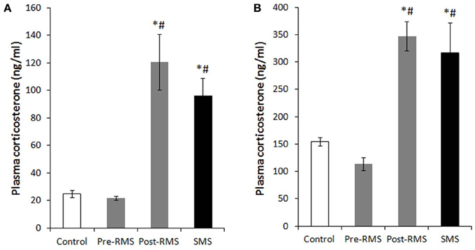
In rodents, there is an unique period during which the HPA axis shows a rapid regression known as the stress hyporesponsive period (SHRP) (Levine, 2001). This period extends from PND4 to PND14 in rats and from PND2 to PND12 in mice. During the course of SHRP, ACTH in increased and baseline plasma glucocorticoid levels are lower than normal (Rosenfeld et al., 1991). Because, during ontogeny, the maintenance of low and stable levels of CORT is necessary for normal growth and development of the central nervous system (CNS), the SHRP is hypothesized to be neuroprotective against stress-induced excessive stimulation of glucocorticoid receptors (GRs) (Sapolsky and Meaney, 1986; Sapolsky, 1996). In rodents, the presence of the mother appears to suppress HPA axis activity, which primarily preserves the SHRP. Indeed, even during the SHRP, MS is a compelling inducer of a stress response. Meaney and his colleagues suggest that the quality of the mother-pup interactions, such as increased maternal licking, grooming, and arched-back nursing, is an important aspect for the preservation of this dampened HPA axis activity (Francis et al., 1999). The disturbance of SHRP induced by MS could cause an excessive exposure of the brain to high concentrations of glucocorticoids and activation of GRs, which may subsequently regulate brain and behavior in later life. Enhanced secretion of stress-induced CORT was observed in pups separated from their mothers for 1 h on PND2 to PND9 (McCormick et al., 1998). Nevertheless, a recent study indicated that repeated MS for 8 h daily from PND3 to PND5 rapidly desensitized the HPA axis activity of neonatal mice (Enthoven et al., 2008). We also reported that repeated MS for 3 h daily from PND1 to PND14 did not elevate a baseline level of CORT on PND14, whereas a single-time MS for 3 h at PND14 raised a baseline CORT level (Figure 1) (Horii-Hayashi et al., 2013). In contrast to the effects of MS on neonatal animals, repeated MS for 3 h daily from PND1 to PND14 significantly raises a CORT level in adulthood, as reported by many studies (Ryu et al., 2008; Jahng et al., 2010; Horii-Hayashi et al., 2013).
FIGURE 1  
http://www.frontiersin.org/files/Articles/87697/fnins-08-00166-HTML/image_m/fnins-08-00166-g001.jpg

Figure 1. Plasma CORT levels of repeated maternal separation (RMS) and single-time maternal separation (SMS) mice on PND14 and PND21 (Horii-Hayashi et al., 2013). The graphs show plasma CORT concentrations of PND14 (A) and PND21 (B) (n = 5–9 for each group). Blood samples were collected before (pre-RMS) and after (post-RMS) the final separation from RMS mice and after the separation from SMS mice. *P < 0.05 vs. control, #P < 0.05 vs. Pre-MS.

Activated Brain Regions Analyzed by c-Fos Expression

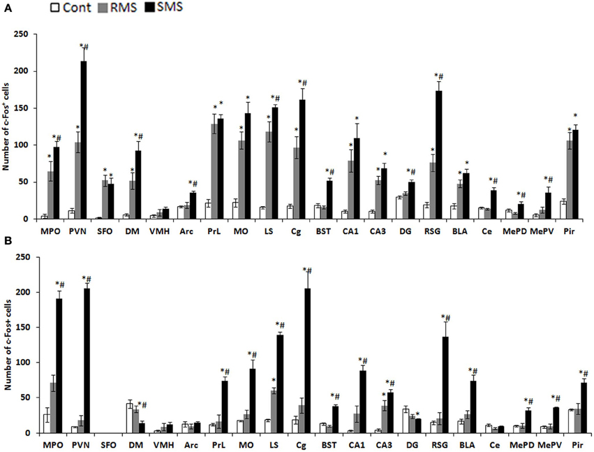
The expression of the immediate early gene product c-Fos is a reliable molecular marker to investigate neuronal activation. The examination of c-Fos expression has revealed that many brain regions are activated by MS, which differs depending on age and the type of stress. We recently analyzed the c-Fos expression induced by repeated MS and single-time MS during different developmental stages and time periods. Mice were exposed to 3 h repeated MS daily from PND1 to PND14 or from PND14 to PND21, or to single-time MS at PND14 or PND21 (Horii-Hayashi et al., 2013). We clarified that MS activated many brain regions and that c-Fos expression patterns changed developmentally (Figure 2). Single-time MS at both ages activated many regions of the hypothalamus and limbic forebrain, while the pattern of c-Fos expression in the repeated MS groups were significantly different on PND14 and PND21. In repeated MS of PND14 mice, the c-Fos expression levels in many regions were markedly increased compared with age-matched controls, excepting the VMH, Arc, BST, DG, Ce, MePV, and MePD. By contrast, in repeated MS on PND21 mice, c-Fos expression was reduced to control levels in all observed brain regions except for the LS and CA3. These findings suggest that repetition of a homotypic stimulus suppresses c-Fos expression by PND21, but that such suppression is barely observed on PND14. Moreover, in animals exposed to repeated homotypic stress during the postnatal period, increase in adrenal CORT secretion does not always associate with increased c-Fos expression in the PVN. Such developmental differences in c-Fos expression detected in the repeated MS groups may be associated with a developmental critical period for stress responses involving the HPA axis, during which animals are more susceptible to MS and other environments. In rodents, the critical period is the first two postnatal weeks. Thus, in early life, a repeated stress will be unlikely to suppress c-Fos expression. In turn, inappropriately activated c-Fos target genes may drastically alter how neurons function in critical neural circuits. Indeed, the suppression of increased c-Fos expression in repeated MS of PND14 mice was observed in specific regions (BST, Ce, MePD, and MePV) that form anatomical neural connections. These regions are referred to as an extended amygdala, which are closely associated with anxiety, fear, and psychiatric disorders (Davis et al., 2010). Therefore, even at PND14, repeated homotypic stress may reduce neural activity in the circuit of the extended amygdala. Moreover, in the SFO, where neurons are influenced by osmolality, calcium, and sodium concentrations in the systemic circulation (Smith and Ferguson, 2010), c-Fos expression was increased in both repeated and single-time MS mice, as compared to controls, on PND14. However, there were no changes in any of the groups on PND21. This difference may reflect the increased resistance of physical growth to the hyperosmolality induced by deprivation of lactation.
FIGURE 2  
http://www.frontiersin.org/files/Articles/87697/fnins-08-00166-HTML/image_m/fnins-08-00166-g002.jpg

Figure 2. c-Fos expression in the hypothalamus and limbic forebrain after MS (Horii-Hayashi et al., 2013). The graphs show the numbers of c-Fos-positive cells on PND14 (A) and PND21 (B) in non-separated control (white bar), RMS (gray bar), and SMS (black bar) mice (n = 4–5 for each group). In both RMS and SMS, the sampling point is just after MS procedure. *P < 0.05 vs. control; #P < 0.05 vs. RMS. MPO, medial preoptic area; PVN, paraventricular nucleus; SFO, subfornical organ; DM, dorsomedial hypothalamic nucleus; VMH, ventromedial hypothalamic nucleus; PrL, prelimbic cortex; MO, medial orbital cortex; LS, lateral septum; Cg, cingulate cortex; BST, bed nucleus of stria terminalis; CA1, hippocampal area CA1; CA3, hippocampal area CA3; DG, dentate gyrus; RSG, retrosplenial granular cortex; La, lateral amygdaloid nucleus; BLA, anterior part of the basolateral amygdaloid nucleus; Ce, central amygdaloid nucleus; MePD, posterodorsal part of the medial amygdaloid nucleus; MePV, posteroventral part of the medial amygdaloid nucleus; Pir, piriform cortex.

Behavioral Changes Induced by MS in Rodents

Early life adverse experiences including MS is one of the greatest contributing factors for mental health problems across life stages (Levine, 2005), relating not only to risk for mental health disorders but also to transdiagnostic features common in many psychological disorders (Glaser et al., 2006). I will introduce some of the behavioral aspects observed in animal model of MS.

Depression- and anxiety-like behaviors

Numerous studies have demonstrated a strong relationship between traumatic events during early life and development of behavioral abnormalities later in life. Early life adversity, such as that induced by MS, child physical, sexual, and emotional abuse, and general neglect has been linked to serious psychiatric impairment in adulthood (MacMillan et al., 2001). Particularly, a stressful life event such as early parental loss is associated with unipolar and bipolar depression, as well as anxiety disorders, beyond familial or genetic factors (Kendler et al., 1992; Agid et al., 1999; Furukawa et al., 1999; Heim and Nemeroff, 2001). Many human studies have reported that major depression and anxiety disorders are frequent in adults with a history of childhood abuse (Stein et al., 1996; Felitti et al., 1998). There have been numerous reports of the behavioral changes induced by MS in animal studies. Neonatal MS induces permanent alterations in the characteristics of the HPA response to stress in the offspring later in life (Ladd et al., 1996; Vazquez et al., 2000). Many studies of repeated MS during the first 2 weeks of neonatal life showed depression- and anxiety-like behaviors in adulthood (Newport et al., 2002; Daniels et al., 2004; Lee et al., 2007; Ryu et al., 2009). In these studies, ambulation and rearing decreased, immobility during a forced swim test increased, and time spent in the closed arms of an elevated plus maze increased.

Fear response

Until recently, no one had investigated how early experiences affected fear retention and extinction development, although these forms of emotional learning could be critically involved in the pathogenesis and treatment of mental health problems. Recent several studies showed that the timing of the maturation of fear learning is not set in static, but can be dynamically regulated by early experiences. Although the exact mechanisms are still unknown, when rats are reared under stressful conditions then they exhibit adult-like fear retention and extinction behaviors at an earlier stage of development (Callaghan et al., 2013). Chocyk et al. reported that MS decreased freezing time in both contextual and auditory fear conditioning in adolescent and adult rats (Chocyk et al., 2014). These results suggest that early life stress may permanently affect fear learning and memory.

Food intake and response to food deprivation

Previous studies showed that repeated MS during the first 2 weeks after birth may not permanently affect food intake and body weight gain of the offspring as long as the pups are reared in a group (Iwasaki et al., 2000; Kalinichev et al., 2002; Ryu et al., 2008). In contrast, post-weaning social isolation promotes food intake and weight gain of adolescent MS pups, with impacts on anxiety-like behaviors (Ryu et al., 2008). Anhedonia to palatable food, one of the major symptoms of depression, was reported in adolescent MS pups with disruption of the mesolimbic dopaminergic activity in response to stress (Noh et al., 2008). Another study showed that sustained hyperphagia observed in the MS pups subjected to a fasting/re-feeding cycle repeated during adolescent period of MS pups induced a binge-like eating disorder, in which increased activity of the HPA axis responding to such metabolic challenges appeared to play a role, at least partly, in mediation with the hypothalamic neuro peptide Y (NPY) (Jahng, 2011).

Gene Expression

Many animal studies, including MS, have improved our knowledge of gene-environment interactions and elucidated the pathways that program an animal in response to its early life experiences (Meaney and Szyf, 2005). Epigenetic mechanisms involving DNA methylation, post-translational modification of histone proteins and non-coding RNAs (most notably micro-RNA) are major candidates for regulating gene expression and integrating intrinsic and environmental signals in the genome (Jaenisch and Bird, 2003). Murgatroyd and colleagues showed that in the parvocellular subdivision of the paraventricular nucleus of the hypothalamus, MS in mice persistently upregulates Avp gene expression associated with reduced DNA methylation of a region in the Avp enhancer. This early life stress-responsive region serves as a binding site for the methyl-CpG binding protein 2, which in turn is regulated through neuronal activity. They also found that the ability of methyl-CpG binding protein 2 to control transcription of the Avp gene and induce DNA methylation occurred by recruiting components of the epigenetic machinery (Murgatroyd et al., 2009; Murgatroyd and Nephew, 2013). Other groups investigated DNA methylation levels at a specific sequence motif upstream of the GR gene (Nr3c1) in the hippocampus of offspring, and found that subjecting pups to a single 24 h MS increases methylation levels (Kember et al., 2012). The epigenetic alterations of these genes suggest that the HPA axis could be dysregulated by MS. Importantly, however, the DNA methylation differences were also often strain specific (Kember et al., 2012). Taken together, these findings demonstrate the importance of investigating environmental effects on a range of genetic backgrounds, emphasizing the need for the further examination of environmental, genetic, and epigenetic interactions.

Conclusions


Adverse environments and experiences during the neonatal period can dramatically affect the development of the HPA axis that underlies adaptive behavioral responses. MS experiments, as a model of early life stress, demonstrate that CORT levels and c-Fos expression change depending upon the different experimental conditions of MS, e.g., age at testing and frequency of repetition. Furthermore, separation conditions (isolation with or without a littermate) could also influence the results of the MS experiments. MS can induce various behavioral changes manifested in later life, which could be caused, at least in part, by alterations in gene expression, particularly through epigenetic mechanisms.

Conflict of Interest Statement

The authors declare that the research was conducted in the absence of any commercial or financial relationships that could be construed as a potential conflict of interest.

Acknowledgments

This work was supported by Grants-in-Aid for Scientific Research (23390040 to Mayumi Nishi and AstraZeneca Research Grant 2009). We thank Dr. Julian G. Mercer, a chief editor of J Neuroendocrinology, for permitting the reuse of our own figures published in J Neuroendocrinology.

Wednesday, July 03, 2013

Understanding PTSD: Researchers Explore Causes, Treatment

This is a good introductory overview of PTSD, with a nice section explaining the impact of trauma on the amygdala and hippocampus (I've formatted this section as an inset for easier access). While the explanation is good, I disagree with the treatment model they propose, for two different reasons.

(1) Exposure therapy may, over time, create habituation to the stimuli that trigger flashbacks or anxiety. This does not address the hypervigilance, the sense that one must be continually alert to possible threats in the environment, or testing people to make sure they are safe. Nor does it address the numbing that can result from trauma, and the desire to numb feelings with addictive behaviors.

(2) Those who experience PTSD, which is only 20-35% of those who experience trauma (depending on the trauma), generally have a history of big T traumas (physical, emotional, or sexual abuse; severe neglect; natural disaster; refugee status; death of parent) or a collection of small T traumas (bullying, shaming and humiliation, emotionally distant caregivers, etc). It is the reduction in resilience created by these earlier traumas that make one vulnerable to PTSD. Part of recovery has to be addressing and resolving these prior traumas, and the model presented in the article.

With that, here's the article.

Understanding PTSD: Researchers explore causes, treatment


by Pete Zrioka

Eric Batory, pictured during his time as a special operations medic, assigned to the U.S. Army's 1st Ranger Battalion, 75th Ranger Regiment. Batory went through two years of extensive medical and special forces training to qualify for the position. Credit: Eric Batory


(Medical Xpress)—In 2005, ASU student Eric Batory was a long way from Arizona. He was in Mosul, Iraq on his first deployment as a special operations medic with the U.S. Army's 1st Ranger Battalion, 75th Ranger Regiment.

During a night raid to extract a high-value target, his unit came under fire as they were entering a compound. The squad leader was shot on his right side, where the round glanced off his body armor and came to rest near his heart. The only medic on the scene, Batory began rendering aid to the Ranger in the midst of a heated firefight, feeling the concussive force of grenades and gunfire all around him.

"That's what really rung my bell," says Batory, who is now a student in ASU's College of Liberal Arts and Sciences. "I'm in a closed room, with an LED light on this guy, all this stuff blowing up, concrete raining down."

Batory's patient survived and Batory was awarded the Bronze Star with Combat 'V' for his actions, the fifth-highest combat decoration. After that, Batory says he started to feel a little bit "off." He became withdrawn from his platoon, obsessing over every possible medical scenario. He distanced himself from his fellow Rangers, afraid he'd lose someone as a result of his actions.

Two years and two deployments later, he left the Army as a sergeant, joining the scores of veterans living with cognitive and psychological wounds from the Global War on Terror.

Six years and a world away from the battlegrounds of Iraq and Afghanistan, Batory still lives with post-traumatic stress disorder (PTSD) from his time in combat.
Any traumatic experience, such as combat, violent assault or natural disaster, can cause PTSD. Symptoms can include flashbacks to the event, nightmares, avoiding triggering situations, numbness and withdrawal, fear and increased emotional arousal. 
Why do these kinds of experiences create such a painful and long-lasting effect? The answer lies in very old structures in the human brain, called the amygdala and the hippocampus. These are nestled within the temporal lobe, which manages sensory input, speech and language, and memory formation and association. Memories are formed, stored and contextualized by the hippocampus. The amygdala is tied to our emotions, particularly those felt in fearful, life-or-death situations. 
"The hippocampus' job is to remember the context of the situation and the amygdala ensures the longevity of that memory, especially the emotional memory," says Harold Burke, a faculty associate in ASU's College of Health Solutions. "It's not just the context of the visual and audio cues, but the emotional experience of fear." 
In addition to branding these memories in the brain, the amygdala also regulates part of the sympathetic nervous system, commonly known as the fight or flight system. 
"It's a very old part of brains from an evolutionary standpoint," says Burke, who teaches courses in ASU's Doctor of Behavioral Health program. "It's been designed over millions of years of evolution to trigger in an emergency situation to save our lives." 
In these emergency situations, like the heated combat Batory experienced, the sympathetic nervous system pumps out norepinephrine. 
Norepinephrine increases heart rate as well as oxygen supply to the brain, allows us to breathe faster and deeper, dilates pupils to take in more visual information and triggers the release of glucose stores for extra energy. 
"It also activates the upper part of the brain, namely the cerebral cortex, so you can be very alert and make quick decisions," says Burke. 
A secondary system, which takes longer to kick in, lasts longer and aids the body's recovery. The hypothalamic-pituitary-adrenal axis, or HPA axis, is a conglomerate of structures throughout the body that responds to highly stressful situations by releasing the hormone cortisol. 
Cortisol is incredibly beneficial to both the body and mind in small, measured doses. But with chronically stressful situations, cortisol becomes more of a problem than a solution. 
Back in the hippocampus, there are neurons that act as a thermostat that regulates cortisol production. But prolonged or repeated release of cortisol kills the very neurons that help inhibit it, resulting in a negative feedback loop of more cortisol and fewer neurons to stop its release. 
"It's like smashing the thermostat," Burke explains. 
Furthermore, repeated exposure to similar high-stress trauma not only kills neurons and can inhibit cognition, but can also result in the strengthening of the memories being encoded in the hippocampus and amygdala. 
"Here's the issue: with PTSD, the activation of the amygdala and HPA axis is so intense that the synapses, the various connections between the neurons, how well they communicate, are strengthened as a product of that intensity and it can last a very, very long time," says Burke. 
It's like tracing a picture over and over again. The more you go over those lines, the clearer and more distinct an image becomes, just as repeated exposure to trauma enhances the emotional memory and deeply encodes a fear response. 
To make matters worse, the job of the hippocampus and amygdala together is to not just remember that exact, specific situation, but to remember things that are similar to it. 
"The brain generalizes the stimuli, so that if anything else is even somewhat similar it will trigger the same response," says Burke. "And that's actually a good thing. We want to be able to generalize our learning to different contexts, but the catch with PTSD is that then other things that are pretty innocent actually trigger the same kind of memories or re-experiencing."
For instance, some veterans who spent time on convoys have difficulty driving if there's trash or debris in the road, as hiding improvised explosive devices in garbage is a common insurgent tactic. In the case of Batory, there's a gamut of different stimuli that trigger an intense fear response for him back in the civilian world.

"Dogs barking, alarms or phones ringing. Anything beeping or with a high pitch," says Batory, listing the things that send his heart racing. "Noise in the dark, or a lot of light at night ... I don't like bright lights at night, I feel like you're exposed."

The sound and concussive force of fireworks has sent him diving to the ground before.

"Even at Fourth of July, knowing what it was, just feeling the concussions or hearing the blasts gets my heart rate up," says Batory.

Batory's struggle with PTSD and its lingering effects aren't limited to barking dogs or fireworks. When he was honorably discharged from the Army in 2007, he was bored. After deploying three times as a special operations medic and working through two years of intensive schooling to qualify for the coveted position, a desk job left him restless.

"I was so dissatisfied with feelings of insignificance and lack of stimulation, compared to what I was used to," says Batory.

Distracted, depressed and disillusioned with civilian life, Batory joined the Army Reserves, which only seemed to exacerbate his symptoms. Batory had trouble sleeping, and when he did, he was sleepwalking. He started to fear social events and public places like movie theatres, malls or sporting events.

"I started to get overwhelming anxiety being around groups of people," says Batory. "Whenever I was around large crowds, I'd want to leave because I'd feel overwhelmed, because there was no structure and no order. That all started to compound until I started having dissociative episodes, getting in car accidents where I would zone out and think about Iraq or Afghanistan. I'd hit a median or rear end someone. Started getting a lot of tickets and got into financial trouble."

Eventually, Batory's car was repossessed and he became homeless, all in the midst of trying to attain his undergraduate degree. He put in a disability claim with the VA and sought additional help there, receiving neuropsychological testing. He found some relief with medication, but he wasn't happy about it.

"It's disheartening to know that you have this pile of pills in front of you and you're dependent on it to feel human," says Batory.

Batory sought out additional resources available to ASU students. Fortunately for him, ASU offers a range of counseling services and has been named a Military Friendly School four years running.

He began therapy with Andrea Hekler, a clinical psychologist at ASU Health Services - NP Healthcare, located near the Downtown Phoenix campus. Hekler provides two types of evidence-based psychotherapies for PTSD patients: cognitive processing therapy and prolonged exposure therapy. Both are a class of therapies called cognitive behavioral therapies.

"Cognitions refer to the thoughts we have and how we perceive life through a certain lens," says Hekler, who has previously worked with the National Center for PTSD. "When individuals experience a trauma – be it combat, Hurricane Sandy, 9/11 in New York or being sexually assaulted – having those traumatic experiences leads us to perceive the world in different ways. People tend to start thinking the world is a much more dangerous place than most people believe and they're not able to handle and cope with the world."

Cognitive processing therapy focuses on the assumptions that PTSD victims have as a result of their trauma. Prolonged exposure therapy homes in on a victim's behavior.

Cognitive processing therapy teaches patients how their thoughts are connected to how they behave and feel.

"It's essentially using logic and rational thinking to help them understand the assumptions they have and challenge those assumptions," says Hekler.

Prolonged exposure therapy can be more challenging as it's based on the premise that PTSD victims haven't processed their trauma or even talked about it, according to Hekler.

This therapy breaks down into four parts: education on what PTSD is and its symptoms, breathing exercises to relax the patient and two types of exposure. In the first, imaginal exposure, patients close their eyes and describe their trauma as if it were happening in the moment, not the past. This process is repeated, recorded and listened to outside of the sessions as well.

"Through this rehearsal, they come to something called habituation," says Hekler. "Essentially, stimuli that once had a very strong intensity, over time reduces that intensity. Each time, when someone comes back to therapy they recite the event again and typically what happens is they don't become as emotionally distressed or upset about it. Eventually it becomes a memory, not one that you want to revisit all the time, but without the same emotional intensity."

The second type of exposure is in vivo exposure, where patients list all of the situations they avoid and then insert themselves in them. This fosters a similar process of habituation, where the patient learns to manage the crippling fear or anxiety they experience in situations they avoid.

"At the heart of PTSD is avoidance," says Hekler. "Avoidance is really good in the short term because it makes anxiety go away – what it's really doing is telling your brain that this is terrifying and we need to get away."

In other words, it's only serving to strengthen the fear conditioning in the amygdala and hippocampus. The process of habituation doesn't seek to overwrite those incredibly intense emotional memories, but to create a separate set of circuits that associate the same stimuli with a safe emotional response.

"Cognitive behavior therapy literally sets up and strengthens circuits to inhibit fear," says Burke. "You don't get rid of the original trauma, but you overlay it with other behavior."

For Batory, in vivo exposure proved to be a beneficial form of therapy.

"It was effective not because it changed what I feel or think, but because I learned to deal," says Batory. "Even though I feel anxiety, it's not as severe and I know how to keep it in check."

While both therapy and medication can be effective, Burke believes that PTSD demands a holistic approach to treatment in which different health professionals work closely together.

"I think ASU is on the cutting edge of integrated health solutions," says Burke. "We can have Dr. A doing one thing to a person and Dr. B doing something else to the same person across town, especially when Dr. A is just physical medicine and Dr. B is just psychological. Those two groups have not been talking nearly enough over the last decades, and that's got to change. And ASU is right on the cusp of it. The Doctorate of Behavioral Health program is the first of its kind in the whole country."

Batory now lives with a group of other veterans and is on track to graduate with a bachelor's degree in microbiology in December 2013. After spending years of attaching negative associations to his military experience, even when saving lives, he has newfound perspective and direction. He is applying to medical schools in the coming months, saying he wants to get back to helping people.

"I've taken up an interest in medicine again, and I think it's a good measure of the return of myself and my life's purpose," says Batory. "But I really had to rebuild myself to get back to that point."

To others suffering from PTSD, Batory offers pretty straightforward advice: "You're not weak. You're not any less of a person for acknowledging you're struggling, and the sooner you do that, the sooner you'll be on the road to recovering."

Friday, June 07, 2013

Trauma's Physical Effects Persist for Years - Dr. Phebe M. Tucker

This feels like another piece of "well, duh!" news about trauma. But it's important that this information become more widely known. The author suggests that the neurobiological effects of trauma might help survivors better handle future trauma or increase their risk of cardiovascular disease and other problems, however, my experience and the trauma literature seems to suggest that those who experience early trauma (abuse, neglect, molestation) are LESS able to handle future trauma (their brains are not as resilient) and are often much more likely to experience other similar traumas as teens and adults.

They are correct that trauma increases risks for health issues, including cardiovascular disease, irritable bowel syndrome, autoimmune disorders, ulcerative colitis, and other issues.

We need to make it more widely known that while kids are very resilient, sometimes their brains are less so, especially when exposed to frequent or repeated trauma.


Trauma's physical effects persist for years


By: SHERRY BOSCHERT, Clinical Psychiatry News Digital Network
06/03/13

SAN FRANCISCO – Neurobiological effects of trauma persist for years and might help survivors better handle future trauma or increase their risk of cardiovascular disease and other problems, three studies suggest.

One study assessed 34 adult survivors of Hurricane Katrina who were relocated to Oklahoma 22 months after the hurricane, and compared them with 34 control participants in Oklahoma who matched the survivors’ characteristics. A second study assessed nine adolescent survivors who were relocated 22 months after Hurricane Katrina and nine matched controls. The third study compared 60 adults who directly experienced the Oklahoma City bombing (84% of whom were injured) with matched controls 7 years after the bombing.

Dr. Phebe M. Tucker

The results showed that autonomic, neuroendocrine, and immune system changes from trauma might last for years, even after emotional wounds have healed, Dr. Phebe M. Tucker reported in a press briefing and a poster presentation at the annual meeting of the American Psychiatric Association.

The survivors and controls differed in mean arterial blood pressure, heart rate, variability of heart rate, and levels of cortisol, a regulatory substance that promotes the fight-or-flight response; interleukin-2 (IL-2), which protects against infection; and interleukin 6 (IL-6), which promotes inflammation).

Some of these changes might enhance a person’s fight-or-flight response, and so could prepare survivors for future disasters, but the health implications are unclear, she said. Previous studies have linked trauma to increased cardiovascular and other health problems, such as a tripling in the myocardial infarction rate at Tulane University in New Orleans after Hurricane Katrina. The physiologic changes seen in the current studies might contribute to that.

The current studies also found more short-term and long-term neurobiological changes in survivors with depression or posttraumatic stress disorder (PTSD), compared with survivors without depression or PTSD or control participants.

In the study of adult survivors of Hurricane Katrina, 35% of survivors and 12% of controls had PTSD. Baseline heart rates were significantly higher among survivors (81 beats per minute), compared with controls (75 beats per minute). Survivors with or without PTSD had significantly higher levels of IL-6, compared with control participants who did not have PTSD, reported Dr. Tucker, chair of psychiatry at the University of Oklahoma Health Sciences Center, Oklahoma City. She conducted the studies with Dr. David H. Tiller, also of the university.

Survivors’ baseline sympathetic (fight-or-flight) heart rate variability was significantly higher – approximately double – that of the control group. The protective, parasympathetic heart rate variability at rest was significantly lower than in controls. When participants were exposed to reminders of the hurricane, the controls showed a significantly greater reaction in the parasympathetic heart rate variability, compared with a flat response among survivors, she said.

"Overall, the adult Katrina survivors’ higher heart rates, decreased protective heart rate variability, and increased inflammatory IL-6 may increase their risk for heart disease," Dr. Tucker said.

The pilot study of 18 adolescent survivors and controls (average age 15 years) found significantly higher rates of symptoms for PTSD or depression among survivors. As might be expected from previous studies of trauma and PTSD, the survivors had lower levels of cortisol, and IL-2 levels correlated with cortisol levels, suggesting that survivors might have reduced immune protection and could be more susceptible to infection, she reported.

In contrast with the adult findings, however, higher PTSD symptoms in the adolescents correlated with lower levels of the inflammatory cytokine IL-6. This might be because the youths lacked the inflammatory changes seen in adults after trauma or the youths were more resilient in some ways, she speculated.

In the third study of the bombing survivors, mean PTSD and depression symptom severity scores were below clinically relevant levels 7 years after the bombing. The handful of survivors who still had PTSD had significantly higher cortisol levels, compared with non–PTSD survivors and controls.

When exposed to reminders of the bombing, the survivors showed greater increases in heart rate, systolic and diastolic blood pressures, and mean arterial pressure. "Autonomic reactivity may be a generalized long-term response" to trauma that’s independent of PTSD, she said.

Dr. Tucker reported having no financial disclosures.

sboschert@frontlinemedcom.com

Twitter @sherryboschert

Monday, April 02, 2012

Elissa Epel - Emotions Stress and Rate of Telomere Shortening: Are Our Cells Listening to Us?


From UCTV,  a lecture on the role of stress and emotions in cell damage (telomere shortening). Research has shown that middle-aged people who were physically active have longer telomere's than their couch-potato peers - in fact, they have the telomeres of much younger people.

A more recent study looked at telomere length in people who are depressed and who suffer chronic stress - "telomere lengths were shortest for both depressed and healthy participants who were showing chronic stress." The culprit is likely cortisol, since both stress and depression are associated with impaired cortisol function.

More below the video.


Does stress speed up the aging process at a genetic level? Our cells are constantly aging. When a cell divides some telomere is lost, but if it becomes too short, that cell can no longer replicate and eventually dies. Elissa Epel, UCSF department of psychiatry, explores the affects of stress and emotions on the process of telomere shortening. Series: "UCSF Osher Mini Medical School for the Public" [4/2012]
This brief article is from at the National Institute for the Clinical Application of Behavioral Medicine.

Stress, Depression, and Telomeres: A Brain Health Update


One way to measure how much age has fatigued an individual is to measure the length of their telomeres.

Since telomeres naturally shorten with time many researchers use telomere length to determine cell age, and give a picture of overall brain health

What are telomeres?

They act as protective caps on the end of chromosomes to keep them from deteriorating. When cells replicate (think aging), telomeres are cut and become increasingly shorter. If the telomere becomes too short, it dies or at the very least, becomes dormant.
bike riding for brain health

I reported last year that researchers had found that middle-aged people who were physically active not only had higher aerobic capacities, but also longer telomeres than those who were sedentary.  They had telomere lengths that were similar to people much younger than they were.

This is compared to people who were middle-aged and sedentary. Their telomeres were about 40% shorter, on average than those of younger folks.

Now a new study in the journal Biological Psychiatry looked at the role of stress and depression on telomere length.

Karl-Fredrik Norrback, PhD and his colleagues at Umeå University (Sweden) took 91 patients with recurrent depression and 451 healthy controls.

Telomere length and stress levels were measured. They measured stress in two ways: cortisol levels were measured and participants also completed a questionnaire on stress.

The findings?  Telomere lengths were shortest for both depressed and healthy participants who were showing chronic stress.

Many of the depressed participants exhibited disturbed cortisol regulation, which may explain why they had a higher overall probability of having shorter telomere lengths.

Stress and depression are frequently linked by researchers, including Norrback and his crew, as are depression and shortened telomeres.

This current research adds to the results of earlier research linking depression and shorter telomere lengths.

Depression and stress are two things that can help derail brain health, and actually, some day soon, researchers will be studying the relationship between depression, telemere length and lifespan.
And when they do, I’ll be here at this blog telling you about it.

But depression and stress certainly aren’t the only factors that hurt the brain.  We know now that our brains can change, for better or worse, by what we eat, how we think, and what we do (or don’t do).