Showing posts with label emotions. Show all posts

Thursday, November 06, 2014

Our Brain Dissociates Emotional Response from Explicit Memory in Fearful Situations

Anyone who has experienced trauma, or works with survivors of trauma, already that one of the most common neuromechanisms for dealing overwhelming fear is to dissociate the emotional impact from the narrative memory of the event(s). Maybe you have had the experience of someone telling you about a horrible thing that happened to him, and all the while he is smiling or even laughing. As a therapist, I immediately think, "affect incongruity." This is a clear indication that the person in question has dissociated the emotions from the experience.

The reason for this, as far as we know, is that part of what the thalamus does is assemble all of the sense data (sight, sound, touch, emotions, etc.) when encoding a memory into a coherent package, but it does not function correctly under conditions of extreme stress, so the memory ends of being fragmented. Often the emotional content gets separated out so that the person can describe the event with no related affect being visible. On the other hand, a sound associated with the trauma may trigger a flashback of the emotional experience.

We are finally beginning to understand the neuroscience behind how the brain processes trauma.

Our Brain Dissociates Emotional Response from Explicit Memory in Fearful Situations


Neuroscience News
November 6, 2014

Researchers at the Cognition and Brain Plasticity Group of the Bellvitge Biomedical Research Institute (IDIBELL) and the University of Barcelona have been tracking the traces of implicit and explicit memories of fear in human. The study was published in the journal Neurobiology of Learning and Memory; it describes how, in a context of fear, our brain differently encodes contextual memory of a negative event (the place, what we saw, etc) and the emotional response associated.

The study measures electrodermal activity of eighty-six individuals in a fearful context generated in the laboratory and in a neutral context in which they have to learn a list of words. One week and two weeks after the experiment they were tested to see which words they remembered.

“In both contexts —explains Pau Packard, author of the study—, forgetting curve was normal. Over time, they forgot all the words, the explicit trace. Moreover, in the fearful context the electrodermal activity —the emotional implicit response— was exactly the same, much higher than in the neutral context”.

This image shows a man's face in red with fearful images accompanying.
In traumatic events, it seems that, over time, there is a portion of memory that is erased or inaccesible; we forget the details but still maintaining the emotional reaction. This image is for illustrative purposes only. Credit geralt.

In traumatic events, it seems that, over time, there is a portion of memory that is erased or inaccesible; we forget the details but still maintaining the emotional reaction. The imprint is divided into two separate paths. According to Packard, “the brain dissociates the explicit memory of a negative event from the emotional response”.

This may help to understand why in pathological situations of post-traumatic stress disorders, the uncontrolled emotional response linked to the negative event is generated without knowing what causes it.

Lluís Fuentemilla, project coordinator, emphasises that “the study helps to explain how the processing of fearful memories can lead to post-traumatic stress disorder”. Furthermore, it opens the door to the investigation of new therapeutic strategies for these disorders because “the implicit memory trace in a fearful context does not loose over time and can be detected through electrodermal measures”.

About this memory research

Contact: Press Office – University of Barcelona
Source: University of Barcelona press release
Image Source: The image is credited to geralt and is in the public domain

Original Research:
Packard, P. A., Rodríguez Fornells, A., Stein, L. M., Nicolás, B., and Fuentemilla, Ll. (2014, Dec). Tracking explicit and implicit long-lasting traces of fearful memories in humans. Neurobiology of Learning and Memory; 116: 96–104. Published online September 26 2014 doi:10.1016/j.nlm.2014.09.004
* * * * *

Here is the original abstract from Neurobiology of Learning and Memory.

Tracking explicit and implicit long-lasting traces of fearful memories in humans


Pau Alexander Packarda, Antoni Rodríguez-Fornellsa, Lilian Milnitsky Stein, Berta Nicolás, Lluís Fuentemilla,

Highlights

 
• We track implicit and explicit memory traces for fearful episodes over time.
• SCR at test was higher for verbatim words encoded in a fearful context.
• Subjects were unable to report words’ emotional encoding context.
•Implicit memory traces of a fearful context was dissociated from the gist.


Abstract


Recent accounts of Posttraumatic Stress Disorder (PTSD) suggest that the encoding of an episode within a fearful context generates different implicit and explicit memory representations. Whilst implicit memory traces include the associated emotional states, explicit traces include a recoding into an abstract or gist-based structural context of the episode. Theoretically, the long-term preservation of implicit memory traces may facilitate the often untreatable memory intrusions in PTSD. Here, we tracked in two experiments how implicit and explicit memory traces for fearful episodes dissociate and evolve over time. Subjects (N = 86) were presented with semantically-related word-lists in a contextual fear paradigm and tested for explicit memories either immediately (i.e., 30 min) or after a delay (i.e., 1 or 2 weeks) with a verbal recognition task. Skin Conductance Response (SCR) was used to assess implicit memory responses.

Subjects showed high memory accuracy for words when tested immediately after encoding. At test, SCR was higher during the presentation of verbatim but not gist-based words encoded in a fearful context, and remained unchanged after 2 weeks, despite subjects being unaware of words’ encoding context. We found no clear evidence of accurate explicit memory traces for the fearful or neutral contexts of words presented during encoding, either 30 min or 2 weeks afterwards. These findings indicate that the implicit, but not the explicit, memory trace of a fearful context of an episode can be detected at long-term through SCR and is dissociated from the gist-based memory. They may have implications towards the understanding of how the processing of fearful memories could lead to PTSD.

Sunday, September 28, 2014

The Science of Emotions: Jaak Panksepp at TEDxRainier

http://ecx.images-amazon.com/images/I/41TIr4mBuJL._SY344_BO1,204,203,200_.jpg

Awesome - Jaak Panksepp is one of the founders of affective neuroscience, and author of Affective Neuroscience: The Foundations of Human and Animal Emotions (2004) and The Archaeology of Mind: Neuroevolutionary Origins of Human Emotions (2012).

The Science of Emotions: Jaak Panksepp at TEDxRainier

Published on Jan 13, 2014


Given an inherent subjective nature, emotions have long been a nearly impenetrable topic for scientific research. Affective neuroscientist Jaak Panksepp explains a modern approach to emotions, and how taking seriously the emotions of other animals might soon improve the lives of millions.

Jaak Panksepp introduced the concept of Affective Neuroscience in 1990, consisting of an overarching vision of how mammalian brains generate experienced affective states in animals, as effective models for fathoming the primal evolutionary sources of emotional feelings in human beings. This work has implications for further developments in Biological Psychiatry, ranging from an understanding of the underlying brain disorders, to new therapeutic strategies. Panksepp is a Ph.D. Professor and Baily Endowed Chair of Animal Well-Being Science, College of Veterinary Medicine, Washington State University. His scientific contributions include more than 400 papers devoted to the study of basic emotional and motivational processes of the mammalian brain. He has conducted extensive research on brain and bodily mechanisms of feeding and energy-balance regulation, sleep physiology, and most importantly the study of emotional processes, including associated feelings states, in other animals.

This talk was given November 9, 2013 in Seattle at TEDxRainier, a local TEDx event, produced independently of the TED Conferences.
* * * * *

Jaak Panksepp - Animal Emotion Researcher and Neuroscientist


Speaker Bio 

Given an inherent subjective nature, emotions have long been a nearly impenetrable topic for scientific research. Affective neuroscientist Jaak Panksepp explains a modern approach to emotions, and how taking seriously the emotions of other animals might soon improve the lives of millions.

Jaak Panksepp, Ph.D. Professor and Baily Endowed Chair of Animal Well-Being Science, College of Veterinary Medicine, Washington State University. His scientific contributions include more than 400 papers devoted to the study of basic emotional and motivational processes of the mammalian brain. He has conducted extensive research on brain and bodily mechanisms of feeding and energy-balance regulation, sleep physiology, and most importantly the study of emotional processes, including associated feelings states, in other animals. He introduced the concept of Affective Neuroscience in 1990, consisting of an overarching vision of how mammalian brains generate experienced affective states in animals, as effective models for fathoming the primal evolutionary sources of emotional feelings in human beings. This work has implications for further developments in Biological Psychiatry, ranging from an understanding of the underlying brain disorders, to new therapeutic strategies.

His recent work has focused primarily on the subcortical brain mechanisms of sadness (brain PANIC or separation distress circuitry) and joy (brain PLAY and animal laughter circuitry), work that has implications for the treatment of autism, ADHD, and depression. His work is informed by exploring the consequences of basic knowledge about emotional endophenotypes (natural brain states) for better understanding of human mental health. His monograph Affective Neuroscience (Oxford, 1998) outlined ways to understand brain affective processes neuroscientifically; his Textbook of Biological Psychiatry (Wiley, 2004) focused on how elucidation of emotional processes can facilitate psychiatric practice; and Archaeology of Mind (Norton, 2012) summarized how such knowledge can inform psychotherapeutic practice.

This work has led to three novel antidepressant concepts that are currently being evaluated in human. But perhaps equally importantly, this work provides a novel vision for how consciousness emerged in human brains, which can only be clarified through the scientific study of the phenomenal-experiential-emotional capacities of that exist in the brains/minds of other animals.

Friday, September 26, 2014

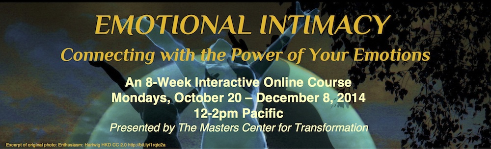
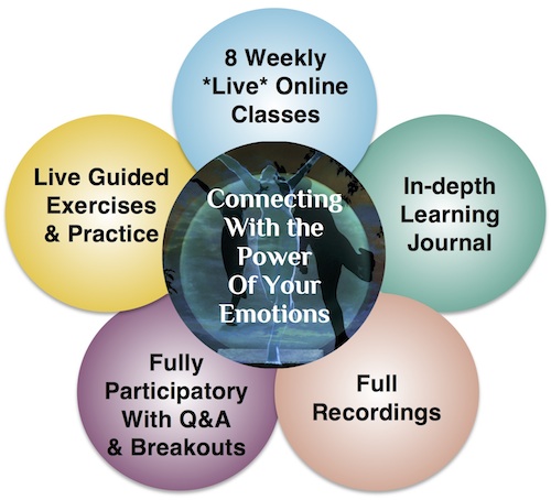
Emotional Intimacy: Connecting with the Power of Your Emotions - 8-Week Online Course

http://ecx.images-amazon.com/images/I/41QPJKox-3L._SY344_BO1,204,203,200_.jpg

This 8-week course is based on Robert Augustus Masters' excellent book, Emotional Intimacy: A Comprehensive Guide for Connecting with the Power of Your Emotions (August, 2013). If this were not a program based on Masters' book and work, and taught by one of his certified students, I would not be recommending it.

The program costs $169.00 (+ $5.22 charge) if you register by Sept. 30; $199.00 (+ $5.97 charge) if you register by Oct, 10; and $269.00 (+ $7.72 charge) after Oct. 10. The course begins on Monday, October, 20.

Here is the description:

Join us for Emotional Intimacy: Connecting with the Power of Your Emotions


Posted by admin on Sept 20, 2014


Embark on an 8-week inner quest that can transform the way you experience your emotions and revolutionize your life and relationships.


What benefits can a course on Emotional Intimacy bring to your life?
  • Deepen your understanding of the major emotions in a way that gives you skills for transformation
  • Increase your emotional intelligence–a skill that correlates with better relationships, improved social supports, and less conflict with others
  • Break free of old emotional patterns that hold you back
  • Reduce reactivity allowing you to experience more ease, connection, and emotional freedom
  • Discover the surprising role challenging emotions play in your wellbeing
  • Become more intimate with your undiscovered emotional self
  • Improve your relationships through greater emotional competence and self-knowledge
  • Learn to get off the rollercoaster of toxic emotional interactions that create conflict and pain
  • Discover more joy, resilience, and inner ease
Our Journey…


Enter a unique experiential learning environment held with deep consciousness and compassion. Connecting With the Power of Your Emotions includes original content, intimate Q&A, guided meditations and solo practices, as well as partner exercises.


We will learn together in eight weekly 2-hour teleclasses (Mondays, October 20, 27, November 3, 10, 17, 24, December 1 and 8 at 12:00 pm Pacific Time). Additional live 60-minute Q&A and practice sessions will be held on Thursday, November 13, 5:00pm Pacific, Saturday, November 22, 4:00pm, Pacific, and Thursday, December 4, 5:00pm, Pacific to accommodate those who may be unable to attend at the regular course time.

All classes will be recorded and made available within 48-72 hours of the class for those who are unable to attend live.

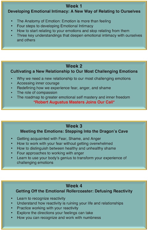
Each week you will receive a Learning Journal specifically designed to support you in getting the most out of our time together. With guided pre- and post- class reflection questions and homework exercises, the Learning Journal will allow those who want to go deeper additional opportunities to get in touch with the emotions and skills we will be working on in each class.

Taught by Iyeshka Farmer, RPC, Senior Teacher/Trainer with the Masters Center for Transformation. The course will utilize and expand upon the book, “Emotional Intimacy: A Comprehensive Guide for Connecting With the Power of Your Emotions,” by Robert Augustus

Masters, PhD. Robert Augustus Masters will make special guest appearances during two of our classes (Oct. 27 and Nov. 24) for live sessions of discussion and Q&A. This is a rare and precious opportunity to speak with Dr. Masters directly.

Thursday, September 18, 2014

Conversations on Compassion with Paul Ekman, Ph.D.


Here is another video talk from Stanford's CCARE (Center for Compassion and Altruism Research and Education). This one features Dr. Paul Ekman, a psychologist who pioneered the study of emotions and their relation to facial expressions. He has created an 'atlas of emotions' with more than ten thousand facial expressions, and has gained a reputation as "the best human lie detector in the world" (according to Wikipedia).

Dr. Ekman is the author of many books, including Unmasking the Face: A Guide to Recognizing Emotions From Facial Expressions (2003), Emotions Revealed, Second Edition: Recognizing Faces and Feelings to Improve Communication and Emotional Life (2007), Emotional Awareness: Overcoming the Obstacles to Psychological Balance and Compassion (2009, with the Dalai Lama), and Telling Lies: Clues to Deceit in the Marketplace, Politics, and Marriage (2009).

Conversations on Compassion with Paul Ekman, Ph.D.

Published on Sep 2, 2014


Paul Ekman, Professor Emeritus in Psychology at UCSF, is the researcher and author best known for furthering our understanding of nonverbal behavior, encompassing facial expressions and gestures. In addition to his own distinguished academic career, Ekman has authored more than 100 published articles and holds several honorary doctoral degrees. A pre-eminent psychologist and co-discoverer of micro expressions with Friesen, Haggard and Isaacs, Ekman was named by the American Psychological Association as one of the most influential psychologists of the 20th century, and TIME Magazine (2009) hailed him as one of the 100 most influential people in the world.

Tuesday, September 16, 2014

New Reseach on Schizophrenia - 5 Recent Studies

https://farm3.staticflickr.com/2680/4276710116_5aa7a2e980_z.jpg?zz=1

Presented below are five new studies on schizophrenia from the last week or two. The first three come from Science Daily, which posts press releases from researchers and journals about new studies. All three of these studiers are behind paywalls, and they cannot be accessed to assess their validity.

The last two articles are from Frontiers in Psychiatry: Schizophrenia and are open access articles.

I have added some comments to a few of these.

Schizophrenia not a single disease but multiple genetically distinct disorders

Date: September 15, 2014
Source: Washington University in St. Louis
Summary:
Schizophrenia isn’t a single disease but a group of eight genetically distinct disorders, each with its own set of symptoms, research shows. The finding could be a first step toward improved diagnosis and treatment for the debilitating psychiatric illness.

***

Cloninger, the Wallace Renard Professor of Psychiatry and Genetics, and his colleagues matched precise DNA variations in people with and without schizophrenia to symptoms in individual patients. In all, the researchers analyzed nearly 700,000 sites within the genome where a single unit of DNA is changed, often referred to as a single nucleotide polymorphism (SNP). They looked at SNPs in 4,200 people with schizophrenia and 3,800 healthy controls, learning how individual genetic variations interacted with each other to produce the illness.

In some patients with hallucinations or delusions, for example, the researchers matched distinct genetic features to patients' symptoms, demonstrating that specific genetic variations interacted to create a 95 percent certainty of schizophrenia. In another group, they found that disorganized speech and behavior were specifically associated with a set of DNA variations that carried a 100 percent risk of schizophrenia.

"What we've done here, after a decade of frustration in the field of psychiatric genetics, is identify the way genes interact with each other, how the 'orchestra' is either harmonious and leads to health, or disorganized in ways that lead to distinct classes of schizophrenia," Cloninger said.
Although individual genes have only weak and inconsistent associations with schizophrenia, groups of interacting gene clusters create an extremely high and consistent risk of illness, on the order of 70 to 100 percent. That makes it almost impossible for people with those genetic variations to avoid the condition. In all, the researchers identified 42 clusters of genetic variations that dramatically increased the risk of schizophrenia.
Full Citation:
Javier Arnedo, Dragan M. Svrakic, Coral del Val, Rocío Romero-Zaliz, Helena Hernández-Cuervo, Ayman H. Fanous, Michele T. Pato, Carlos N. Pato, Gabriel A. de Erausquin, C. Robert Cloninger, Igor Zwir. Uncovering the Hidden Risk Architecture of the Schizophrenias: Confirmation in Three Independent Genome-Wide Association Studies. American Journal of Psychiatry, 2014; DOI: 10.1176/appi.ajp.2014.14040435

Of course, the article is behind a paywall.

I remain skeptical of these results. As has long been the case in studies of schizophrenia and its genetic origins, the researchers do not account for HOW those single nucleotide polymorphisms (SNPs) get triggered in the first place. There is a LOT of research demonstrating powerful correlations between childhood abuse and neglect and epigenetic changes in the brain which can lea to schizophrenia and other psychoses.

* * * * *

Is the pattern of brain folding a 'fingerprint' for schizophrenia?

Date: September 11, 2014
Source: Elsevier
 

Summary:
Anyone who has seen pictures or models of the human brain is aware that the outside layer, or cortex, of the brain is folded in an intricate pattern of “hills”, called gyri, and “valleys”, called sulci. It turns out that the patterns of cortical folding are largely consistent across healthy humans, broadly speaking. However, disturbances in cortical folding patterns suggest deeper disturbances in brain structure and function.


Anyone who has seen pictures or models of the human brain is aware that the outside layer, or cortex, of the brain is folded in an intricate pattern of "hills," called gyri, and "valleys," called sulci.

It turns out that the patterns of cortical folding are largely consistent across healthy humans, broadly speaking. However, disturbances in cortical folding patterns suggest deeper disturbances in brain structure and function..

A new study published in the current issue of Biological Psychiatry suggests that schizophrenia is associated with reductions in the complexity of the cortical folding pattern that may reflect deficits in the structural connections between brain regions.

"The cortical folding pattern itself may not be so important, but the disturbances in connections between brain regions implicated by the changes in cortical folding could provide critical clues to deficits in the integrity of brain circuits that contribute to symptoms and functional impairment in schizophrenia," commented Dr. John Krystal, Editor of Biological Psychiatry.
Full Citation:
Pranav Nanda, Neeraj Tandon, Ian T. Mathew, Christoforos I. Giakoumatos, Hulegar A. Abhishekh, Brett A. Clementz, Godfrey D. Pearlson, John Sweeney, Carol A. Tamminga, Matcheri S. Keshavan. Local Gyrification Index in Probands with Psychotic Disorders and Their First-Degree Relatives. Biological Psychiatry, 2014; 76 (6): 447 DOI: 10.1016/j.biopsych.2013.11.018

* * * * *

Brain development in schizophrenia strays from normal path

Date: September 15, 2014
Source: Elsevier
 

Summary:
Schizophrenia is generally considered to be a disorder of brain development and it shares many risk factors, both genetic and environmental, with other neurodevelopmental disorders such as autism and intellectual disability. The normal path for brain development is determined by the combined effects of a complex network of genes and a wide range of environmental factors. However, longitudinal brain imaging studies in both healthy and patient populations are required in order to map the disturbances in brain structures as they emerge, researchers say.


***

A new study by an international, collaborative group of researchers has measured neurodevelopment in schizophrenia, by studying brain development during childhood and adolescence in people with and without this disorder. With access to new statistical approaches and long-term follow-up with participants, in some cases over more than a decade, the researchers were able to describe brain development patterns associated with schizophrenia.

"Specifically, this paper shows that parts of the brain's cortex develop differently in people with schizophrenia," said first author Dr. Aaron F. Alexander-Bloch, from the National Institute of Mental Health.

"The mapping of the path that the brain follows in deviating from normal development provides important clues to the underlying causes of the disorder," said Dr. John Krystal, Editor of Biological Psychiatry.

The findings were derived by investigating the trajectory of cortical thickness growth curves in 106 patients with childhood-onset schizophrenia and a comparison group of 102 healthy volunteers.

Each participant, ranging from 7-32 years of age, had repeated imaging scans over the course of several years. Then, using over 80,000 vertices across the cortex, the researchers modeled the effect of schizophrenia on the growth curve of cortical thickness.

This revealed differences that occur within a specific group of highly-connected brain regions that mature in synchrony during typical development, but follow altered trajectories of growth in schizophrenia.
Full Citation:
Aaron F. Alexander-Bloch, Philip T. Reiss, Judith Rapoport, Harry McAdams, Jay N. Giedd, Ed T. Bullmore, Nitin Gogtay. Abnormal Cortical Growth in Schizophrenia Targets Normative Modules of Synchronized Development. Biological Psychiatry, 2014; 76 (6): 438 DOI: 10.1016/j.biopsych.2014.02.010

It wold be interesting (i.e., essential) to know what types of environmental factors played into this atypical developmental patterns. Decreased cortical thickness has long been associated with adverse childhood experiences (neglect, abuse, molestation, etc.).

* * * * *

Deficits in agency in schizophrenia, and additional deficits in body image, body schema, and internal timing, in passivity symptoms

Kyran T. Graham [1,2], Mathew T. Martin-Iverson [1,2], Nicholas P. Holmes [3], Assen Jablensky [4] and Flavie Waters [2,4]
1. Pharmacology, Pharmacy and Anaesthesiology Unit, School of Medicine and Pharmacology, Faculty of Medicine, Dentistry and Health Sciences, The University of Western Australia, Perth, WA, Australia
2. Statewide Department of Neurophysiology and Clinical Research Centre, Graylands Hospital, North Metropolitan Health Services – Mental Health, Perth, WA, Australia
3. Centre for Integrative Neuroscience and Neurodynamics, School of Psychology and Clinical Language Sciences, University of Reading, Reading, UK
4. Centre for Clinical Research in Neuropsychiatry, School of Psychiatry and Clinical Neurosciences, The University of Western Australia, Perth, WA, Australia
Abstract

Individuals with schizophrenia, particularly those with passivity symptoms, may not feel in control of their actions, believing them to be controlled by external agents. Cognitive operations that contribute to these symptoms may include abnormal processing in agency as well as body representations that deal with body schema and body image. However, these operations in schizophrenia are not fully understood, and the questions of general versus specific deficits in individuals with different symptom profiles remain unanswered. Using the projected-hand illusion (a digital video version of the rubber-hand illusion) with synchronous and asynchronous stroking (500 ms delay), and a hand laterality judgment task, we assessed sense of agency, body image, and body schema in 53 people with clinically stable schizophrenia (with a current, past, and no history of passivity symptoms) and 48 healthy controls. The results revealed a stable trait in schizophrenia with no difference between clinical subgroups (sense of agency) and some quantitative (specific) differences depending on the passivity symptom profile (body image and body schema). Specifically, a reduced sense of self-agency was a common feature of all clinical subgroups. However, subgroup comparisons showed that individuals with passivity symptoms (both current and past) had significantly greater deficits on tasks assessing body image and body schema, relative to the other groups. In addition, patients with current passivity symptoms failed to demonstrate the normal reduction in body illusion typically seen with a 500 ms delay in visual feedback (asynchronous condition), suggesting internal timing problems. Altogether, the results underscore self-abnormalities in schizophrenia, provide evidence for both trait abnormalities and state changes specific to passivity symptoms, and point to a role for internal timing deficits as a mechanistic explanation for external cues becoming a possible source of self-body input.

Full Citation: 
Graham KT, Martin-Iverson MT, Holmes NP, Jablensky A and Waters F. (2014, Sep 10). Deficits in agency in schizophrenia, and additional deficits in body image, body schema, and internal timing, in passivity symptoms. Front. Psychiatry 5:126. doi: 10.3389/fpsyt.2014.00126

* * * * *

Social cognition in schizophrenic patients: the effect of semantic content and emotional prosody in the comprehension of emotional discourse


Perrine Brazo [1,2], Virginie Beaucousin [3], Laurent Lecardeur [1,2], Annick Razafimandimby [2] and Sonia Dollfus [1,2]
1. Service de Psychiatrie, Centre Hospitalier Universitaire de Caen, Caen, France
2. UMR6301 Imagerie et Stratégies Thérapeutiques des Pathologies Cérébrales et Tumorales (ISTCT), ISTS Team, Université de Caen Basse-Normandie, Caen, France
3. Laboratoire de Psychopathologie et Neuropsychologie, Université de Paris 8, Saint Denis, France
Abstract

Background: The recognition of the emotion expressed during conversation relies on the integration of both semantic processing and decoding of emotional prosody. The integration of both types of elements is necessary for social interaction. No study has investigated how these processes are impaired in patients with schizophrenia during the comprehension of an emotional speech. Since patients with schizophrenia have difficulty in daily interactions, it would be of great interest to investigate how these processes are impaired. We tested the hypothesis that patients present lesser performances regarding both semantic and emotional prosodic processes during emotional speech comprehension compared with healthy participants.
Methods: The paradigm is based on sentences built with emotional (anger, happiness, or sadness) semantic content uttered with or without congruent emotional prosody. The study participants had to decide with which of the emotional categories each sentence corresponded.
Results: Patients performed significantly worse than their matched controls, even in the presence of emotional prosody, showing that their ability to understand emotional semantic content was impaired. Although prosody improved performances in both groups, it benefited the patients more than the controls.
Conclusion: Patients exhibited both impaired semantic and emotional prosodic comprehensions. However, they took greater advantage of emotional prosody adjunction than healthy participants. Consequently, focusing on emotional prosody during carrying may improve social communication.

Full Citation: 
Brazo P, Beaucousin V, Lecardeur L, Razafimandimby A and Dollfus S. (2014, Sep 10). Social cognition in schizophrenic patients: the effect of semantic content and emotional prosody in the comprehension of emotional discourse. Front. Psychiatry 5:120. doi: 10.3389/fpsyt.2014.00120

This is an interesting finding - it confirms one of the beliefs I am developing in working with clients who exhibit schizophrenic symptoms. My belief is that this inability to recognize and/or experience emotions is part of the genesis of the symptoms. For the clients I have seen, their emotions and the emotions of others are unbearable,  overwhelming, or simply incomprehensible.

The disconnect from the emotional (and therefore the sotmatic) self results, in my opinion, in the majority of symptoms, including the thought disorders and delusional beliefs. In this sense, schizophrenia is the defense mechanism of last resort, and the most extreme of all of the defense mechanisms. If we approach it that way in treatment, and slowly move the client into their emotions, I suspect there is a much better chance of a positive outcome.

Friday, September 12, 2014

Unraveling Emotional Mysteries: Insights From Studies of Couples, Cultures, Aging, and Patients

http://fibromialgiamelilla.files.wordpress.com/2012/12/cerebro-humano-r.jpg

At the 2014 APS Annual Convention, APS Past President Robert W. Levenson, a 2014 APS William James Fellow recipient, described some of the enduring mysteries about emotion that have driven his research over the years.


Unraveling Emotional Mysteries: Insights From Studies of Couples, Cultures, Aging, and Patients
Friday, May 23, 2014
  

Robert W. Levenson - University of California, Berkeley

Emotions are the threads that make up the fabric of our lives. They protect us from danger, create and break bonds with others, help us navigate our way in the social world, and powerfully influence whether we are ultimately happy or unhappy, healthy or sick. In this William James award address, I will describe some of the enduring mysteries about emotion that have driven my research over the years (some of which trace back to James’ writings over a century ago). These include questions about what emotions are and what they do, the sources of emotional differences among individuals and their consequences, how emotions are organized in the nervous system, and how emotions are influenced by normal aging and disease. I will report on progress made toward unraveling some of these mysteries using laboratory methods for eliciting emotions, studies of dyadic interaction, cross-cultural comparisons, longitudinal designs, and work with neurological patients.

Robert W. Levenson is a recipient of the Association for Psychological Science (APS) William James Fellow Award for his lifetime of significant intellectual contributions to the basic science of psychology.

Wednesday, September 10, 2014

Frans de Waal - Do Animals Have Morals?

Frans de Waal is one of the foremost researchers in primate emotions, especially prosocial behaviors. He is the author of several books, including Primates and Philosophers: How Morality Evolved (2006), The Age of Empathy: Nature's Lessons for a Kinder Society (2010), and The Bonobo and the Atheist: In Search of Humanism Among the Primates (2014).

In this TED Talk he discusses the morality of animals.


Do Animals Have Morals?


by NPR/TED Staff
September 05, 2014
Frans de Waal explains his work regarding morality and animals. LeahAndMark.com

About Frans de Waal's TED Talk

Empathy, cooperation and fairness seem like distinctly human traits. But biologist Frans de Waal explains why other animals might share those same qualities.

About Frans de Waal

Dr. Frans de Waal is a biologist and primatologist known for his work on the behavior and social intelligence of primates. His first book, Chimpanzee Politics, compared the schmoozing and scheming of chimps to that of human politicians. He is a professor of psychology at Emory University and director of the Living Links Center at the Yerkes National Primate Center.

Thursday, August 28, 2014

Researchers Investigate Novel Approaches to Reducing Negative Memories

Two new studies hit the news this on Wednesday, both of which involve changing the emotional impact of memories.

The first was a joint project between MIT and Howard Hughes Medical Institute researchers. We'll start with the press release from MIT, a study that uses optogenetics (light stimulation) to alter emotional connections with memories:

Neuroscientists reverse memories' emotional associations: Brain circuit that links feelings to memories manipulated

Date: August 27, 2014
Source: Massachusetts Institute of Technology

Summary:
Most memories have some kind of emotion associated with them: Recalling the week you just spent at the beach probably makes you feel happy, while reflecting on being bullied provokes more negative feelings. A new study from neuroscientists reveals the brain circuit that controls how memories become linked with positive or negative emotions.

This image depicts the injection sites and the expression of the viral constructs in the two areas of the brain studied: the Dentate Gyrus of the hippocampus (middle) and the Basolateral Amygdala (bottom corners). Credit: Image courtesy of the researchers

Most memories have some kind of emotion associated with them: Recalling the week you just spent at the beach probably makes you feel happy, while reflecting on being bullied provokes more negative feelings.

A new study from MIT neuroscientists reveals the brain circuit that controls how memories become linked with positive or negative emotions. Furthermore, the researchers found that they could reverse the emotional association of specific memories by manipulating brain cells with optogenetics -- a technique that uses light to control neuron activity.

The findings, described in the Aug. 27 issue of Nature, demonstrated that a neuronal circuit connecting the hippocampus and the amygdala plays a critical role in associating emotion with memory. This circuit could offer a target for new drugs to help treat conditions such as post-traumatic stress disorder, the researchers say.

"In the future, one may be able to develop methods that help people to remember positive memories more strongly than negative ones," says Susumu Tonegawa, the Picower Professor of Biology and Neuroscience, director of the RIKEN-MIT Center for Neural Circuit Genetics at MIT's Picower Institute for Learning and Memory, and senior author of the paper.

The paper's lead authors are Roger Redondo, a Howard Hughes Medical Institute postdoc at MIT, and Joshua Kim, a graduate student in MIT's Department of Biology.

Shifting memories

Memories are made of many elements, which are stored in different parts of the brain. A memory's context, including information about the location where the event took place, is stored in cells of the hippocampus, while emotions linked to that memory are found in the amygdala.

Previous research has shown that many aspects of memory, including emotional associations, are malleable. Psychotherapists have taken advantage of this to help patients suffering from depression and post-traumatic stress disorder, but the neural circuitry underlying such malleability is not known.

In this study, the researchers set out to explore that malleability with an experimental technique they recently devised that allows them to tag neurons that encode a specific memory, or engram. To achieve this, they label hippocampal cells that are turned on during memory formation with a light-sensitive protein called channelrhodopsin. From that point on, any time those cells are activated with light, the mice recall the memory encoded by that group of cells.

Last year, Tonegawa's lab used this technique to implant, or "incept," false memories in mice by reactivating engrams while the mice were undergoing a different experience. In the new study, the researchers wanted to investigate how the context of a memory becomes linked to a particular emotion. First, they used their engram-labeling protocol to tag neurons associated with either a rewarding experience (for male mice, socializing with a female mouse) or an unpleasant experience (a mild electrical shock). In this first set of experiments, the researchers labeled memory cells in a part of the hippocampus called the dentate gyrus.

Two days later, the mice were placed into a large rectangular arena. For three minutes, the researchers recorded which half of the arena the mice naturally preferred. Then, for mice that had received the fear conditioning, the researchers stimulated the labeled cells in the dentate gyrus with light whenever the mice went into the preferred side. The mice soon began avoiding that area, showing that the reactivation of the fear memory had been successful.

The reward memory could also be reactivated: For mice that were reward-conditioned, the researchers stimulated them with light whenever they went into the less-preferred side, and they soon began to spend more time there, recalling the pleasant memory.

A couple of days later, the researchers tried to reverse the mice's emotional responses. For male mice that had originally received the fear conditioning, they activated the memory cells involved in the fear memory with light for 12 minutes while the mice spent time with female mice. For mice that had initially received the reward conditioning, memory cells were activated while they received mild electric shocks.

Next, the researchers again put the mice in the large two-zone arena. This time, the mice that had originally been conditioned with fear and had avoided the side of the chamber where their hippocampal cells were activated by the laser now began to spend more time in that side when their hippocampal cells were activated, showing that a pleasant association had replaced the fearful one. This reversal also took place in mice that went from reward to fear conditioning.

Altered connections

The researchers then performed the same set of experiments but labeled memory cells in the basolateral amygdala, a region involved in processing emotions. This time, they could not induce a switch by reactivating those cells -- the mice continued to behave as they had been conditioned when the memory cells were first labeled.

This suggests that emotional associations, also called valences, are encoded somewhere in the neural circuitry that connects the dentate gyrus to the amygdala, the researchers say. A fearful experience strengthens the connections between the hippocampal engram and fear-encoding cells in the amygdala, but that connection can be weakened later on as new connections are formed between the hippocampus and amygdala cells that encode positive associations.

"That plasticity of the connection between the hippocampus and the amygdala plays a crucial role in the switching of the valence of the memory," Tonegawa says.

These results indicate that while dentate gyrus cells are neutral with respect to emotion, individual amygdala cells are precommitted to encode fear or reward memory. The researchers are now trying to discover molecular signatures of these two types of amygdala cells. They are also investigating whether reactivating pleasant memories has any effect on depression, in hopes of identifying new targets for drugs to treat depression and post-traumatic stress disorder.

David Anderson, a professor of biology at the California Institute of Technology, says the study makes an important contribution to neuroscientists' fundamental understanding of the brain and also has potential implications for treating mental illness.

"This is a tour de force of modern molecular-biology-based methods for analyzing processes, such as learning and memory, at the neural-circuitry level. It's one of the most sophisticated studies of this type that I've seen," he says.

The research was funded by the RIKEN Brain Science Institute, Howard Hughes Medical Institute, and the JPB Foundation.

Story Source:
The above story is based on materials provided by Massachusetts Institute of Technology. The original article was written by Anne Trafton. Note: Materials may be edited for content and length.

Journal Reference:
Redondo RL, Kim J, Arons AL, Ramirez S, Liu X, Tonegawa S. (2014, Aug 27). Bidirectional switch of the valence associated with a hippocampal contextual memory engram. Nature; DOI: 10.1038/nature13725

* * * * *

Here is the abstract for the Nature article, which is pay-walled, of course.

Bidirectional switch of the valence associated with a hippocampal contextual memory engram

Roger L. Redondo, Joshua Kim, Autumn L. Arons, Steve Ramirez, Xu Liu & Susumu Tonegawa

Nature (2014) doi:10.1038/nature13725 Published online 27 August 2014

The valence of memories is malleable because of their intrinsic reconstructive property1. This property of memory has been used clinically to treat maladaptive behaviours2. However, the neuronal mechanisms and brain circuits that enable the switching of the valence of memories remain largely unknown. Here we investigated these mechanisms by applying the recently developed memory engram cell- manipulation technique3, 4. We labelled with channelrhodopsin-2 (ChR2) a population of cells in either the dorsal dentate gyrus (DG) of the hippocampus or the basolateral complex of the amygdala (BLA) that were specifically activated during contextual fear or reward conditioning. Both groups of fear-conditioned mice displayed aversive light-dependent responses in an optogenetic place avoidance test, whereas both DG- and BLA-labelled mice that underwent reward conditioning exhibited an appetitive response in an optogenetic place preference test. Next, in an attempt to reverse the valence of memory within a subject, mice whose DG or BLA engram had initially been labelled by contextual fear or reward conditioning were subjected to a second conditioning of the opposite valence while their original DG or BLA engram was reactivated by blue light. Subsequent optogenetic place avoidance and preference tests revealed that although the DG-engram group displayed a response indicating a switch of the memory valence, the BLA-engram group did not. This switch was also evident at the cellular level by a change in functional connectivity between DG engram-bearing cells and BLA engram-bearing cells. Thus, we found that in the DG, the neurons carrying the memory engram of a given neutral context have plasticity such that the valence of a conditioned response evoked by their reactivation can be reversed by re-associating this contextual memory engram with a new unconditioned stimulus of an opposite valence. Our present work provides new insight into the functional neural circuits underlying the malleability of emotional memory.

References:
  1. Pavlov, I. P. Conditioned Reflexes: An Investigation of the Physiological Activity of the Cerebral Cortex (Oxford Univ. Press, 1927)
  2. Wolpe, J. Psychotherapy by Reciprocal Inhibition (Stanford Univ. Press, 1958)
  3. Liu, X. et al. Optogenetic stimulation of a hippocampal engram activates fear memory recall. Nature 484, 381–385 (2012)
  4. Ramirez, S. et al. Creating a false memory in the hippocampus. Science 341, 387–391 (2013)
* * * * *

The second study comes from researchers at Harvard University who are using Xenon gas to remove the emotional context from traumatic memories. This is a murine study, but the results suggest further research will be coming.

Xenon gas is already being used for general anesthetic with fewer side effects and actually providing some cardioprotection and neuorprotection. From Wikipedia:
Xenon is a high-affinity glycine-site NMDA receptor antagonist.[129] However, xenon distinguishes itself from other clinically used NMDA receptor antagonists in its lack of neurotoxicity and its ability to inhibit the neurotoxicity of ketamine and nitrous oxide.[130][131] Unlike ketamine and nitrous oxide, xenon does not stimulate a dopamine efflux from the nucleus accumbens.[132]
First up the press release from Harvard (the Harvard Gazette) and then the abstract and introduction from PLOS ONE, the open access publication platform for science.

Erasing traumatic memories

Xenon exposure may be potential new treatment for people with PTSD

August 27, 2014 | Editor's Pick


By Scott O’Brien, McLean Hospital Communications

Researchers at Harvard-affiliated McLean Hospital are reporting that xenon gas, used in humans for anesthesia and diagnostic imaging, has the potential to become a treatment for post-traumatic stress disorder (PTSD) and other memory-related disorders.

“In our study, we found that xenon gas has the capability of reducing memories of traumatic events,” said Edward G. Meloni, assistant psychologist at McLean and an assistant professor of psychiatry at Harvard Medical School (HMS). “It’s an exciting breakthrough.”

In the study, published in the current issue of PLOS ONE, Meloni and HMS Associate Professor of Psychiatry Marc J. Kaufman, director of the Translational Imaging Laboratory at McLean, examined whether a low concentration of xenon gas could interfere with a process called reconsolidation — a state in which reactivated memories become susceptible to modification. “We know from previous research that each time an emotional memory is recalled, the brain actually re-stores it as if it were a new memory. With this knowledge, we decided to see whether we could alter the process by introducing xenon gas immediately after a fear memory was reactivated,” explained Meloni.


Statistics show an increase in PTSD diagnoses among the military. Harvard researchers are investigating a potential breakthrough that would treat symptoms associated with PTSD. Credit: Congressional Research Service PTSD data/McLean Hospital
The investigators used an animal model of PTSD called fear conditioning to train rats to be afraid of environmental cues that were paired with brief foot shocks. Reactivating the fearful memory was done by exposing the rats to those same cues and measuring their freezing response as a readout of fear. “We found that a single exposure to the gas, which is known to block NMDA receptors involved in memory formation in the brain, dramatically and persistently reduced fear responses for up to two weeks. It was as though the animals no longer remembered to be afraid of those cues,” said Meloni.

Meloni points out that the inherent properties of a gas such as xenon make it especially attractive for targeting dynamic processes like memory reconsolidation. “Unlike other drugs or medications that may also block NMDA receptors involved in memory, xenon gets in and out of the brain very quickly. This suggests that xenon could be given at the exact time the memory is reactivated, and for a limited amount of time, which may be key features for any potential therapy used in humans.”

“The fact that we were able to inhibit remembering of a traumatic memory with xenon is very promising because it is currently used in humans for other purposes, and thus it could be repurposed to treat PTSD,” added Kaufman.

For these investigators, several questions remain to be addressed with further testing. “From here we want to explore whether lower xenon doses or shorter exposure times would also block memory reconsolidation and the expression of fear. We’d also like to know if xenon is as effective at reducing traumatic memories from past events, so-called remote memories, versus the newly formed ones we tested in our study.”

Meloni and Kaufman indicate that future studies are planned to test whether the effects of xenon in rats that they saw in their study translate to humans. Given that intrusive re-experiencing of traumatic memories — including flashbacks, nightmares, and distress and physiological reactions induced by with trauma reminders — is a hallmark symptom for many who suffer from PTSD, a treatment that alleviates the impact of those painful memories could provide welcome relief.

The study may be viewed on the PLOS ONE website.
* * * * *

Xenon Impairs Reconsolidation of Fear Memories in a Rat Model of Post-Traumatic Stress Disorder (PTSD)


Edward G. Meloni, Timothy E. Gillis, Jasmine Manoukian, Marc J. Kaufman

Abstract

Xenon (Xe) is a noble gas that has been developed for use in people as an inhalational anesthestic and a diagnostic imaging agent. Xe inhibits glutamatergic N-methyl-D-aspartate (NMDA) receptors involved in learning and memory and can affect synaptic plasticity in the amygdala and hippocampus, two brain areas known to play a role in fear conditioning models of post-traumatic stress disorder (PTSD). Because glutamate receptors also have been shown to play a role in fear memory reconsolidation – a state in which recalled memories become susceptible to modification – we examined whether Xe administered after fear memory reactivation could affect subsequent expression of fear-like behavior (freezing) in rats. Male Sprague-Dawley rats were trained for contextual and cued fear conditioning and the effects of inhaled Xe (25%, 1 hr) on fear memory reconsolidation were tested using conditioned freezing measured days or weeks after reactivation/Xe administration. Xe administration immediately after fear memory reactivation significantly reduced conditioned freezing when tested 48 h, 96 h or 18 d after reactivation/Xe administration. Xe did not affect freezing when treatment was delayed until 2 h after reactivation or when administered in the absence of fear memory reactivation. These data suggest that Xe substantially and persistently inhibits memory reconsolidation in a reactivation and time-dependent manner, that it could be used as a new research tool to characterize reconsolidation and other memory processes, and that it could be developed to treat people with PTSD and other disorders related to emotional memory.
Full Citation: 
Meloni EG, Gillis TE, Manoukian J, Kaufman MJ. (2014. Aug 27). Xenon Impairs Reconsolidation of Fear Memories in a Rat Model of Post-Traumatic Stress Disorder (PTSD). PLoS ONE 9(8): e106189. doi:10.1371/journal.pone.0106189

Introduction

Mitigation of persistent, intrusive, traumatic memories experienced by people with post-traumatic stress disorder (PTSD) remains a key therapeutic challenge [1]. Behavioral treatments such as extinction training – administered alone or in combination with cognitive-enhancing drugs (e.g. d-cycloserine) – attempt to inhibit underlying traumatic memories by facilitating a new set of learning contingencies, but often achieve limited success [2]. Another learning and memory phenomenon known as reconsolidation, a process by which reactivated (retrieved) memories temporarily enter a labile state (the reconsolidation window), has been studied to determine whether drug or behavioral interventions can prevent a traumatic memory trace from being re-incorporated back into the neural engram, inhibiting the memory [3]–[6]. Several chemical agents have been found to inhibit fear memory reconsolidation in animals [7] but unfortunately do not translate well to humans, limiting their clinical use. They either are toxic (e.g. protein synthesis inhibitors), induce unwanted side effects, are slow acting such that brain drug concentrations peak outside of the reconsolidation window, or are slowly eliminated such that they interfere with later onset memory processes including extinction [8]. A recent human study documented that a single electroconvulsive therapy (ECT) treatment administered to unipolar depressed subjects immediately after emotional memory reactivation disrupted reconsolidation, confirming that reconsolidation occurs in humans and that it can be inhibited by a brief treatment [9]. While ECT is indicated for therapeutic use in people with treatment-resistant major depression, it may not be a viable treatment for other clinical populations. Thus, there is a significant unmet need for a minimally invasive, safe and well-tolerated treatment that can be used clinically to inhibit fear memory reconsolidation in people with PTSD.

The noble gas xenon (Xe) inhibits glutamatergic N-methyl-D-aspartate (NMDA) receptors [10] known to play a role in memory reconsolidation [11]. Xe reduces NMDA-mediated synaptic currents and neuronal plasticity in the basolateral amygdala and CA1 region of the hippocampus [12], [13]; these brain areas are involved in Pavlovian fear conditioning, an animal model of PTSD used to elucidate learning and memory processes, including reconsolidation [14]–[16]. Xe already is used in humans at high concentration (>50%) as an anesthetic and at subsedative concentration (28%) as a diagnostic imaging agent; in both applications, Xe has excellent safety/side effect profiles and is well tolerated [17]–[19]. Further, NMDA receptor glycine antagonists like Xe [10] do not appear to have significant abuse liability and do not induce psychosis [20], [21], consistent with clinical experience [18], [19]. Thus, Xe has a number of favorable properties that might be beneficial for treating fear memory disorders. As fear memory reconsolidation is an “evolutionarily conserved memory-update mechanism” [5], we evaluated in rats whether administering a subsedative concentration of Xe (maximum concentration 25%, 1 h) via inhalation following conditioned fear memory reactivation could reduce subsequent expression of fear-like behavior. Here, we report that Xe impaired reconsolidation of fear memory demonstrated as a reduction in conditioned freezing, a behavioral readout used to measure fear in animals.

Saturday, August 16, 2014

Elizabeth Loftus - The Memory Factory


Dr. Elizabeth Loftus teaches at UC Irvine, and previously was at the University of Washington and several other schools. Loftus's primary field is the study of human memory.

Her experiments reveal how memories can be changed by things that we are told. Facts, ideas, suggestions and other post-event information can modify our memories. The legal field, so reliant on memories and "eyewitnesses," has been central in the memory research.

An outcome of her work, and the one that has made her most well-known, is her rejection of "repressed memories," generally in the realm of childhood abuse. Her 1996 book, The Myth of Repressed Memory: False Memories and Allegations of Sexual Abuse, helped put a lid on the satanic abuse hysteria of the 1980s and early 1990s. Unfortunately, it also marginalized the reality that trauma memories ARE sometimes repressed.

Loftus has made a LOT of her papers available online for free, a sample of which I will share below the video. She is also the author of Witness for the Defense: The Accused, the Eyewitness and the Expert Who Puts Memory on Trial (1992).

The Memory Factory

Published on Aug 11, 2014


One of the biggest myths in the history of psychology is that memory is like a video tape that can be played back for everyone to see what "really happened." In this lecture, presented at The Amaz!ng Meeting 2014 in Las Vegas, Dr. Elizabeth Loftus, one of the world's leading experts on memory, shows how we all edit our memories from the moment they are formed to the last time we recall them. That editing process is based on a number of emotional, psychological, and social factors that shape our memories.
* * * * *

Here are some of her publications - there are many more at her site (and here).

CLICK ON LINKS BELOW TO SEE or DOWNLOAD SELECTED PUBLICATIONS   (if asked for password just click OK three times)

SELECTED PUBLICATIONS SINCE 2010