Showing posts with label optogenetics. Show all posts

Sunday, October 12, 2014

Manipulating Memory with Light: Scientists Erase Specific Memories in Mice

I have no doubt that this will eventually be used to target traumatic memories, but I am also quite sure that is not a good thing. We form memories for a reason - the goal should be to reduce the emotional impact of the memories, not erase them.

Manipulating memory with light: Scientists erase specific memories in mice

Date: October 9, 2014
Source: University of California - Davis  
Summary:
Neuroscientists have used light to erase a specific memory in mice, showing how the hippocampus and cortex work together to retrieve memories.

During memory retrieval, cells in the hippocampus connect to cells in the brain cortex.
Credit: Photo illustration by Kazumasa Tanaka and Brian Wiltgen/UC Davis

Just look into the light: not quite, but researchers at the UC Davis Center for Neuroscience and Department of Psychology have used light to erase specific memories in mice, and proved a basic theory of how different parts of the brain work together to retrieve episodic memories.

Optogenetics, pioneered by Karl Diesseroth at Stanford University, is a new technique for manipulating and studying nerve cells using light. The techniques of optogenetics are rapidly becoming the standard method for investigating brain function.

Kazumasa Tanaka, Brian Wiltgen and colleagues at UC Davis applied the technique to test a long-standing idea about memory retrieval. For about 40 years, Wiltgen said, neuroscientists have theorized that retrieving episodic memories -- memories about specific places and events -- involves coordinated activity between the cerebral cortex and the hippocampus, a small structure deep in the brain.

"The theory is that learning involves processing in the cortex, and the hippocampus reproduces this pattern of activity during retrieval, allowing you to re-experience the event," Wiltgen said. If the hippocampus is damaged, patients can lose decades of memories.

But this model has been difficult to test directly, until the arrival of optogenetics.

Wiltgen and Tanaka used mice genetically modified so that when nerve cells are activated, they both fluoresce green and express a protein that allows the cells to be switched off by light. They were therefore able both to follow exactly which nerve cells in the cortex and hippocampus were activated in learning and memory retrieval, and switch them off with light directed through a fiber-optic cable.

They trained the mice by placing them in a cage where they got a mild electric shock. Normally, mice placed in a new environment will nose around and explore. But when placed in a cage where they have previously received a shock, they freeze in place in a "fear response."

Tanaka and Wiltgen first showed that they could label the cells involved in learning and demonstrate that they were reactivated during memory recall. Then they were able to switch off the specific nerve cells in the hippocampus, and show that the mice lost their memories of the unpleasant event. They were also able to show that turning off other cells in the hippocampus did not affect retrieval of that memory, and to follow fibers from the hippocampus to specific cells in the cortex.

"The cortex can't do it alone, it needs input from the hippocampus," Wiltgen said. "This has been a fundamental assumption in our field for a long time and Kazu’s data provides the first direct evidence that it is true."

They could also see how the specific cells in the cortex were connected to the amygdala, a structure in the brain that is involved in emotion and in generating the freezing response.

Co-authors are Aleksandr Pevzner, Anahita B. Hamidi, Yuki Nakazawa and Jalina Graham, all at the Center for Neuroscience. The work was funded by grants from the Whitehall Foundation, McKnight Foundation, Nakajima Foundation and the National Science Foundation.


Story Source:
The above story is based on materials provided by University of California - Davis. Note: Materials may be edited for content and length.

Journal Reference:
Kazumasa Z. Tanaka, Aleksandr Pevzner, Anahita B. Hamidi, Yuki Nakazawa, Jalina Graham, Brian J. Wiltgen. (2014). Cortical Representations Are Reinstated by the Hippocampus during Memory Retrieval. Neuron;  DOI: 10.1016/j.neuron.2014.09.037


* * * * *

Cortical Representations Are Reinstated by the Hippocampus during Memory Retrieval


Highlights

  • Neurons active during context fear learning can be selectively tagged with H2B-GFP
  • When tagged CA1 neurons are silenced, memory retrieval is impaired
  • CA1 silencing disrupts the activity of tagged neurons in cortex and amygdala
  • CA1 reinstates representations in cortex and amygdala during memory retrieval

Summary


The hippocampus is assumed to retrieve memory by reinstating patterns of cortical activity that were observed during learning. To test this idea, we monitored the activity of individual cortical neurons while simultaneously inactivating the hippocampus. Neurons that were active during context fear conditioning were tagged with the long-lasting fluorescent protein H2B-GFP and the light-activated proton pump ArchT. These proteins allowed us to identify encoding neurons several days after learning and silence them with laser stimulation. When tagged CA1 cells were silenced, we found that memory retrieval was impaired and representations in the cortex (entorhinal, retrosplenial, perirhinal) and the amygdala could not be reactivated. Importantly, hippocampal inactivation did not alter the total amount of activity in most brain regions. Instead, it selectively prevented neurons that were active during learning from being reactivated during retrieval. These data provide functional evidence that the hippocampus reactivates specific memory representations during retrieval.

Thursday, August 28, 2014

Researchers Investigate Novel Approaches to Reducing Negative Memories

Two new studies hit the news this on Wednesday, both of which involve changing the emotional impact of memories.

The first was a joint project between MIT and Howard Hughes Medical Institute researchers. We'll start with the press release from MIT, a study that uses optogenetics (light stimulation) to alter emotional connections with memories:

Neuroscientists reverse memories' emotional associations: Brain circuit that links feelings to memories manipulated

Date: August 27, 2014
Source: Massachusetts Institute of Technology

Summary:
Most memories have some kind of emotion associated with them: Recalling the week you just spent at the beach probably makes you feel happy, while reflecting on being bullied provokes more negative feelings. A new study from neuroscientists reveals the brain circuit that controls how memories become linked with positive or negative emotions.

This image depicts the injection sites and the expression of the viral constructs in the two areas of the brain studied: the Dentate Gyrus of the hippocampus (middle) and the Basolateral Amygdala (bottom corners). Credit: Image courtesy of the researchers

Most memories have some kind of emotion associated with them: Recalling the week you just spent at the beach probably makes you feel happy, while reflecting on being bullied provokes more negative feelings.

A new study from MIT neuroscientists reveals the brain circuit that controls how memories become linked with positive or negative emotions. Furthermore, the researchers found that they could reverse the emotional association of specific memories by manipulating brain cells with optogenetics -- a technique that uses light to control neuron activity.

The findings, described in the Aug. 27 issue of Nature, demonstrated that a neuronal circuit connecting the hippocampus and the amygdala plays a critical role in associating emotion with memory. This circuit could offer a target for new drugs to help treat conditions such as post-traumatic stress disorder, the researchers say.

"In the future, one may be able to develop methods that help people to remember positive memories more strongly than negative ones," says Susumu Tonegawa, the Picower Professor of Biology and Neuroscience, director of the RIKEN-MIT Center for Neural Circuit Genetics at MIT's Picower Institute for Learning and Memory, and senior author of the paper.

The paper's lead authors are Roger Redondo, a Howard Hughes Medical Institute postdoc at MIT, and Joshua Kim, a graduate student in MIT's Department of Biology.

Shifting memories

Memories are made of many elements, which are stored in different parts of the brain. A memory's context, including information about the location where the event took place, is stored in cells of the hippocampus, while emotions linked to that memory are found in the amygdala.

Previous research has shown that many aspects of memory, including emotional associations, are malleable. Psychotherapists have taken advantage of this to help patients suffering from depression and post-traumatic stress disorder, but the neural circuitry underlying such malleability is not known.

In this study, the researchers set out to explore that malleability with an experimental technique they recently devised that allows them to tag neurons that encode a specific memory, or engram. To achieve this, they label hippocampal cells that are turned on during memory formation with a light-sensitive protein called channelrhodopsin. From that point on, any time those cells are activated with light, the mice recall the memory encoded by that group of cells.

Last year, Tonegawa's lab used this technique to implant, or "incept," false memories in mice by reactivating engrams while the mice were undergoing a different experience. In the new study, the researchers wanted to investigate how the context of a memory becomes linked to a particular emotion. First, they used their engram-labeling protocol to tag neurons associated with either a rewarding experience (for male mice, socializing with a female mouse) or an unpleasant experience (a mild electrical shock). In this first set of experiments, the researchers labeled memory cells in a part of the hippocampus called the dentate gyrus.

Two days later, the mice were placed into a large rectangular arena. For three minutes, the researchers recorded which half of the arena the mice naturally preferred. Then, for mice that had received the fear conditioning, the researchers stimulated the labeled cells in the dentate gyrus with light whenever the mice went into the preferred side. The mice soon began avoiding that area, showing that the reactivation of the fear memory had been successful.

The reward memory could also be reactivated: For mice that were reward-conditioned, the researchers stimulated them with light whenever they went into the less-preferred side, and they soon began to spend more time there, recalling the pleasant memory.

A couple of days later, the researchers tried to reverse the mice's emotional responses. For male mice that had originally received the fear conditioning, they activated the memory cells involved in the fear memory with light for 12 minutes while the mice spent time with female mice. For mice that had initially received the reward conditioning, memory cells were activated while they received mild electric shocks.

Next, the researchers again put the mice in the large two-zone arena. This time, the mice that had originally been conditioned with fear and had avoided the side of the chamber where their hippocampal cells were activated by the laser now began to spend more time in that side when their hippocampal cells were activated, showing that a pleasant association had replaced the fearful one. This reversal also took place in mice that went from reward to fear conditioning.

Altered connections

The researchers then performed the same set of experiments but labeled memory cells in the basolateral amygdala, a region involved in processing emotions. This time, they could not induce a switch by reactivating those cells -- the mice continued to behave as they had been conditioned when the memory cells were first labeled.

This suggests that emotional associations, also called valences, are encoded somewhere in the neural circuitry that connects the dentate gyrus to the amygdala, the researchers say. A fearful experience strengthens the connections between the hippocampal engram and fear-encoding cells in the amygdala, but that connection can be weakened later on as new connections are formed between the hippocampus and amygdala cells that encode positive associations.

"That plasticity of the connection between the hippocampus and the amygdala plays a crucial role in the switching of the valence of the memory," Tonegawa says.

These results indicate that while dentate gyrus cells are neutral with respect to emotion, individual amygdala cells are precommitted to encode fear or reward memory. The researchers are now trying to discover molecular signatures of these two types of amygdala cells. They are also investigating whether reactivating pleasant memories has any effect on depression, in hopes of identifying new targets for drugs to treat depression and post-traumatic stress disorder.

David Anderson, a professor of biology at the California Institute of Technology, says the study makes an important contribution to neuroscientists' fundamental understanding of the brain and also has potential implications for treating mental illness.

"This is a tour de force of modern molecular-biology-based methods for analyzing processes, such as learning and memory, at the neural-circuitry level. It's one of the most sophisticated studies of this type that I've seen," he says.

The research was funded by the RIKEN Brain Science Institute, Howard Hughes Medical Institute, and the JPB Foundation.

Story Source:
The above story is based on materials provided by Massachusetts Institute of Technology. The original article was written by Anne Trafton. Note: Materials may be edited for content and length.

Journal Reference:
Redondo RL, Kim J, Arons AL, Ramirez S, Liu X, Tonegawa S. (2014, Aug 27). Bidirectional switch of the valence associated with a hippocampal contextual memory engram. Nature; DOI: 10.1038/nature13725

* * * * *

Here is the abstract for the Nature article, which is pay-walled, of course.

Bidirectional switch of the valence associated with a hippocampal contextual memory engram

Roger L. Redondo, Joshua Kim, Autumn L. Arons, Steve Ramirez, Xu Liu & Susumu Tonegawa

Nature (2014) doi:10.1038/nature13725 Published online 27 August 2014

The valence of memories is malleable because of their intrinsic reconstructive property1. This property of memory has been used clinically to treat maladaptive behaviours2. However, the neuronal mechanisms and brain circuits that enable the switching of the valence of memories remain largely unknown. Here we investigated these mechanisms by applying the recently developed memory engram cell- manipulation technique3, 4. We labelled with channelrhodopsin-2 (ChR2) a population of cells in either the dorsal dentate gyrus (DG) of the hippocampus or the basolateral complex of the amygdala (BLA) that were specifically activated during contextual fear or reward conditioning. Both groups of fear-conditioned mice displayed aversive light-dependent responses in an optogenetic place avoidance test, whereas both DG- and BLA-labelled mice that underwent reward conditioning exhibited an appetitive response in an optogenetic place preference test. Next, in an attempt to reverse the valence of memory within a subject, mice whose DG or BLA engram had initially been labelled by contextual fear or reward conditioning were subjected to a second conditioning of the opposite valence while their original DG or BLA engram was reactivated by blue light. Subsequent optogenetic place avoidance and preference tests revealed that although the DG-engram group displayed a response indicating a switch of the memory valence, the BLA-engram group did not. This switch was also evident at the cellular level by a change in functional connectivity between DG engram-bearing cells and BLA engram-bearing cells. Thus, we found that in the DG, the neurons carrying the memory engram of a given neutral context have plasticity such that the valence of a conditioned response evoked by their reactivation can be reversed by re-associating this contextual memory engram with a new unconditioned stimulus of an opposite valence. Our present work provides new insight into the functional neural circuits underlying the malleability of emotional memory.

References:
  1. Pavlov, I. P. Conditioned Reflexes: An Investigation of the Physiological Activity of the Cerebral Cortex (Oxford Univ. Press, 1927)
  2. Wolpe, J. Psychotherapy by Reciprocal Inhibition (Stanford Univ. Press, 1958)
  3. Liu, X. et al. Optogenetic stimulation of a hippocampal engram activates fear memory recall. Nature 484, 381385 (2012)
  4. Ramirez, S. et al. Creating a false memory in the hippocampus. Science 341, 387391 (2013)
* * * * *

The second study comes from researchers at Harvard University who are using Xenon gas to remove the emotional context from traumatic memories. This is a murine study, but the results suggest further research will be coming.

Xenon gas is already being used for general anesthetic with fewer side effects and actually providing some cardioprotection and neuorprotection. From Wikipedia:
Xenon is a high-affinity glycine-site NMDA receptor antagonist.[129] However, xenon distinguishes itself from other clinically used NMDA receptor antagonists in its lack of neurotoxicity and its ability to inhibit the neurotoxicity of ketamine and nitrous oxide.[130][131] Unlike ketamine and nitrous oxide, xenon does not stimulate a dopamine efflux from the nucleus accumbens.[132]
First up the press release from Harvard (the Harvard Gazette) and then the abstract and introduction from PLOS ONE, the open access publication platform for science.

Erasing traumatic memories

Xenon exposure may be potential new treatment for people with PTSD

August 27, 2014 | Editor's Pick


By Scott O’Brien, McLean Hospital Communications

Researchers at Harvard-affiliated McLean Hospital are reporting that xenon gas, used in humans for anesthesia and diagnostic imaging, has the potential to become a treatment for post-traumatic stress disorder (PTSD) and other memory-related disorders.

“In our study, we found that xenon gas has the capability of reducing memories of traumatic events,” said Edward G. Meloni, assistant psychologist at McLean and an assistant professor of psychiatry at Harvard Medical School (HMS). “It’s an exciting breakthrough.”

In the study, published in the current issue of PLOS ONE, Meloni and HMS Associate Professor of Psychiatry Marc J. Kaufman, director of the Translational Imaging Laboratory at McLean, examined whether a low concentration of xenon gas could interfere with a process called reconsolidation — a state in which reactivated memories become susceptible to modification. “We know from previous research that each time an emotional memory is recalled, the brain actually re-stores it as if it were a new memory. With this knowledge, we decided to see whether we could alter the process by introducing xenon gas immediately after a fear memory was reactivated,” explained Meloni.


Statistics show an increase in PTSD diagnoses among the military. Harvard researchers are investigating a potential breakthrough that would treat symptoms associated with PTSD. Credit: Congressional Research Service PTSD data/McLean Hospital
The investigators used an animal model of PTSD called fear conditioning to train rats to be afraid of environmental cues that were paired with brief foot shocks. Reactivating the fearful memory was done by exposing the rats to those same cues and measuring their freezing response as a readout of fear. “We found that a single exposure to the gas, which is known to block NMDA receptors involved in memory formation in the brain, dramatically and persistently reduced fear responses for up to two weeks. It was as though the animals no longer remembered to be afraid of those cues,” said Meloni.

Meloni points out that the inherent properties of a gas such as xenon make it especially attractive for targeting dynamic processes like memory reconsolidation. “Unlike other drugs or medications that may also block NMDA receptors involved in memory, xenon gets in and out of the brain very quickly. This suggests that xenon could be given at the exact time the memory is reactivated, and for a limited amount of time, which may be key features for any potential therapy used in humans.”

“The fact that we were able to inhibit remembering of a traumatic memory with xenon is very promising because it is currently used in humans for other purposes, and thus it could be repurposed to treat PTSD,” added Kaufman.

For these investigators, several questions remain to be addressed with further testing. “From here we want to explore whether lower xenon doses or shorter exposure times would also block memory reconsolidation and the expression of fear. We’d also like to know if xenon is as effective at reducing traumatic memories from past events, so-called remote memories, versus the newly formed ones we tested in our study.”

Meloni and Kaufman indicate that future studies are planned to test whether the effects of xenon in rats that they saw in their study translate to humans. Given that intrusive re-experiencing of traumatic memories — including flashbacks, nightmares, and distress and physiological reactions induced by with trauma reminders — is a hallmark symptom for many who suffer from PTSD, a treatment that alleviates the impact of those painful memories could provide welcome relief.

The study may be viewed on the PLOS ONE website.
* * * * *

Xenon Impairs Reconsolidation of Fear Memories in a Rat Model of Post-Traumatic Stress Disorder (PTSD)


Edward G. Meloni, Timothy E. Gillis, Jasmine Manoukian, Marc J. Kaufman

Abstract

Xenon (Xe) is a noble gas that has been developed for use in people as an inhalational anesthestic and a diagnostic imaging agent. Xe inhibits glutamatergic N-methyl-D-aspartate (NMDA) receptors involved in learning and memory and can affect synaptic plasticity in the amygdala and hippocampus, two brain areas known to play a role in fear conditioning models of post-traumatic stress disorder (PTSD). Because glutamate receptors also have been shown to play a role in fear memory reconsolidation – a state in which recalled memories become susceptible to modification – we examined whether Xe administered after fear memory reactivation could affect subsequent expression of fear-like behavior (freezing) in rats. Male Sprague-Dawley rats were trained for contextual and cued fear conditioning and the effects of inhaled Xe (25%, 1 hr) on fear memory reconsolidation were tested using conditioned freezing measured days or weeks after reactivation/Xe administration. Xe administration immediately after fear memory reactivation significantly reduced conditioned freezing when tested 48 h, 96 h or 18 d after reactivation/Xe administration. Xe did not affect freezing when treatment was delayed until 2 h after reactivation or when administered in the absence of fear memory reactivation. These data suggest that Xe substantially and persistently inhibits memory reconsolidation in a reactivation and time-dependent manner, that it could be used as a new research tool to characterize reconsolidation and other memory processes, and that it could be developed to treat people with PTSD and other disorders related to emotional memory.
Full Citation: 
Meloni EG, Gillis TE, Manoukian J, Kaufman MJ. (2014. Aug 27). Xenon Impairs Reconsolidation of Fear Memories in a Rat Model of Post-Traumatic Stress Disorder (PTSD). PLoS ONE 9(8): e106189. doi:10.1371/journal.pone.0106189

Introduction

Mitigation of persistent, intrusive, traumatic memories experienced by people with post-traumatic stress disorder (PTSD) remains a key therapeutic challenge [1]. Behavioral treatments such as extinction training – administered alone or in combination with cognitive-enhancing drugs (e.g. d-cycloserine) – attempt to inhibit underlying traumatic memories by facilitating a new set of learning contingencies, but often achieve limited success [2]. Another learning and memory phenomenon known as reconsolidation, a process by which reactivated (retrieved) memories temporarily enter a labile state (the reconsolidation window), has been studied to determine whether drug or behavioral interventions can prevent a traumatic memory trace from being re-incorporated back into the neural engram, inhibiting the memory [3][6]. Several chemical agents have been found to inhibit fear memory reconsolidation in animals [7] but unfortunately do not translate well to humans, limiting their clinical use. They either are toxic (e.g. protein synthesis inhibitors), induce unwanted side effects, are slow acting such that brain drug concentrations peak outside of the reconsolidation window, or are slowly eliminated such that they interfere with later onset memory processes including extinction [8]. A recent human study documented that a single electroconvulsive therapy (ECT) treatment administered to unipolar depressed subjects immediately after emotional memory reactivation disrupted reconsolidation, confirming that reconsolidation occurs in humans and that it can be inhibited by a brief treatment [9]. While ECT is indicated for therapeutic use in people with treatment-resistant major depression, it may not be a viable treatment for other clinical populations. Thus, there is a significant unmet need for a minimally invasive, safe and well-tolerated treatment that can be used clinically to inhibit fear memory reconsolidation in people with PTSD.

The noble gas xenon (Xe) inhibits glutamatergic N-methyl-D-aspartate (NMDA) receptors [10] known to play a role in memory reconsolidation [11]. Xe reduces NMDA-mediated synaptic currents and neuronal plasticity in the basolateral amygdala and CA1 region of the hippocampus [12], [13]; these brain areas are involved in Pavlovian fear conditioning, an animal model of PTSD used to elucidate learning and memory processes, including reconsolidation [14][16]. Xe already is used in humans at high concentration (>50%) as an anesthetic and at subsedative concentration (28%) as a diagnostic imaging agent; in both applications, Xe has excellent safety/side effect profiles and is well tolerated [17][19]. Further, NMDA receptor glycine antagonists like Xe [10] do not appear to have significant abuse liability and do not induce psychosis [20], [21], consistent with clinical experience [18], [19]. Thus, Xe has a number of favorable properties that might be beneficial for treating fear memory disorders. As fear memory reconsolidation is an “evolutionarily conserved memory-update mechanism” [5], we evaluated in rats whether administering a subsedative concentration of Xe (maximum concentration 25%, 1 h) via inhalation following conditioned fear memory reactivation could reduce subsequent expression of fear-like behavior. Here, we report that Xe impaired reconsolidation of fear memory demonstrated as a reduction in conditioned freezing, a behavioral readout used to measure fear in animals.

Neuroscience’s New Toolbox - Optogenetics

https://www.technologyreview.com/sites/default/files/images/toolboxx960.jpg

New technology, like optogenetics, is making brain imaging much more precise. I remain unconvinced that pretty pictures of the brain, even highly detailed 3-dimensional images, will reveal the secrets of emotions or consciousness, what it is like to experience red, or what it feels like to be a bat. The technology and the pictures are pretty cool, though.

Via the MIT Technology Review.

Neuroscience’s New Toolbox

With the invention of optogenetics and other technologies, researchers can investigate the source of emotions, memory, and consciousness for the first time.


By Stephen S. Hall on June 17, 2014
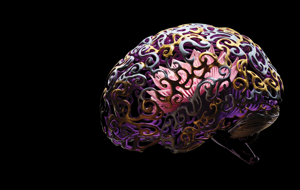
Sculpture by Joshua Harker
Why It Matters
A better understanding of how memories, emotions, and cognition work in the brain could lead to ways to improve and manipulate such functions.
What might be called the “make love, not war” branch of behavioral neuroscience began to take shape in (where else?) California several years ago, when researchers in David J. Anderson’s laboratory at Caltech decided to tackle the biology of aggression. They initiated the line of research by orchestrating the murine version of Fight Night: they goaded male mice into tangling with rival males and then, with painstaking molecular detective work, zeroed in on a smattering of cells in the hypothalamus that became active when the mice started to fight.

The hypothalamus is a small structure deep in the brain that, among other functions, coördinates sensory inputs—the appearance of a rival, for example—with instinctual behavioral responses. Back in the 1920s, Walter Hess of the University of Zurich (who would win a Nobel in 1949) had shown that if you stuck an electrode into the brain of a cat and electrically stimulated certain regions of the hypothalamus, you could turn a purring feline into a furry blur of aggression. Several interesting hypotheses tried to explain how and why that happened, but there was no way to test them. Like a lot of fundamental questions in brain science, the mystery of aggression didn’t go away over the past century—it just hit the usual empirical roadblocks. We had good questions but no technology to get at the answers.

By 2010, Anderson’s Caltech lab had begun to tease apart the underlying mechanisms and neural circuitry of aggression in their pugnacious mice. Armed with a series of new technologies that allowed them to focus on individual clumps of cells within brain regions, they stumbled onto a surprising anatomical discovery: the tiny part of the hypothalamus that seemed correlated with aggressive behavior was intertwined with the part associated with the impulse to mate. That small duchy of cells—the technical name is the ventromedial hypothalamus—turned out to be an assembly of roughly 5,000 neurons, all marbled together, some of them seemingly connected to copulating and others to fighting.

“There’s no such thing as a generic neuron,” says Anderson, who estimates that there may be up to 10,000 distinct classes of neurons in the brain. Even tiny regions of the brain contain a mixture, he says, and these neurons “often influence behavior in different, opposing directions.” In the case of the hypothalamus, some of the neurons seemed to become active during aggressive behavior, some of them during mating behavior, and a small subset—about 20 percent—during both fighting and mating.

That was a provocative discovery, but it was also a relic of old-style neuroscience. Being active was not the same as causing the behavior; it was just a correlation. How did the scientists know for sure what was triggering the behavior? Could they provoke a mouse to pick a fight simply by tickling a few cells in the hypothalamus?

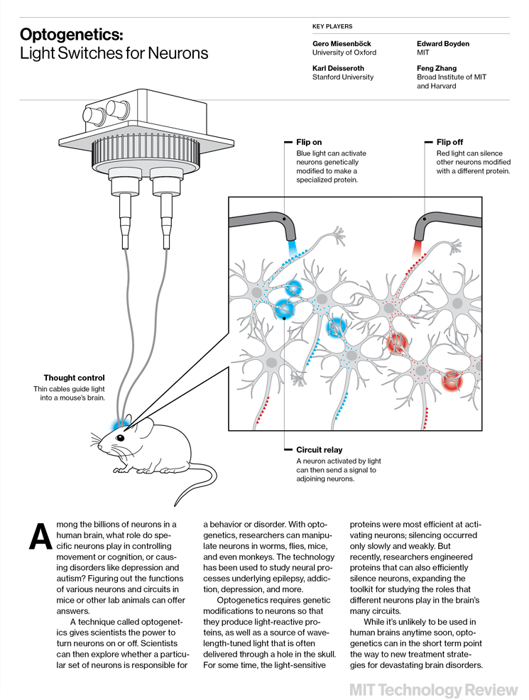
A decade ago, that would have been technologically impossible. But in the last 10 years, neuroscience has been transformed by a remarkable new technology called optogenetics, invented by scientists at Stanford University and first described in 2005. The Caltech researchers were able to insert a genetically modified light-sensitive gene into specific cells at particular locations in the brain of a living, breathing, feisty, and occasionally canoodling male mouse. Using a hair-thin fiber-optic thread inserted into that living brain, they could then turn the neurons in the hypothalamus on and off with a burst of light.


Optogenetics: Light Switches for Neurons

Anderson and his colleagues used optogenetics to produce a video dramatizing the love-hate tensions deep within rodents. It shows a male mouse doing what comes naturally, mating with a female, until the Caltech researchers switch on the light, at which instant the murine lothario flies into a rage. When the light is on, even a mild-mannered male mouse can be induced to attack whatever target happens to be nearby—his reproductive partner, another male mouse, a castrated male (normally not perceived as a threat), or, most improbably, a rubber glove dropped into the cage.

“Activating these neurons with optogenetic techniques is sufficient to activate aggressive behavior not only toward appropriate targets like another male mouse but also toward inappropriate targets, like females and even inanimate objects,” Anderson says. Conversely, researchers can inhibit these neurons in the middle of a fight by turning the light off, he says: “You can stop the fight dead in its tracks.”

Moreover, the research suggests that lovemaking overrides war-making in the calculus of behavior: the closer a mouse was to consummation of the reproductive act, the more resistant (or oblivious) he became to the light pulses that normally triggered aggression. In a paper published in Biological Psychiatry, titled “Optogenetics, Sex, and Violence in the Brain: Implications for Psychiatry,” Anderson noted, “Perhaps the imperative to ‘make love, not war’ is hard-wired into our nervous system, to a greater extent than we have realized.” We may be both lovers and fighters, with the slimmest of neurological distances separating the two impulses.

No one is suggesting that we’re on the verge of deploying neural circuit breakers to curb aggressive behavior. But, as Anderson points out, the research highlights a larger point about how a new technology can reinvent the way brain science is done. “The ability of optogenetics to turn a largely correlational field of science into one that tests causation has been transformative,” he says.

What’s radical about the technique is that it allows scientists to perturb a cell or a network of cells with exquisite precision, the key to sketching out the circuitry that affects various types of behavior. Whereas older technologies like imaging allowed researchers to watch the brain in action, optogenetics enables them to influence that action, tinkering with specific parts of the brain at specific times to see what happens.

And optogenetics is just one of a suite of revolutionary new tools that are likely to play leading roles in what looks like a heyday for neuroscience. Major initiatives in both the United States and Europe aspire to understand how the human brain—that tangled three-pound curd of neurons, connective tissue, and circuits—gives rise to everything from abstract thought to basic sensory processing to emotions like aggression. Consciousness, free will, memory, learning—they are all on the table now, as researchers use these tools to investigate how the brain achieves its seemingly mysterious effects (see “Searching for the “Free Will” Neuron”).

Connections
More than 2,000 years ago, Hippocrates noted that if you want to understand the mind, you must begin by studying the brain. Nothing has happened in the last two millennia to change that imperative—except the tools that neuroscience is bringing to the task.

The history of neuroscience, like the history of science itself, is often a story of new devices and new technologies. ­Luigi Galvani’s first accidental electrode, which provoked the twitch of a frog’s muscle, has inspired every subsequent electrical probe, from ­Walter Hess’s cat prod to the current therapeutic use of deep brain stimulation to treat Parkinson’s disease (approximately 30,000 people worldwide now have electrodes implanted in their brains to treat this condition). The patch clamp allowed neuroanatomists to see the ebb and flow of ions in a neuron as it prepares to fire. And little did Paul Lauterbur realize, when he focused a strong magnetic field on a single hapless clam in his lab at the State University of New York at Stony Brook in the early 1970s, that he and his colleagues were laying the groundwork for the magnetic resonance imaging (MRI) machines that have helped reveal the internal landscape and activity of a living brain.


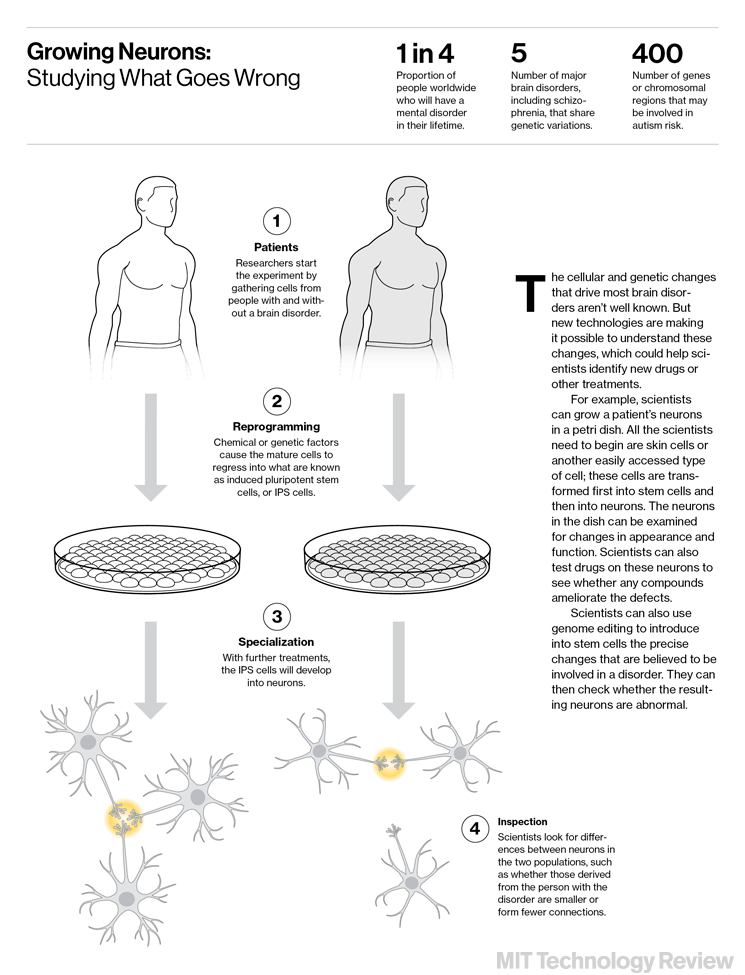
Growing Neurons: Studying What Goes Wrong

But it is the advances in genetics and genomic tools during the last few years that have truly revolutionized neuroscience. Those advances made the genetic manipulations at the heart of optogenetics possible. Even more recent genome-editing methods can be used to precisely alter the genetics of living cells in the lab. Along with optogenetics, these tools mean scientists can begin to pinpoint the function of the thousands of different types of nerve cells among the roughly 86 billion in the human brain.

Nothing testifies to the value of a new technology more than the number of scientists who rapidly adopt it and use it to claim new scientific territories. As Edward Boyden, a scientist at MIT who helped develop optogenetics, puts it, “Often when a new technology comes out, there’s a bit of a land grab.”

And even as researchers grab those opportunities in genomics and optogenetics, still other advances are coming on the scene. A new chemical treatment is making it possible to directly see nerve fibers in mammalian brains; robotic microelectrodes can eavesdrop on (and perturb) single cells in living animals; and more sophisticated imaging techniques let researchers match up nerve cells and fibers in brain slices to create a three-dimensional map of the connections. Using these tools together to build up an understanding of the brain’s activity, scientists hope to capture the biggest of cognitive game: memory, decision-­making, consciousness, psychiatric illnesses like anxiety and depression, and, yes, sex and violence.

In January 2013, the European Commission invested a billion euros in the launch of its Human Brain Project, a 10-year initiative to map out all the connections in the brain. Several months later, in April 2013, the Obama administration announced an initiative called Brain Research through Advanced Innovative Neurotechnologies (BRAIN), which is expected to pour as much as $1 billion into the field, with much of the early funding earmarked for technology development. Then there is the Human Connectome Project, which aims to use electron microscope images of sequential slices of brain tissue to map nerve cells and their connections in three dimensions. Complementary connectome and mapping initiatives are getting under way at the Howard Hughes Medical Institute in Virginia and the Allen Institute for Brain Science in Seattle. They are all part of a large global effort, both publicly and privately funded, to build a comprehensive picture of the human brain, from the level of genes and cells to that of connections and circuits.

Last December, as an initial step in the BRAIN Initiative, the National Institutes of Health solicited proposals for $40 million worth of projects on technology development in the neurosciences. “Why is the BRAIN Initiative putting such a heavy emphasis on technology?” says Cornelia Bargmann, the Rockefeller University neuroscientist who co-directs the planning process for the project. “The real goal is to understand how the brain works, at many levels, in space and time, in many different neurons, all at once. And what’s prevented us from understanding that is limitations in technology.”

Eavesdropping

Optogenetics had its origins in 2000, in late-night chitchat at Stanford University. There, neuroscientists Karl Deisseroth and Edward Boyden began to bounce ideas back and forth about ways to identify, and ultimately manipulate, the activity of specific brain circuits. Deisseroth, who had a PhD in neuroscience from Stanford, longed to understand (and someday treat) the mental afflictions that have vexed humankind since the era of Hippocrates, notably anxiety and depression (see “Shining Light on Madness”). Boyden, who was pursuing graduate work in brain function, had an omnivorous curiosity about neurotechnology. At first they dreamed about deploying tiny magnetic beads as a way to manipulate brain function in intact, living animals. But at some point during the next five years, a different kind of light bulb went off.

Since the 1970s, microbiologists had been studying a class of light-sensitive molecules known as rhodopsins, which had been identified in simple organisms like bacteria, fungi, and algae. These proteins act like gatekeepers along the cell wall; when they detect a particular wavelength of light, they either let ions into a cell or, conversely, let ions out of it. This ebb and flow of ions mirrors the process by which a neuron fires: the electrical charge within the nerve cell builds up until the cell unleashes a spike of electrical activity flowing along the length of its fiber (or axon) to the synapses, where the message is passed on to the next cell in the pathway. Scientists speculated that if you could smuggle the gene for one of these light-sensitive proteins into a neuron and then pulse the cell with light, you might trigger it to fire. Simply put, you could turn specific neurons in a conscious animal on—or off—with a burst of light.

In 2004, Deisseroth successfully inserted the gene for a light-sensitive molecule from algae into mammalian neurons in a dish. Deisseroth and ­Boyden went on to show that blue light could induce the neurons to fire. At about the same time, a graduate student named Feng Zhang joined Deisseroth’s lab. Zhang, who had acquired a precocious expertise in the techniques of both molecular biology and gene therapy as a high school student in Des Moines, Iowa, showed that the gene for the desired protein could be introduced into neurons by means of genetically engineered viruses. Again using pulses of blue light, the Stanford team then demonstrated that it could turn electrical pulses on and off in the virus-modified mammalian nerve cells. In a landmark paper that appeared in Nature Neuroscience in 2005 (after, Boyden says, it was rejected by Science), Deisseroth, Zhang, and Boyden described the technique. (No one would call it “optogenetics” for another year.)

Neuroscientists immediately seized on the power of the technique by inserting light-sensitive genes into living animals. Researchers in Deisseroth’s own lab used it to identify new pathways that control anxiety in mice, and both ­Deisseroth’s team and his collaborators at Mount Sinai Hospital in New York used it to turn depression on and off in rats and mice. And Susumu Tonegawa’s lab at MIT recently used optogenetics to create false memories in laboratory animals.

When I visited Boyden’s office at MIT’s Media Lab last December, the scientist called up his favorite recent papers involving optogenetics. In a rush of words as rapid as his keystrokes, Boyden described second-generation technologies already being developed. One involves eavesdropping on single nerve cells in anesthetized and conscious animals in order to see “the things roiling underneath the sea of activity” within a neuron when the animal is unconscious. Boyden said, “It literally sheds light on what it means to have thoughts and awareness and feelings.”

Boyden’s group had also just sent off a paper reporting a new twist on optogenetics: separate, independent neural pathways can be perturbed simultaneously with red and blue wavelengths of light. The technique has the potential to show how different circuits interact with and influence each other. His group is also working on “insanely dense” recording probes and microscopes that aspire to capture whole-brain activity. The ambitions are not modest. “Can you record all the cells in the brain,” he says, “so that you can watch thoughts or decisions or other complex phenomena emerge as you go from sensation to emotion to decision to action site?”


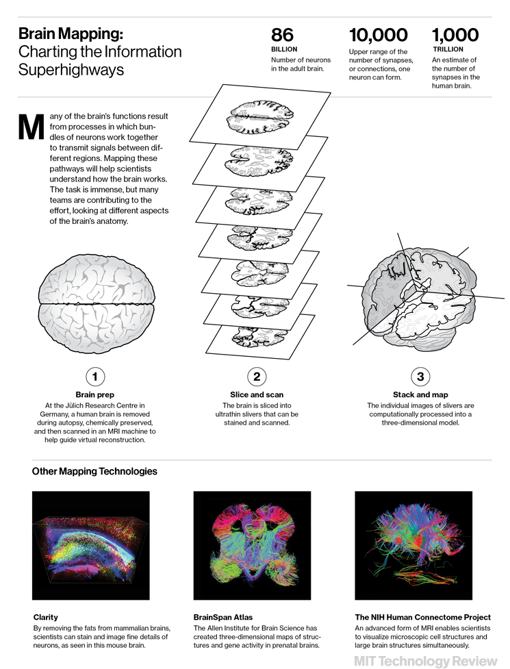
Brain Mapping: Charting the Information Superhighways

A few blocks away, Feng Zhang, who is now an assistant professor at MIT and a faculty member at the Broad Institute, listed age-old neuroscience questions that might now be attacked with the new technologies. “Can you do a memory upgrade and increase the capacity?” he asked. “How are neural circuits genetically encoded? How can you reprogram the genetic instructions? How do you fix the genetic mutations that cause miswiring or other malfunctions of the neural system? How do you make the old brain younger?”

In addition to helping to invent optogenetics, Zhang played a central role in developing a gene-editing technique called CRISPR (see “10 Breakthrough Technologies: Genome Editing,” May/June). The technology allows scientists to target a gene—in neurons, for example—and either delete or modify it. If it’s modified to include a mutation known or suspected to cause brain disorders, scientists can study the progression of those disorders in lab animals. Alternatively, researchers can use CRISPR in the lab to alter stem cells that can then be grown into neurons to see the effects.

Transparency

Back at Stanford, when he’s not seeing patients with autism spectrum disorders or depression in the clinic, Deisseroth continues to invent tools that he and others can use to study these conditions. Last summer, his lab reported a new way for scientists to visualize the cables of nerve fibers, known as “white matter,” that connect distant precincts of the brain. The technique, called Clarity, first immobilizes biomolecules such as protein and DNA in a plastic-like mesh that retains the physical integrity of a postmortem brain. Then researchers flush a kind of detergent through the mesh to dissolve all the fats in brain tissue that normally block light. The brain is rendered transparent, suddenly exposing the entire three-­dimensional wiring pattern to view.

Together, the new tools are transforming many conventional views in neuroscience. For example, as Deisseroth noted in a review article published earlier this year in Nature, optogenetics has challenged some of the ideas underlying deep brain stimulation, which has been widely used to treat everything from tremors and epilepsy to anxiety and obsessive-­compulsive disorder. No one knows just why it works, but the operating assumption has been that its therapeutic effects derive from electrical stimulation of very specific brain regions; neurosurgeons exert extraordinary effort to place electrodes with the utmost precision.

In 2009, however, Deisseroth and colleagues showed that specifically stimulating the white matter, the neural cables that happen to lie near the electrodes, produced the most robust clinical improvement in symptoms of Parkinson’s disease. In other words, it wasn’t the neighborhood of the brain that mattered so much as which neural highways happened to pass nearby. Scientists often employ words like “surprising” and “unexpected” to characterize such recent results, reflecting the impact that optogenetics has had on the understanding of psychiatric illness.

In the same vein, Caltech’s Anderson points out that the public and scientific infatuation with functional MRI studies over the last two decades has created the impression that certain regions of the brain act as “centers” of neural activity—that the amygdala is the “center” of fear, for example, or the hypothalamus is the “center” of aggression. But he likens fMRI to looking down on a nighttime landscape from an airplane at 30,000 feet and “trying to figure out what is going on in a single town.” Optogenetics, by contrast, has provided a much more detailed view of that tiny subdivision of cells in the hypothalamus, and thus a much more complex and nuanced picture of aggression. Activating specific neurons in that little town can tip an organism to make war, but activating the neurons next door can nudge it to make love.

The new techniques will give scientists the first glimpses of human cognition in action—a look at how thoughts, feelings, forebodings, and dysfunctional mental activity arise from the neural circuitry and from the activity of particular types of cells. Researchers are just beginning to gain these insights, but given the recent pace of technology development, the picture might emerge sooner than anyone dreamed possible when the light of optogenetics first flickered on a few years ago.

~ Stephen S. Hall is a science writer and author in New York City. His last feature for MIT Technology Review was “Repairing Bad Memories.”

Wednesday, October 02, 2013

Vaughan Bell - Changing Brains: Why Neuroscience Is Ending the Prozac Era

Last week in The Observer (UK), Vaughan Bell took a brief but important look at the quiet shift occurring right now in the neuroscience world in relation to treating mental illness. The idea of being able to fix anything and everything with a magic bullet pill is ending. Now scientists are looking more closely at brain networks, hoping to learn more about how the brain is functioning in depression, or schizophrenia, or some other symptom collection.

The idea is that cannot understand a dysfunction until we understand the range of "normal" functioning. With this shift comes the Research Domain Criteria or the RDoC Project, which will sooner or later replace the current DSM model used by psychiatrists.


This is a step in the right direction - but it also is a step further away from the real source of nearly ALL mental illness symptoms - relational dysfunction in the family of origin, among caretakers, or in some other form. Everything else is life - sometimes it sucks and we feel like hell, but it's not a mental illness, it's grief, or loss, or sadness.

Changing brains: Why neuroscience is ending the Prozac era


The big money has moved from developing psychiatric drugs to manipulating our brain networks


Vaughan Bell
The Observer, Saturday 21 September 2013


Mice are used in research into optogenetics, which suggests that even finer control of the brain may be possible. Photograph: John B Carnett/Popular Science via Getty Images

The psychiatric drug age may have reached its peak. Although mind-altering medications are being prescribed in record numbers, signs of a radically new approach to understanding and treating mental illness are emerging from the deep waters of neuroscience. No longer focused on developing pills, a huge research effort is now devoted to altering the function of specific neural circuits by physical intervention in the brain.

The starkest indication that drugs are increasingly being thought of as yesterday's cutting-edge comes from the little mentioned fact that almost all the major drug companies have closed or curtailed their drug discovery programmes for mental and neurological disorders. The realisation that there has been little in the way of genuine innovation since the major classes of psychiatric drugs were discovered in the 1950s has made future sales look bleak. New drugs have regularly appeared since then, often with fewer side effects, but most are little better in terms of effectiveness.

This is largely because these drugs tend not to be very specific in their effects on the brain. For example, the medication fluoxetine (better known as Prozac) alters levels of the neurotransmitter serotonin in brain networks related to mood, but it has the same effect in brain networks involved in sexual response, leading to the common side effect of difficulty with orgasm. The pharmaceutical holy grail has been to develop drugs that are more selective in their effects, but this multibillion dollar dream has largely been ditched by Big Pharma as too difficult.

In its place is a science focused on understanding the brain as a series of networks, each of which supports a different aspect of our experience and behaviour. By this analysis, the brain is a bit like a city: you can't make sense of the bigger picture without knowing how everything interacts. Relatively few residents of Belfast who live in the Shankill spend their money in the Falls Road and this tells us much more about the city – as these are the key loyalist and republican areas – than knowing that the average income of each area is much the same. Similarly, knowing that key brain areas interact differently when someone gets depressed tells us something important that a measure of average brain activity would miss.

The idea is that we can better understand complex human emotion and behaviour by understanding neural networks. This is where a new wave of interest is beginning to rise within neuroscience. The surge of interest is not with the concepts, which, if truth be told, became common currency in the mid-20th century, but in the extent to which research and treatment are being driven by a desire to identify and modify key brain circuits.

Big money has already been committed. The Obama White House has promised $3bn to develop technology to help identify brain circuits, while the National Institute of Mental Health has promised to move its seven-figure funding away from research into conditions such as schizophrenia and depression towards a system that looks at how brain networks contribute to difficulties that are shared across diagnoses. This project, given the unspectacular name Research Domain Criteria or the RDoC Project, is being cited as an eventual replacement for the diagnostic system used by current-day psychiatrists.

Perhaps more surprising for some is the explosion in deep brain stimulation procedures, where electrodes are implanted in the brains of patients to alter electronically the activity in specific neural circuits. Medtronic, just one of the manufacturers of these devices, claims that its stimulators have been used in more than 100,000 patients. Most of these involve well-tested and validated treatments for Parkinson's disease, but increasingly they are being trialled for a wider range of problems. Recent studies have examined direct brain stimulation for treating pain, epilepsy, eating disorders, addiction, controlling aggression, enhancing memory and for intervening in a range of other behavioural problems.

New technologies such as optogenetics suggest that even finer control of brain circuits may be possible. While deep brain stimulation involves stimulating the brain with electrical currents, optogenetics involves injecting neurons with a benign virus that contains the genetic information for light-sensitive proteins. The brain cells then become light sensitive themselves and their activity can be controlled with millisecond flashes of light sent through embedded fibre optic cables. Until now, this has only been demonstrated in animals but there are high hopes that it could lead to precisely controlled treatments in humans that intervene only in carefully selected brain circuits.

Let's make this clear. The scientific revolution in identifying and manipulating brain circuits is already under way. Additionally, with billions committed to research over the next 10 years, the medical revolution is likely to follow in the decades after. But a more important change will occur. Advances in neuroscience are not just discoveries, they also shape, as they always have done, how we view ourselves. As the Prozac nation fades, the empire of the circuit-based human will rise, probably to the point where dinner party chatter will include the misplaced jargon of systems neuroscience. But these are tools to help us understand humanity, not our humanity itself. Grief remains the loss of a loved one, joy a fulfilment of life's desires, and neither could be explained just by neural circuits. Life will still stretch beyond the confines of our inner worlds.