Showing posts with label University of Arizona. Show all posts

Friday, March 27, 2015

2014 Nobel Prize Winners Speak at the University of Arizona

Thursday afternoon, Edvard Moser, May-Britt Moser, and John O'Keefe, the three 2014 Nobel Prize winners for Physiology or Medicine, along with Eleanor Maguire, the winner of the 2003 Ig Nobel Prize for her "London taxi driver" study on hippocampal plasticity, spoke at a public forum at the University of Arizona.

Maquire's research on London taxi drivers, before and after going through two to three years of training required to learn and memorize 25,000 streets, revealed that their hippocampus grew as they memorized London's maze of streets. This was one of the first studies that demonstrated hippocampal neuroplasticity. She was awarded a 2003 Ig Nobel Prize for this study.

According to Improbable Research, the bestowers of the Ig Nobel Prize, "The Ig Nobel Prizes honor achievements that make people LAUGH, and then THINK. The prizes are intended to celebrate the unusual, honor the imaginative — and spur people's interest in science, medicine, and technology."

The Mosers and O'Keefe won their Nobel Prize in 2014 for their discoveries of specialized cells in the brain that together act as a navigation system. The Mosers discovered neurons that function as grid cells, found in the entorhinal cortex, while O'Keefe discovered neurons that function as place cells, found in the hippocampus.
"All memories are attached in some way to where you are, and in that way, the hippocampus acts as an anchor for remembering yourself within your experience," said Carol Barnes, Regents' Professor in the Departments of Psychology, Neurology and Neuroscience, the Evelyn F. McKnight Endowed Chair for Learning and Memory in Aging.
The hippocampus, according to Wikipedia, "belongs to the limbic system and plays important roles in the consolidation of information from short-term memory to long-term memory and spatial navigation." If a person were to suffer severe damage to the hippocampus (both hippocampi, as one exists in each brain hemisphere), it's likely they may acquire anterograde amnesia, an inability to form and/or retain new memories (episodic or autobiographical memory, which are forms of declarative memory). In Alzheimer's Disease, the hippocampus is one of the first brain regions to experience damage, resulting in the disorientation and loss of recognition so common in the disease. However, damage to the hippocampus does not inhibit the ability to learn new skills, such as riding a bicycle (procedural memory).
 
 Spatial firing patterns of 8 place cells recorded from the CA1 layer of a rat. The rat ran back and forth along an elevated track, stopping at each end to eat a small food reward. Dots indicate positions where action potentials were recorded, with color indicating which neuron emitted that action potential. [via Wikipedia]
Below, I have included the honored guests as well as the first half of the press release about the public forum (which is heavy on the U of A is awesome rhetoric).
Guests

Edvard Moser
, Professor and Director, Kavli Institute of Systems Neuroscience; Co-Director, Centre of Neural Computation, Norwegian University of Science and Technology


May-Britt Moser, Professor and Director, Centre of Neural Computation; Co-Director, Kavli Institute of Systems Neuroscience, Norwegian University of Science and Technology

The Moser’s received the 2014 Nobel Prize in Medicine or Physiology (along with J. O’Keefe) for this discovery of grid cells.

John O’Keefe, Professor, Institute of Cognitive Neuroscience and Department of Anatomy, University College London. Dr. O’Keefe received the 2014 Nobel Prize in Medicine or Physiology (along with the Moser’s) for his discovery of place cells.

Eileen O’Keefe
(John’s wife), Emeritus Professor, Public Health, London Metropolitan University


Eleanor Maguire, Professor of Cognitive Neuroscience, University College Dublin Dr. Maguire received the 2003 Ig Nobel Prize for Medicine for her ‘London taxi driver’ student on hippocampal plasticity.
And here is the beginning of the press release . . . 

Nobel Laureates Say UA Scientists Paved Way

By Daniel Stolte, University Relations - Communications | March 27, 2015
 
UA researcher Carol Barnes says the new Center for Innovation in Brain Science will serve as a hub of transdisciplinary research. (Photo: John de Dios/UANews)

 Four internationally renowned brain scholars visit campus and describe the UA as "one of the centers of neuroscience." The new Center for Innovation in Brain Science will foster transdisciplinary research, with the goal of better diagnostics and treatments for disorders such as Alzheimer's disease.

Nobel Prize laureate John O'Keefe, with May-Britt Moser (left) and Eleanor Maguire, says the UA's Carol Barnes "has told us as much about how the brain ages as anyone else." (Photo: John de Dios/UANews)

Take it from several Nobel laureates: Brain researchers at the University of Arizona are poised to make important contributions to finding better diagnoses and possibly treatments for brain disorders such as Alzheimer's disease.

To help commemorate three milestones in brain science research at the UA, four internationally renowned brain scholars — including three who shared the latest Nobel Prize in Physiology or Medicine — visited the UA campus this week to speak about their scientific careers and reflect on the tight connections they have shared with UA colleagues over many years.

This year marks the 25th anniversary of the UA Arizona Research Laboratories Division of Neural Systems, Memory and Aging, or NSMA; the 10th anniversary of the Evelyn F. McKnight Brain Institute at the UA; and the fifth anniversary of the UA School of Mind, Brain and Behavior.

UA President Ann Weaver Hart has named neuroscience as a research priority under the UA's strategic Never Settle plan. The BIO5 Institute and the UA Health Sciences Center have goals of supporting transdisciplinary neuroscience research in partnership with institutions across the state — from the molecular underpinnings of brain-cell health to the translation of this biological knowledge into treatments for neurological disease. The College of Science and the Office for Research and Discovery also are involved in supporting these efforts through the School of Mind, Brain and Behavior; NSMA; and the Evelyn F. McKnight Brain Institute.

During a public forum on Thursday, the UA welcomed the four guests to share their stories of discoveries in neuroscience with UA students, members of the public and the media. The visitors were John O'Keefe and Edvard and May-Britt Moser, who shared the 2014 Nobel Prize in Physiology or Medicine, and Eleanor Maguire, who received the Ig Nobel Prize for Medicine in 2003.

O'Keefe and the Mosers received the prize for their discoveries of specialized cells in the brain that together act like a navigation system.

"All memories are attached in some way to where you are, and in that way, the hippocampus acts as an anchor for remembering yourself within your experience," said Carol Barnes, who organized the visit along with two other UA brain researchers: Lynn Nadel, a Regents' Professor of Psychology and Cognitive Science and chair of the UA faculty; and Mary Peterson, professor of psychology and chair of the School of Mind, Brain and Behavior executive committee; director of the Cognitive Science Program; and chair of the Cognitive Science Graduate Interdisciplinary Program.

Edvard Moser said that some important work leading up to the Nobel Prize was done at the UA — for example, developing the technology for recording the activity of many brain cells at the same time, and developing ideas of how memory is generated in the hippocampus.

"The UA is one of the centers of neuroscience," O'Keefe said. "As one of the world's experts on the aging brain, Carol has told us as much about how the brain ages by looking at the hippocampus as anyone else.
Read the whole report on the event.

Tuesday, April 08, 2014

Searching for the Self: Morality Makes You Who You Are

This comes from the U of Arizona via the UA News blog. Shaun Nichols, a professor in the U of A’s Department of Philosophy, conducted a meta-study (looked at five other studies to find patterns) of how morality and sense of self are connected and found that personal morality is the most important part of defining individual identity.

Interesting.

They also found, in another study, that people's beliefs about whether or not the self changes over time affects their actions.

Also interesting.

Searching for the Self: Morality Makes You Who You Are

Personal moral traits are considered the most important part of individual identity, according to a new study co-authored by a UA philosophy professor.


By Shelley Littin, University Communications | April 4, 2014


What makes you who you are? Your experiences? Your memories? Your favorite foods?

None of these truly define us. What people believe really make us individuals are our personal moral traits, according to a new study co-authored by Shaun Nichols, a professor in the University of Arizona’s Department of Philosophy.

The research moves beyond the tradition in philosophy, which has focused on memory as the most important factor in defining the individual self, and introduces a broader perspective on the question of what makes you who you are.

“The philosophical tradition has emphasized the importance of memory - that you need to have the same memories to be the same person,” said Nichols, who co-authored the study with Nina Strohminger, a psychologist at Duke University.

“Previous studies had focused on the presence versus absence of memory,” Nichols said. “This paper looks at a variety of different dimensions, including morality, personality and intelligence, and every result came out with morality as the most important factor in defining the self. We found that memory was second place to morality by far.”

Morality, in this case, refers to personal moral traits such as the tendency to be kind or the tendency to be selfish, Nichols said.

“When you imagine yourself losing all of your memories that seems terrible,” he said. “But we found that for most people, the idea that somebody who used to be nice is now cruel would be considered a much more profound influence on their behavior than just forgetting various things.”

The paper, published in March in the journal Cognition, documents the results of five independent studies involving over 800 U.S. participants, all showing overwhelmingly that personal morality is considered the most important trait in defining the self.

The researchers compared moral personality traits, such as the tendency to be kind, with non-moral personality traits, such as the tendency to be shy.

“We found that the moral personality traits really were much more important to the way that people thought about the true self,” Nichols said.

“If you’re shy or artistic or a slow learner and those traits change, people don’t think that’s as much of a change in who you are,” Nichols said. “But if you go from being callous to being kind, people think that’s a big change.”

Nichols and Strohminger did not investigate whether an individual’s personal morality is likely to change over the course of their lifetime, but they did find that people’s belief about whether they will change affects their actions.

In an earlier study, also published in the journal Cognition, Nichols, along with Daniel Bartels of the University of Chicago and then UA graduate student Trevor Kvaran, investigated how perceptions of whether the self changes over time influences people’s actions. They found that people were more likely to give money to charity in the long term if they believed the self would change over time.

This is predicted by Buddhism, Nichols said. If people come to appreciate that they won’t be the same person over time, then they may be less concerned about how their life turns out in the end.

Now Nichols is gearing up for a new investigation into whether individual beliefs about whether or not the self will change over time affect attitudes about death in Christian, Hindu and Buddhist populations. That investigation is being supported by a $249,000 grant from the John Templeton Foundation. Nichols is leading one of 10 research teams selected for the Immortality Project, based at the University of California, Riverside.

Contacts


UANews Contact:
Shelley Littin
319-541-1482
littin@email.arizona.edu

Researcher Contact:
Shaun Nichols
520-626-0616
sbn@email.arizona.edu

Monday, February 17, 2014

You Can Teach a Damaged Brain New Tricks

Researchers at the University of Arizona (here in Tucson) have been identifying where language centers lie in the brain and how they can formulate therapies to restore function after those areas are damaged in stroke (or otherwise).

Pélagie Beeson, professor and head of Dept. of Speech Language and Hearing at the University of Arizona, will be speaking tonight as part of the UA College of Science’s series “The Evolving Brain.” These talks are FREE and therefore quite popular - get there early for good seating.

You can teach a damaged brain new tricks


Courtesy UA - Pélagie Beeson, professor and head of Dept. of Speech Language and Hearing at the University of Arizona.

February 11, 2014


Like much of what we know about the brain, knowledge of the areas involved in spoken and written language comes mainly from studying the loss of those abilities to trauma or disease.

Pélagie Beeson, who studies the neural substrates of written language, will talk Monday about “The Literate Brain.” She will describe where those language centers lie and how she and her colleagues formulate therapies to restore function after those areas are damaged.

The lecture, part of the UA College of Science’s series “The Evolving Brain,” is at 7 p.m. in Centennial Hall on the University of Arizona Mall. [See below for more info.]

Beeson is a professor and head of the Department of Speech, Language and Hearing Sciences at the University of Arizona, with a joint appointment in the Department of Neurology.

Speech and writing problems usually develop after damage to the left hemisphere of the brain, though a small number of people (usually left-handers) develop those skills in the right hemisphere, she said.

Through years of cataloging symptoms and imaging the brain, specialists such as Beeson and her research group have developed a pretty good understanding of where those centers are and can usually predict, before the brain is imaged, where the damage has occurred, based on symptoms.

Beeson’s Aphasia Research Project sees clients at least six months and sometimes years or decades after loss of speech or writing function.

That six-month period, during which there is ongoing therapy, is also the period in which the brain repairs itself to the extent that it can.

Subjects can continue to improve through behavioral therapy by harnessing other, usually nearby, parts of the brain, to perform functions previously performed by damaged cells, she said.

This is especially true for younger patients, whose “elastic” brains can often develop language capabilities in the right hemisphere, Beeson said. That elasticity vanishes with youth.

Aphasia is the term applied to the acquired impairment of language, usually after stroke or trauma, but sometimes occurring with progressive neurological diseases.

It can be spoken, written or both. The acquired impairment of reading is known as alexia, and the impairment of spelling and writing is called agraphia.

Researchers in the UA’s Aphasia Research Project have developed an algorithm called a “decision tree” that personalizes treatment based on symptoms and performance on a battery of tests.

Functional Magnetic Resonance Imaging of the brain, during which a subject performs reading and writing tasks, further refines the picture of the impairment by allowing researchers to see what parts of the brain “light up” when certain tasks are performed.

Those who suffer a stroke or brain injury have a wide variety of aphasia symptoms — ranging from those who have no understandable speech to those who talk easily and form sentences but “can’t pull up the right word,” Beeson said.

Beeson said she’s never had to advertise for subjects for her research.

She receives a steady stream of referrals from neurologists and therapists in the Tucson area and from elsewhere in the country.

Her department recently kept its No. 5 spot in the annual U.S. News and World Report ranking of speech-language pathology programs.

Part of the center’s charge is to spread the word about new therapies through publishing results and reporting them at conferences, she said.

Promulgation of the latest therapies is part of the charge given by the National Institute on Deafness and Other Communication Disorders, one of the National Institutes of Health, which partly funds the research.

If you go

What: The UA College of Science 2014 lecture series on "The Evolving Brain."
When: Mondays at 7 p.m.
Where: Centennial Hall, 1020 E. University Boulevard 1020 E. University (campus mall east of North Park Ave.)
Admission: Free
Parking: (Fee charged) Tyndall Avenue Garage, between North Tyndall and North Park avenues south of University Boulevard.
Information: Call 520-621-4090621-4090cq
  • Feb 17 (Monday): "The Literate Brain" by Pélagie M. Beeson, professor and head of Speech, Language and Hearing Science
  • March 3: "The Ancestors in Our Brains" by Dr. Katalin M. Gothard, associate professor of Physiology, Neurobiology, and the Evelyn F. McKnight Brain Institute
  • March 10: "More Perfect Than We Think" by William Bialek, the John Archibald Wheeler/Battelle Professor in Physics, Princeton University

Monday, October 14, 2013

Development of Neuronal Connections (Synapses) in the Brain - 2 New Studies

Two new studies last week offer insights into how neurons connect and form synapses, as well as providing some insight into neuroplasticity. This may seems pretty elementary on the surface, but keep in mind we drug the brain, operate on the brain, electroshock the brain, silence the brain, and manipulate it in many other ways, all the while being ignorant of the very basic process of how neurons get connected to each other to form synapses.

First up, a pair of researchers from the University of Arizona - this is a segment that aired on Arizona Public Media (NPR, PBS) - discuss the research their team conducted to bring a new understanding to how axons and dendrites are created.

UA Researchers Discover How Nerve Cells Make Connections

Story by Mary Olivas
October 1, 2013


University of Arizona researchers have made a discovery about how nerve cells make connections. This discovery is an important step towards curing conditions like spinal cord injuries and Alzheimer's.

Developed nerves have a short, truncated branch called an axon and long branch called a dendrite.

Sourav Ghosh, UA assistant professor of cellular and molecular medicine, said that this structure determines how nerves send signals.

"When nerves fire and communicate information, it's direction," Ghosh said. "It's sort of the basis for directionality of function."

Ghosh and UA doctoral candidate Sara Parker have been studying the polarity of neurons in their lab by closely examining a family of proteins and watching the neurons go through their development process.

Parker said she would knock down the levels of a protein or introduce excessive amounts of the protein to try and influence the establishment of a neuron's polarity.

"What we found is that when I disrupt this short form, I end up creating neurons developing multiple axons," Parker explained.

She added that usually neurons only have one axon.

This new understanding of how axons and dendrites are created could possibly help those with spinal injuries or neurodegenerative diseases.

"In traumatic injury what happens is that the axon is severed, but the cell is still alive and if we understand this mechanism and play with it to make the cell come up with a new axon, that would be exciting," Ghosh said.

Here is a more in-depth report on this study from Science Daily:

How Neurons Get Wired

Aug. 14, 2013 — Two different versions of the same signaling protein tell a nerve cell which end is which, UA researchers have discovered. The findings could help improve therapies for spinal injuries and neurodegenerative diseases.


University of Arizona scientists have discovered an unknown mechanism that establishes polarity in developing nerve cells. Understanding how nerve cells make connections is an important step in developing cures for nerve damage resulting from spinal cord injuries or neurodegenerative diseases such as Alzheimer's.

In a study published on Aug. 12 in the journal Proceedings of the National Academy of Sciences, UA doctoral student Sara Parker and her adviser, assistant professor of cellular and molecular medicine Sourav Ghosh, report that the decision which will be the "plus" and the "minus" end in a newborn nerve cell is made by a long and a short version of the same signaling molecule.

Nerve cells -- or neurons -- differ from many other cells by their highly asymmetric shape: Vaguely resembling a tree, a neuron has one long, trunk-like extension ending in a tuft of root-like bristles. This is called the axon. From the opposite end of the cell body sprout branch-like structures known as dendrites. By connecting the "branches" of their dendrites to the "root tips" of other neurons' axons, nerve cells form networks, which can be as simple as the few connections involved in the knee-jerk reflex or as complex as those in the human brain.

Parker and her team found that embryonic nerve cells manufacture a well-known signaling enzyme called Atypical Protein Kinase C (aPKC) in two varieties: a full-length one and a truncated one. Both varieties compete to bind the same molecular partner, a protein called Par3. If the short form of aPKC pairs up with Par3, it tells the cell to grow a dendrite, and if the long one pairs up with Par3, it will make an axon instead.

When the researchers blocked the production of the short form, the nerve cell grew multiple axons and no dendrites. When they created an artificial abundance of the short form, dendrites formed at the expense of axons. UA undergraduate student Sophie Hapak performed many of the experiments revealing how the two isoforms compete for Par3.

"We show that wiring a neuronal circuit is much more complex than previously thought," said Ghosh. "The process has a built-in robustness that explicitly defines which part of the cell is 'positive' and which is 'negative.'"

"In order to have a functioning neuronal circuit, you have to have receiving and sending ends," Parker said. "Initially, when a neuron is formed, it lacks the polarity it needs once it develops into a part of a circuit. The mechanism we discovered establishes that polarity."

"How the various brain regions are wired is the basis of emotion, memory and all cognitive functions," said Ghosh, who is a member of the UA's BIO5 Institute. "Establishing neuronal polarity in single neurons is absolutely essential for neuronal circuits to form."

"If we understand this mechanism, we could think about methods to spur new axons after the original ones were severed in a traumatic spinal cord injury, for example," Ghosh said.

The findings defy conventional wisdom, which maintains that a developing neuron will make dendrites by default unless instructed by the long form of aPKC to make an axon instead. By cultivating and studying neurons just after they formed, Parker and her group found that both forms of aPKC, long and short, are initially distributed equally throughout the cell. These forms subsequently segregate into different parts of the cell as the neuron matures and establishes polarity.

Because the cells were isolated from rat brains and kept in culture, the researchers could demonstrate that no external clues from other cells are needed to instruct a developing neuron. Whether the establishment of polarity is a random process or whether other signals yet to be identified play a role in regulating the abundance of the two aPKC varieties is not known.

Funded by the National Institutes of Health and a scholarship awarded to Parker by the Achievement Rewards for College Scientists Foundation, Inc., this research resulted from a collaboration of three UA departments -- cellular and molecular medicine, pharmacology and physiology.
The full article is behind a paywall, but here is the full citation:

S.S. Parker, E.K. Mandell, S.M. Hapak, I.Y. Maskaykina, Y. Kusne, J.-Y. Kim, J.K. Moy, P.A. St. John, J.M. Wilson, K.M. Gothard, T.J. Price, S. Ghosh. Competing molecular interactions of aPKC isoforms regulate neuronal polarity. Proceedings of the National Academy of Sciences, 2013; DOI: 10.1073/pnas.1301588110
 * * * * *

The second article looks at how synapses are formed - the original article is open access and will be partially presented below. Bur first, here is a summary press release posted at Science Codex.

New theory of synapse formation in the brain


Jülich, 10 October 2013 – The human brain keeps changing throughout a person's lifetime. New connections are continually created while synapses that are no longer in use degenerate. To date, little is known about the mechanisms behind these processes. Jülich neuroinformatician Dr. Markus Butz has now been able to ascribe the formation of new neural networks in the visual cortex to a simple homeostatic rule that is also the basis of many other self-regulating processes in nature. With this explanation, he and his colleague Dr. Arjen van Ooyen from Amsterdam also provide a new theory on the plasticity of the brain – and a novel approach to understanding learning processes and treating brain injuries and diseases.

The brains of adult humans are by no means hard wired. Scientists have repeatedly established this fact over the last few years using different imaging techniques. This so-called neuroplasticity not only plays a key role in learning processes, it also enables the brain to recover from injuries and compensate for the loss of functions. Researchers only recently found out that even in the adult brain, not only do existing synapses adapt to new circumstances, but new connections are constantly formed and reorganized. However, it was not yet known how these natural rearrangement processes are controlled in the brain. In the open-access journal PLOS Computational Biology, Butz and van Ooyen now present a simple rule that explains how these new networks of neurons are formed (DOI: 10.1371/journal.pcbi.1003259).

"It's very likely that the structural plasticity of the brain is the basis for long-term memory formation," says Markus Butz, who has been working at the recently established Simulation Laboratory Neuroscience at the Jülich Supercomputing Centre for the past few months. "And it's not just about learning. Following the amputation of extremities, brain injury, the onset of neurodegenerative diseases, and strokes, huge numbers of new synapses are formed in order to adapt the brain to the lasting changes in the patterns of incoming stimuli."

Activity regulates synapse formation

These results show that the formation of new synapses is driven by the tendency of neurons to maintain a 'pre-set' electrical activity level. If the average electric activity falls below a certain threshold, the neurons begin to actively build new contact points. These are the basis for new synapses that deliver additional input – the neuron firing rate increases. This also works the other way round: as soon as the activity level exceeds an upper limit, the number of synaptic connections is reduced to prevent any overexcitation – the neuron firing rate falls. Similar forms of homeostasis frequently occur in nature, for example in the regulation of body temperature and blood sugar levels.

However, Markus Butz stresses that this does not work without a certain minimal excitation of the neurons: "A neuron that no longer receives any stimuli loses even more synapses and will die off after some time. We must take this restriction into account if we want the results of our simulations to agree with observations." Using the visual cortex as an example, the neuroscientists have studied the principles according to which neurons form new connections and abandon existing synapses. In this region of the brain, about 10 % of the synapses are continuously regenerated. When the retina is damaged, this percentage increases even further. Using computer simulations, the authors succeeded in reconstructing the reorganization of the neurons in a way that conforms to experimental results from the visual cortex of mice and monkeys with damaged retinas.


(Photo Credit: Source: PLOS Computational Biology)

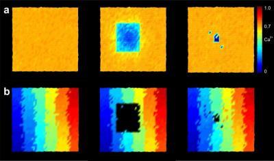
Reorganization in the visual cortex: before (left) and just after damage to the retina (centre), and in a later phase (right). Most of the neurons in the zone where images are projected from the damaged retina can achieve their original activity level again thanks to new connections formed with neighboring cells. The colors in the lower part of the image indicate the points on the retina from which the input to which the neurons show the strongest reaction is received.

The visual cortex is particularly suitable for demonstrating the new growth rule, because it has a property referred to as retinotopy: This means that points projected beside each other onto the retina are also arranged beside each other when they are projected onto the visual cortex, just like on a map. If areas of the retina are damaged, the cells onto which the associated images are projected receive different inputs. "In our simulations, you can see that areas which no longer receive any input from the retina start to build crosslinks, which allow them to receive more signals from their neighbouring cells," says Markus Butz. These crosslinks are formed slowly from the edge of the damaged area towards the centre, in a process resembling the healing of a wound, until the original activity level is more or less restored.

Synaptic and structural plasticity

"The new growth rule provides structural plasticity with a principle that is almost as simple as that of synaptic plasticity," says co-author Arjen van Ooyen, who has been working on models for the development of neural networks for decades. As early as 1949, psychology professor Donald Olding Hebb discovered that connections between neurons that are frequently activated will become stronger. Those that exchange little information will become weaker. Today, many scientists believe that this Hebbian principle plays a central role in learning and memory processes. While synaptic plasticity in involved primarily in short-term processes that take from a few milliseconds to several hours, structural plasticity extends over longer time scales, from several days to months.

Structural plasticity therefore plays a particularly important part during the (early) rehabilitation phase of patients affected by neurological diseases, which also lasts for weeks and months. The vision driving the project is that valuable ideas for the treatment of stroke patients could result from accurate predictions of synapse formation. If doctors knew how the brain structure of a patient will change and reorganize during treatment, they could determine the ideal times for phases of stimulation and rest, thus improving treatment efficiency.

New approach for numerous applications


"It was previously assumed that structural plasticity also follows the principle of Hebbian plasticity. The findings suggest that structural plasticity is governed by the homeostatic principle instead, which was not taken into consideration before," says Prof. Abigail Morrison, head of the Simulation Laboratory Neuroscience at Jülich. Her team is already integrating the new rule into the freely accessible simulation software NEST, which is used by numerous scientists worldwide.

These findings are also of relevance for the Human Brain Project. Neuroscientists, medical scientists, computer scientists, physicists, and mathematicians in Europe are working hand in hand to simulate the entire human brain on high-performance computers of the next generation in order to better understand how it functions. "Due to the complex synaptic circuitry in the human brain, it's not plausible that its fault tolerance and flexibility are achieved based on static connection rules. Models are therefore required for a self-organization process," says Prof. Markus Diesmann from Jülich's Institute of Neuroscience and Medicine, who is involved in the project. He heads Computational and Systems Neuroscience (INM-6), a subinstitute working at the interface between neuroscientific research and simulation technology.

Source: Forschungszentrum Juelich
The full article is open access and appeared at PLoS Computational Biology - below is the abstract, author's summary, and the full citation. Follow the title link to read the whole article.

A Simple Rule for Dendritic Spine and Axonal Bouton Formation Can Account for Cortical Reorganization after Focal Retinal Lesions


Markus Butz, Arjen van Ooyen

Abstract


Lasting alterations in sensory input trigger massive structural and functional adaptations in cortical networks. The principles governing these experience-dependent changes are, however, poorly understood. Here, we examine whether a simple rule based on the neurons' need for homeostasis in electrical activity may serve as driving force for cortical reorganization. According to this rule, a neuron creates new spines and boutons when its level of electrical activity is below a homeostatic set-point and decreases the number of spines and boutons when its activity exceeds this set-point. In addition, neurons need a minimum level of activity to form spines and boutons. Spine and bouton formation depends solely on the neuron's own activity level, and synapses are formed by merging spines and boutons independently of activity. Using a novel computational model, we show that this simple growth rule produces neuron and network changes as observed in the visual cortex after focal retinal lesions. In the model, as in the cortex, the turnover of dendritic spines was increased strongest in the center of the lesion projection zone, while axonal boutons displayed a marked overshoot followed by pruning. Moreover, the decrease in external input was compensated for by the formation of new horizontal connections, which caused a retinotopic remapping. Homeostatic regulation may provide a unifying framework for understanding cortical reorganization, including network repair in degenerative diseases or following focal stroke.

Author Summary


The adult brain is less hard-wired than traditionally thought. About ten percent of synapses in the mature visual cortex is continually replaced by new ones (structural plasticity). This percentage greatly increases after lasting changes in visual input. Due to the topographically organized nerve connections from the retina in the eye to the primary visual cortex in the brain, a small circumscribed lesion in the retina leads to a defined area in the cortex that is deprived of input. Recent experimental studies have revealed that axonal sprouting and dendritic spine turnover are massively increased in and around the cortical area that is deprived of input. However, the driving forces for this structural plasticity remain unclear. Using a novel computational model, we examine whether the need for activity homeostasis of individual neurons may drive cortical reorganization after lasting changes in input activity. We show that homeostatic growth rules indeed give rise to structural and functional reorganization of neuronal networks similar to the cortical reorganization observed experimentally. Understanding the principles of structural plasticity may eventually lead to novel treatment strategies for stimulating functional reorganization after brain damage and neurodegeneration.
Full Citation:  
Butz M, van Ooyen A. (2013, Oct 10). A Simple Rule for Dendritic Spine and Axonal Bouton Formation Can Account for Cortical Reorganization after Focal Retinal Lesions. PLoS Comput Biol 9(10): e1003259. doi:10.1371/journal.pcbi.1003259

Friday, July 26, 2013

Putting the Brakes on Inflammation - T-Cells and Protein S

Current research has linked heart disease, diabetes, depression, Alzheimer's Disease, stroke, and cancer to a common denominator - chronic inflammation. 

While inflammation is one of the body's defense mechanisms in response to injury and exogenous irritants, too much inflammation - resulting from an unhealthy diet (especially sugars and trans-fats), smoking, excessive body fat, or even constant stress or sleep deprivation - can cause the immune system to become over-activated and increase the risk for disease. When inflammation reaches chronic levels, it can damage the heart, damage brain cells, cause strokes, increase insulin resistance (primary cause of diabetes), and we now know that chronic inflammation is associated with the development of cancer.

Researchers led by the University of Arizona's Sourav Ghosh, an assistant professor in the department of cellular and molecular medicine at the UA College of Medicine, as well as a member of the University of Arizona Cancer Center, have identified a cellular signal that prevents the immune system from "spinning out of control."

Here is the press release about the study, from Daniel Stolte in the University Communications office.

Putting the Brakes on Inflammation

By Daniel Stolte, University Communications | July 22, 2013


Researchers have uncovered a signal that prevents the immune system from spinning out of control. The findings could help develop more effective therapies for autoimmune disorders, allergies, chronic inflammation and cancer.


In this microscopic image, three cells of the innate immune response (red with blue cell nuclei) are seen next to two T-cells. T-cells appear blue when they’re not active. Only when they make contact with and are activated by the innate cells do the T-cells turn on Protein S, causing them to glow green. (Photo: Lidia Bosurgi)
A team led by a University of Arizona researcher has discovered a previously unknown mechanism that prevents the immune system from going into overdrive, shedding light not only on how our body controls its response to pathogens but on conditions such as autoimmune diseases, allergies and chronic inflammation as well.

The group found a protein previously believed to only play a role in blood clotting acts as a negative feedback signal, telling defense cells to calm down, thereby preventing an immune reaction from spiraling out of control. The results, which could lead to new therapeutics for a variety of disorders caused by a faulty immune response, are published in the scientific journal Immunity. 
The researchers discovered a negative feedback loop preventing our immune system from overreacting: Dendritic cells (DC) activate T-cells, which, once activated, trigger a signal telling the dendritic cells to stop. (Illustration by Cynthia Orensztajn)
When pathogens such as viruses or bacteria invade our body, the immune system reacts by producing a flurry of chemical signals, known as chemokines that act as a bugle call recruiting specialized defender cells to the scene, such as macrophages, which devour the intruders. This first line of defense is known as inflammation.

"Inflammation is a necessary defense mechanism – you can't live without it," said Sourav Ghosh, assistant professor in the department of cellular and molecular medicine at the UA College of Medicine and lead author of the study. "On the flip side, if you can't regulate the inflammation, it can damage the body."

To be effective against pathogens, yet prevent collateral damage from the body's own defenses, the immune system has to maintain just the right level of inflammation, explained Ghosh, who is also a member of the University of Arizona Cancer Center and the UA's BIO5 Institute.

"It needs to be not too high and not too low," he said. "The question had always been, how does the immune system maintain that balance? Our discovery explains this."
Using a technique that makes the presence of Protein S visible under the microscope, samples taken from lymph nodes with resting T-cells reveal no sign of the protein (A), while dark staining indicates the presence of Protein S in activated T-cells (B). (Photo: Anthony Perry)
All organisms, even plants, have some kind of immune system at their disposal that acts as an army fighting against the onslaught of microbes, viruses, parasites and other pathogens in the environment. Vertebrates have evolved the most sophisticated arsenal of "soldiers" and "weapons," relying on two powerful lines of defense: a non-specific, or innate, immune response and the specific, or adaptive, immune response.

In the non-specific response, the immune system throws a first wave of countermeasures at the intruders, consisting of – among other things – aggressive chemicals, destructive enzymes and kamikaze-like neutrophils, specialized white blood cells that destroy the attackers by devouring them, killing themselves in the process.

"First you don't know who the enemy is, so you fire everywhere with your eyes closed," Ghosh explained. "But once you know the enemy, you need to shut off this first response firing and bring in the special ops so to speak."

The special ops come in the form of the specific immunity, capable of targeting pathogens very precisely, taking out the enemy in a sniper-like fashion, while sparing friendly microbes and cells belonging to the body. Most importantly, this portion of the immune system contains cells that remember every attacker trying to conquer an organism throughout its lifetime, allowing the immune system to summon the most effective, specialized task force to counter a pathogen it recognizes from a previous battle.

"The innate immune response is necessary to activate the adaptive response," Ghosh said. "But once activated, there has to be a mechanism that prevents the adaptive response from going into overdrive. From previous studies, we knew there had to be some kind of signal that does this, but we didn't know the nature of that signal. Now we do."

Two kinds of immune cells turned out to be the key players in mediating the immune response: the dendritic cells, so called because of the tree-like branches they grow during their development ("dendron" means "tree" in Greek), which belong to the first wave of defense; and the T-cells, so named because they mature in the thymus gland of the second, which are part of the second wave, the specific immune response.

"The dendritic cells activate the T-cells," Ghosh explained. "Only when they're activated, not when they're resting, do the T-cells produce this protein that we knew only from the blood clotting process, called Protein S."

The T-cells display Protein S on their surface, where it makes contact with a receptor the dendritic cells carry on their surface. This triggers a signal telling the dendritic cell to stop switching on T-cells, causing the immune response to slow down.

"We thought about which cells could be the source of that signal," said Carla Rothlin of the School of Medicine at Yale University, who led the study together with Ghosh. "You don't want to put the brakes on from the very beginning, or otherwise the immune response would never amount to anything. But you want to slow it down once it starts going too fast."

"We figured that once the specific response is underway, you don't really need the unspecific response anymore, so the T-cells appeared to be the best candidates for the source of this signal."

To test their hypothesis, the researchers studied the immune response in mice in which the gene coding for Protein S had been deactivated selectively in their T-cells, rendering them unable to communicate with the dendritic cells.

As expected, these mice were unable to regulate their immune response, resulting in higher levels of inflammation compared to their normal counterparts.

To assess the relevance of their findings to humans, Ghosh and his co-workers then studied blood from patients with inflammatory bowel diseases such as ulcerative colitis and Crohn's disease. Consistent with their previous results, patients suffering from increased inflammation had lower levels of Protein S in their blood stream compared to healthy volunteers.

The findings could help scientists and clinicians develop better treatments for inflammatory diseases, for example by designing drugs that substitute for insufficient Protein S. According to Ghosh, patients with inflammatory bowel disease are 20 times more likely to develop colon cancer, further underlining the significance of this study.

Study co-author Dr. Jonathan Leighton reported anecdotal evidence from the clinical practice that is in line with the dual roles Protein S is believed to play.

"Patients with inflammatory bowel disease can develop blood clots if they have active disease," said Leighton, a UA alumnus who holds the Chair of the Division of Gastroenterology at Mayo Clinic in Scottsdale, Ariz. "From a clinical standpoint, we think that three factors predispose to inflammation in inflammatory bowel disease – genetic, environmental and the immune system. This research is exciting because it focuses on the immune system. No one has found a consistent inflammatory pathway that explains all the clinical manifestations, and it may be that different pathways are affected in different patients. We don't understand how it all relates quite yet, but this study is a step toward a better understanding that will ultimately help us treat patients more effectively."
Abstract and Citation:
T Cell-Derived Protein S Engages TAM Receptor Signaling in Dendritic Cells to Control the Magnitude of the Immune Response

Immunity, Volume 39, Issue 1, 160-170, 11 July 2013
Copyright © 2013 Elsevier Inc. All rights reserved.
DOI: 10.1016/j.immuni.2013.06.010

Authors:
Eugenio A. Carrera Silva, Pamela Y. Chan, Leonel Joannas, Andrea E. Errasti, Nicola Gagliani, Lidia Bosurgi, Maurice Jabbour, Anthony Perry, Faye Smith-Chakmakova, Daniel Mucida, Hilde Cheroutre, Tal Burstyn-Cohen, Jonathan A. Leighton, Greg Lemke, Sourav Ghosh, Carla V. Rothlin

Highlights
  • Activated T cells express Protein S (Pros1)
  • T cell-derived Pros1 limits DC activation through the TAM receptor tyrosine kinases
  • Ablation of Pros1 in mouse T cells enhances immune responses
  • PROS1 expression and function is conserved in human T cells
Summary

Dendritic cell (DC) activation is essential for the induction of immune defense against pathogens, yet needs to be tightly controlled to avoid chronic inflammation and exaggerated immune responses. Here, we identify a mechanism of immune homeostasis by which adaptive immunity, once triggered, tempers DC activation and prevents overreactive immune responses. T cells, once activated, produced Protein S (Pros1) that signaled through TAM receptor tyrosine kinases in DCs to limit the magnitude of DC activation. Genetic ablation of Pros1 in mouse T cells led to increased expression of costimulatory molecules and cytokines in DCs and enhanced immune responses to T cell-dependent antigens, as well as increased colitis. Additionally, PROS1 was expressed in activated human T cells, and its ability to regulate DC activation was conserved. Our results identify a heretofore unrecognized, homeostatic negative feedback mechanism at the interface of adaptive and innate immunity that maintains the physiological magnitude of the immune response.

Friday, July 19, 2013

Dr. Anne E. Cress Named Interim Director of University of Arizona Cancer Center

I want to give a shout out and congratulations to my friend and personal training client, Dr. Anne Cress, the new Interim Director of the University of Arizona Cancer Center. They are lucky to have her wisdom, intelligence, creativity, and dedication to integrity - traits I have enjoyed in her for more than six years.

She is without doubt one of my most dedicated and hard-working clients - and she makes me laugh and think, sometimes simultaneously, in every session.

Deep bow of respect.

Anne E. Cress Named Interim Director of UA Cancer Center

By Sara Hammond, University of Arizona Cancer Center | July 19, 2013

Cress succeeds Dr. David S. Alberts, who served as cancer center director for more than eight years until his recent retirement


Anne E. Cress, professor of cellular and molecular medicine and radiation oncology at the University of Arizona College of Medicine, has been named interim director of the University of Arizona Cancer Center. The appointment was made by Dr. Joe G.N. "Skip" Garcia, incoming UA senior vice president for health sciences, and Dr. Steve Goldschmid, dean of the UA College of Medicine.

Cress, who also is deputy dean for research affairs at the UA College of Medicine, succeeds Dr. David S. Alberts, who served as cancer center director for more than eight years until his recent retirement. Alberts remains at the UA Cancer Center to conduct research.

Cress was named a research associate in 1980 and became a research assistant professor in 1981, both in the UA's division of radiation oncology. She was named an associate professor in 1990 and attained full professorship with tenure in 1996. She was appointed professor of cell biology and anatomy (now cellular and molecular medicine) in 1999. She has been a UACC member since 1987. She also served as the College of Medicine's associate dean for research and academic affairs for six years. Her research has continuously been supported since 1981 by extramural research grants from the National Institutes of Health, the Department of Defense, and the American Cancer Society.

"Anne Cress is one of the top cancer biology scientists in the world," said Garcia. "Her breadth of knowledge about translational approaches to cancer will be crucial as she leads the UA's comprehensive cancer center through this time of transition."

Goldschmid added: "Dr. Cress's service to the University of Arizona, especially within the College of Medicine, is unparalled. Between her extensive research credentials and her administrative leadership, she is unquestionably the right person to take the reins of our cancer center at this critical time."

Cress's research is focused on stopping tumor cells "in their tracks" and preventing tumor metastasis. She is examining how cell surface molecules, called integrins, behave abnormally in tumor cells, enabling their transit along vessels and nerves to their final destination in bone.

Through her collaborative research, Cress is helping to design strategies that will prevent these harmful and painful processes from occurring and at the same time providing new ways for physicians to determine if a patient's cancer is likely to spread.

Cress has a 30-year history of contributions to interdisciplinary science through more than 100 original research publications, several major federal and foundation grant and patent awards, and national study section service chairmanships, including those organized by the National Institutes of Health, Department of Defense, and the American Cancer Society.

She received her bachelor of science in microbiology/chemistry and a doctorate in biochemistry/molecular biology from the UA. She completed postdoctoral work in cancer biology at the UA and served as a visiting professor in biochemistry at Stanford University, and completed visiting scholar sabbaticals at the Drug Development and Design Center in Brisbane, Australia, and with the Developmental Biology Group at Netherlands Cancer Institute in Amsterdam.

Her awards and honors include: Outstanding Research Investigator Award, Moffitt Cancer Center, 1999; Arizona Health Sciences Center Founder's Day Award, 2005; Elkin Award for Cancer Biology Research, Emory University, 2005; UA Cancer Center's Sydney E. Salmon, MD, Senior Investigator Award, 2008.

Sunday, March 10, 2013

Super-Change Agent - Susan Stryker Is Changing the Conversation on Transgender Issues


From the Tucson Weekly, a very cool article on Susan Stryker and her work with transgender issues at the University of Arizona Institute of LGBT Studies. Here is a little biography from Wikipedia:
Susan O'Neal Stryker is a US American professor, author, filmmaker, and theorist of gender and sexuality. She is currently an associate professor of Gender and Women's Studies at the University of Arizona, and is the director of the university's Institute for LGBT Studies.[3][4] She has served as a visiting professor at Harvard University, University of California, Santa Cruz, and Simon Fraser University.[5] Stryker is an openly lesbian trans woman who has written extensively about transgenderism and queer culture.[6]

Stryker received a bachelor's degree in Letters from University of Oklahoma in 1983. She earned a Ph.D. in United States History at the University of California, Berkeley in 1992;[7] the doctoral thesis she presented was Making Mormonism: A Critical and Historical Analysis of Cultural Formation.[8] She began to transition from man to woman shortly after earning her doctorate.[9] In 1994, her essay "My Words to Victor Frankenstein Above the Village of Chamounix" became the first article to be published in a peer-reviewed academic journal by an openly transgender author.[10]

Stryker was later awarded a postdoctoral research fellowship in human sexuality studies at Stanford University, sponsored by the Social Science Research Council and the Ford Foundation.[5] From 1999 to 2003, she was the executive director of the GLBT Historical Society.
This is a nice profile of her work.

Super-Change Agent

Susan Stryker is changing the conversation on transgender issues and more at UA Institute of LGBT Studies


CURTIS RYAN - Susan Stryker at the Institute for the LGBT Studies
at the University of Arizona.


If you missed viewing excerpts from Susan Stryker's film Christine in the Cutting Room at downtown's Playground Bar & Lounge, I feel your pain.

After sitting in Stryker's office and listening to her talk about her project, which focuses on the world's most famous transsexual, Christine Jorgensen, I'm kicking myself for not being there.

Stryker, associate professor of gender and women's studies and director of the Institute for LGBT Studies at the UA, says she wants to change how we think about queer media with this project. Jorgensen wasn't the first person to change genders. But in 1952 she became a celebrity because of that decision and the times in which her transition took place.

The clip Stryker plays on her office computer practically jumps off the screen: a narrator speaks from Jorgensen's point of view, with film and photos of Jorgensen mixed in with clips that highlight the tensions of the era, such as fear of communism, fear of the unknown and fear of the nuclear bomb. It's what Stryker describes in the clip as "the Age of Anxiety."

Stryker says the project, when complete, won't be a typical documentary. It's not, for instance, like her previous film, Screaming Queens: The Riot at Compton's Cafeteria, which won a 2006 Emmy for telling the story of what's considered the first militant queer resistance to police harassment in San Francisco, three years before Stonewall in New York.

"This is a multimedia experience. It's going to be more about using Christine Jorgensen's story to explore the relationship between embodiment and image, and between cinema and surgery, than just a typical bio-pic. Jorgensen is a really interesting case," Stryker says.

"She was trained as a photographer and film editor before she became a celebrity for being transsexual. What I see when I look at her is somebody who knew how to make images whichever side of the camera she was on. Some of her appearance-making practices involved cutting film, and some involved cutting her body. I want to look at the relationship between those practices in my media project, and make people conscious of the different 'cutting' practices going on in her work through how I present her story. I want people to interact with the media in lots of different ways, not just seeing an image on a screen."

During two sit-downs with Stryker, the word change comes up often in our conversations. There's no doubt that this historian, academician, filmmaker and activist is a change agent through her work, and she's also amazingly persistent.

"If we talked 20 years ago about what I was going to do, if I said, 'Help create a field in transgender studies, teach graduate students, create a graduate program, help create a whole new field,' you'd have ..." Stryker pauses and feigns a yawn. "OK, maybe you would have rolled your eyes."

Back then, the ideas of marriage equality and of gays and lesbians serving openly in the military were barely a dream in the LGBT community. So, yeah, Stryker is probably right. People would have rolled their eyes, or at least been patronizingly nice.

But Stryker, a transsexual lesbian woman, decided 20 years ago it was time to change the conversation on transgender people, and that's exactly what she's done.

"I decided 20 some years ago that—partly out a sense of outrage—I was going to change things. Not in a kind of a heroic sort of way and certainly not feeling like I'm the only person who ever felt like this. There's a real social movement of people working in many different fronts on this," Stryker says.

"I just decided, look, what do I have to bring to the table in this social justice struggle? I am a historian. I'm an academic. What do I do? I change how people think ..."

Stryker has attained many of her goals. In fact, she's accomplishing more of them right now, such as with the release of The Transgender Studies Reader 2, which she co-edited and contributed to; the creation of a scholarly transgender studies journal; and teaching transgender studies to graduate students.

Stryker was the keynote speaker at Wingspan's annual dinner last year. She outlined a career that started in the world of academia when she received her doctorate at the University of California, Berkeley. Her dissertation was titled Marking Mormonism: A Critical and Historical Analysis of Cultural Formation. Stryker also began to transition from man to woman soon after she got her Ph.D.

Stryker did postdoctoral research on human sexuality at Stanford University, and then forged a life as a visiting professor, including at Harvard University; the University of California, Santa Cruz; and Simon Fraser University in British Columbia. She also spent time away from academia, working from 1999 to 2003 as executive director of the GLBT Historical Society in San Francisco.

Her first book, published in 1996, was Gay by the Bay: A History of Queer Culture in the San Francisco Bay Area. It was followed by Queer Pulp: Perverted Passions from the Golden Age of the Paperback in 2001, and Transgender History, published in 2008. There have been dozens of scholarly papers and other works in between.

Stryker left her job as associate professor of gender studies at Indiana University to come to Arizona in 2011, replacing Eithne Luibheid, founding director of the Institute for LGBT Studies. Stryker says the institute's origins stretch back to the 1990s, when faculty and staff from several UA departments—women's studies, anthropology and others—came together to do more LGBT-centered programming and, eventually, classes.

When the institute was formally created, the UA asked Luibheid to make a five-year commitment, which she fulfilled, returning to full-time faculty life in gender and women's studies when Stryker was hired. During Luibheid's tenure, the institute received its first major gift, from Jim Leos and Clint McCall. Other fund-raising efforts through private donations and the UA president's office helped in the creation of the Miranda Joseph Endowed Lecture Series, which continues to bring LGBT scholars to the UA.

Under Luibheid's direction, the institute funded research projects and helped create five courses in LGBT studies at the UA. Other projects included a public forum with Derechos Humanos on the needs of LGBT immigrants and refugees.

During her keynote talk at the Wingspan dinner, Stryker remarked that she feels lucky that she's found meaningful work in academia, but noted that, "I had to help create the field I work in, in order to get hired to be an expert in this field."

Stryker says she's striving to honor the institute's original vision: focusing on research, class development and bringing in scholars. There's also the work of raising funds. Stryker recently announced another gift from Leos, a Tucsonan and UA alum, who pledged $40,000 a year for the next five years to benefit the institute and the lecture series. When she's not raising funds or promoting the institute, Stryker is focused on teaching. She's currently working with graduate students, and next fall she'll work with undergraduate students. She will be working with gender studies majors, and next spring she will teach a class she created, Sex, Gender and Technology. "It's the kind of class that will get butts in seats," Stryker says, smiling.

There's also the Jorgensen film project and another book she's working on.

"The problem is that there's not enough time in the day to do everything that I want to do," Stryker says. "I like my teaching, and I like having the chance to build a research unit on campus. It was already here when I came, but I like the work of making it bigger, faster, and stronger."

One goal that will soon be accomplished is the publication, starting next year, Transgender Studies Quarterly, a scholarly journal to be published by Duke University Press. Stryker is co-editor.

Stryker turns to a web page dedicated to the journal on the institute's website and notes the tag line: "Changing the way the world thinks about transgender issues."

Then I ask her facetiously, "Is it time to say the world you wanted to change 20 years ago has indeed changed?"

No, Stryker says, there's still a lot that needs to happen. One of her goals is to establish a master's degree program in transgender studies for pre-med students and others who want to work with trans clients.

There's also a project under development at the institute to extend its work to the greater community. "How do we be an engine to make a difference?" Stryker asks. "How do you drive change and how do you do it using a queer perspective?"

What Stryker wants to do, and in many ways already has, is spark conversations—and action—beyond the UA campus. The institute recently hosted Karma R. Chávez of the University of Wisconsin who spoke about queer leadership in the DREAMer immigrant rights movement and its strategy to force the federal government to provide protected legal status to prevent an estimated one million undocumented youth from being deported.

"We are a way for LGBT communities to interact with the university and vice versa, and there is always a divide that has to be overcome, such as those divisions that exist between those who have found a way to access higher education and those who haven't," Stryker says.

It's part of Stryker's work to change how people think, whether its on gay and lesbian issues or trans issues. The LGBT population, she says, remains historically underserved. "We need to do things and bring resources to the table for projects and issues through programming or making donations to the community. Given that we've received significant financial support from members of the Tucson LGBT community, we think it's right to put that money into programs that really serve the community—we've plowed that money back into community events like the Lesbian Looks and the Out in the Desert film programs, doing programs with Fluxx, and Wingspan, and the Southern Arizona AIDS Foundation."

Changing the perspective on LGBT issues has been a long-term process, she says. "I've come a fair ways down that road in 20 years. I don't talk about (my own transition) so much. I just do what I do. I really try to get away from that since it's not the only thing I have to say about myself. We know a lot of gay people who aren't trans, and is the only thing we can say (to them), 'Can you tell me what it was like to come out?' What about, 'Did you like that movie? What are your politics about food? Who do you love?' The coming-out story is not the thing that defines someone."

Stryker says she's not into before-and-after pictures, and she never tells people what her name was before she transitioned. "If someone wants to dig around they could find it."

None of that really matters to her, because Stryker has work to do. She knows she's cast a big net of big dreams. Her life, she says, is about swinging for the fences and dreaming big, although she doesn't expect to hit a home run every time.

"Rather than saying I am going to plot my next move very carefully in a very cautious manner, I might actually start 10 things and eight of them will actually happen," Stryker says.

"It's always been about changing the conversation and changing the way people think. Transgender issues are no longer a small, obscure topic. How we see gender is changing at a global level. I see that in our work here at the UA. Investigating transgender becomes a way to view and study how that system is changing."

Friday, October 07, 2011

Toward a Science of Consciousness Conference Announcement and Call for Abstracts

The 2012 Toward a Science of Consciousness Conference is back in Tucson - and they have just issued the call for abstracts. Here are the details.

Conference Announcement and Call for Abstracts


TOWARD A SCIENCE OF CONSCIOUSNESS
April 9-14, 2012
Loews Ventana Canyon Resort Hotel
Tucson, Arizona


Sponsored by:
The Center for Consciousness Studies,
The University of Arizona
http://www.consciousness.arizona.edu


Toward a Science of Consciousness (TSC) conferences have been held annually since 1994, alternating between Tucson, Arizona and other locations around the world. The tenth biennial 'Tucson Conference', Toward a Science of Consciousness will take place April 9-14, 2012.


Known for rigorous and leading edge approaches to all aspects of the study of conscious experience, TSC includes neuroscience, cognitive science, artificial intelligence, psychology, philosophy, neurobiology, medicine, quantum physics and cosmology as well as art, mind technology and experiential and contemplative approaches. TSC is the largest interdisciplinary gathering probing fundamental questions related to conscious experience. An estimated 500 scientists, philosophers, psychologists, experientialists, artists and students from over 60 countries are due to take part.


For the first time, Toward a Science of Consciousness will be held at the Loews Ventana Canyon Resort Hotel in the Catalina Foothills above Tucson, Arizona (with a special conference rate of 99 dollars/room/night). See: http://www.loewshotels.com/en/Ventana-Canyon-Resort


Plenary/Keynote Session themes will include:
HOT or NOT: Debate on higher-order theories of consciousness
War of the Worldviews: Chopra and Mlodinow on consciousness
Consciousness and Echolocation
Fractal consciousness: Scale-free brain structure and dynamics
Retrocausality and consciousness
Searching for consciousness in coma and anesthesia


Speakers will include:
Daryl Bem
Ned Block
Melanie Boly
Deepak Chopra
Biyu Jade He
Daniel Kish
Victor Lamme
Hakwan Lau
George Mashour
Leonard Mlodinow
David Rosenthal
...and others


As in previous conferences, program sessions will include Plenary and Keynote talks, Concurrent talks, Posters, Art/Science demos and exhibits, Pre-Conference workshops, Side trips and Social events in the Tucson conference tradition. For Information see: http://www.consciousness.arizona.edu/


Abstract Submission
TSC 2012 Conference Abstract Submission System is now open. Abstracts considered for Plenary, Concurrent, Poster and Art/Tech Demo sessions.
See: http://sbs.arizona.edu/project/consciousness/index.php


Schedule of Deadlines -- Tentative
December 10 Abstracts Due
January 5 Decisions
January 15 Early Registration Due
March 1 Final Abstract Edits Due


Call for Pre-Conference Workshop proposals
Toward a Science of Consciousness 2012
April 9-14, 2012
http://www.consciousness.arizona.edu


Proposals for pre-conference workshops are invited in all areas related to understanding conscious experience. Workshops provide in depth, detailed treatments of various methodologies, perspectives, reviews and approaches. Workshops may be solo presentations, or include two or more presenters. Attendance fees are $60 for 4 hour workshops and $80 for full day workshops, split evenly between presenter(s) and the conference which provides the room, A-V, promotion/advertisement and refreshments. A minimum number of registrants will be required.


The conference Plenary program opens Tuesday April 10 at 1:45 pm.
The pre-conference workshops will be held in three 4 hour sessions
1) Monday April 9th, 9 am to 1 pm
2) Monday April 9th, 2 pm to 6 pm
3) Tuesday April 10th, 9 am to 1 pm


Workshop presenters (up to 2 per workshop) also receive free registration for the conference. Please submit a 500 word (or less) abstract/summary and presenter information by email directly to center@email.arizona.edu


Deadline for Workshop proposals is November 1
Submitters will be notified by November 15


On behalf of the Program Committee - Toward a Science of Consciousness 2012.
David Chalmers, Australian National University, Co-Chair
Stuart Hameroff, University of Arizona, Co-Chair
Uriah Kriegel, University of Arizona
Hakwan Lau, Columbia University
Marilyn Schlitz, Institute of Noetic Sciences
Heather Berlin, Mount Sinai Medical Center
Jonathan Schooler, University of California, Santa Barbara
Melanie Boly, University of Liege
Moran Cerf, UCLA/NYU
Abi Behar-Montefiore, conference manager, center@u.arizona.edu


Contact:
Abi Behar-Montefiore, Manager, Center for Consciousness Studies, University of Arizona - center@u.arizona.edu


CONFERENCE LINKS
CCS WEBSITE www.consciousness.arizona.edu
CCS /TSC CONFERENCE REGISTRATION and ABSTRACT SUBMISSION
http://sbs.arizona.edu/project/consciousness/index.php
CONFERENCE HOTEL BOOKING http://www.loewshotels.com/en/Ventana-Canyon-Resort/GroupPages/Consciousness


Follow us:
Facebook http://www.facebook.com/pages/Center-for-Consciousness-Studies/221542854531534
Twitter http://twitter.com/#!/brainyday