Showing posts with label neuroinflammation. Show all posts

Wednesday, March 04, 2015

Psychosocial Stress, Inflammation, and Adhesion Molecules

http://ecx.images-amazon.com/images/I/51egCAixgxL._SY344_BO1,204,203,200_.jpg

I have been re-reading a book by Joseph LeDoux (NYU), Synaptic Self: How Our Brains Become Who We Are (2002), in which he argues that synapses in the brain are the foundation of personality, the basis of our sense of self:

My notion of personality is pretty simple: it‟s that your “self,” the essence of who you are, reflects patterns of interconnectivity between neurons in your brain. Connections between neurons, known as synapses, are the main channels of information flow and storage in the brain. Most of what the brain does is accomplished by synaptic transmission between neurons, and by calling upon the information encoded by past transmission across synapses. (p. 3-4)

This is relevant to some of the work I have been doing for Dr. Cress at the University of Arizona Cancer Center. Dr. Cress has been working for many years on the function of integrins in cancer metastasis, specifically in prostate cancer.

Here is how integrins are defined at Wikipedia, which is as precise as any you will find.
Integrins are transmembrane receptors that are the bridges for cell-cell and cell-extracellular matrix (ECM) interactions. When triggered, integrins in turn trigger chemical pathways to the interior (signal transduction), such as the chemical composition and mechanical status of the ECM, which results in a response (activation of transcription) such as regulation of the cell cycle, cell shape, and/or motility; or new receptors being added to the cell membrane. This allows rapid and flexible responses to events at the cell surface, for example to signal platelets to initiate an interaction with coagulation factors.

There are several types of integrins, and a cell may have several types on its surface. Integrins are found in all metazoa.[3]

Integrins work alongside other receptors such as cadherins, the immunoglobulin superfamily cell adhesion molecules, selectins and syndecans to mediate cell–cell and cell–matrix interaction. Ligands for integrins include fibronectin, vitronectin, collagen, and laminin.
Integrins are heterdimic adhesion receptors, meaning they have two different parts, the α (alpha) and β (beta) subunits. There are at least 18 α and eight β subunits are known in humans and other vertebrates (Takada, Ye, & Simon, 2007).

Dr. Cress has been work with the A6B4 integrin (alpha 6 beta 4) and its role in metastatic prostate cancer. A6 is one of three laminin-binding molecules in humans (the others are A3 and A7), although other researchers suggest there are five laminin-binding integrins, A1, A2, A3, A6, A7 (Alberts, Johnson, & Lewis; 2007; Molecular Biology of the Cell. 5th edition). This graphic explains the functions of the different integrins and their combinations.


Integrins are also expressed in the brain, especially in synapses and in the lamination of axons, which allows information to more quickly and efficiently through the brain (and through neural cells throughout the body). Schwann cells in peripheral nerves interact with axons and extracellular matrix (ECM) as part of their work in ensheathing and myelinating axons and they express B4 (beta 4 integrin) [Feltri, et al, 1994], and specifically A6B4. 

In digging around for additional information on synapses and brain function, I found a related article by LeDoux - this is the abstract (crucial section is in bold):

STRUCTURAL PLASTICITY AND MEMORY
Raphael Lamprecht and Joseph LeDoux

Much evidence indicates that, after learning, memories are created by alterations in glutamate-dependent excitatory synaptic transmission. These modifications are then actively stabilized, over hours or days, by structural changes at postsynaptic sites on dendritic spines. The mechanisms of this structural plasticity are poorly understood, but recent findings are beginning to provide clues. The changes in synaptic transmission are initiated by elevations in intracellular calcium and consequent activation of second messenger signalling pathways in the postsynaptic neuron. These pathways involve intracellular kinases and GTPases, downstream from glutamate receptors, that regulate and coordinate both cytoskeletal and adhesion remodelling, leading to new synaptic connections. Rapid changes in cytoskeletal and adhesion molecules after learning contribute to short-term plasticity and memory, whereas later changes, which depend on de novo protein synthesis as well as the early modifications, seem to be required for the persistence of long-term memory.
Source: Nature Reviews Neuroscience; January 2004; 5(1):45-54. doi:10.1038/nrn1301 
Rho GTPaseas mediate extracellular stimulation-induced actin cytoskeleton rearrangements. Stimulation of the postsynaptic neuron leads to actin-dependent morphological changes mediated by Rho GTPases105–108. 1) Activation of adhesion molecules, such as integrin or cadherin, which have been shown to be involved in synaptic plasticity, regulates Rho GTPase inactivation by RhoGAPs. 2) Calcium influx through membrane channels can induce activation of tyrosine kinases (TKs), such as the cell adhesion kinase-β/proline-rich tyrosine kinase 2 (CAKβ/Pyk2), that in turn activate Src. The later modulates p190 RhoGAP activity and thereby controls Rho GTPase inactivation. 3) On the other hand, Rho GTPase activators, RhoGEFs, are also regulated by extracellular stimulation. Ephrin A activates, through the receptor tyrosine kinase EphA, a Rho GEF called ephexin. EphA has been implicated in memory formation133. 4) Rho GTPase controls actin polymerization through downstream effectors such as Rho-associated kinase (ROCK). ROCK activates LIM-domain-containing protein kinase (LIMK), which in turn inhibits the actin depolymerizing factor cofilin. This event can contribute to actin polymerization. ROCK, LIMK and cofilin have been shown to be involved in synaptic plasticity. 5) Cdc42 and Rac, other members of the Rho GTPase family, induce actin polymerization by regulating downstream effectors. GAP, GTPase-activating protein; GEF, guanine nucleotide exchange factor; N-WASP, neuronal Wiskott-Aldrich syndrome; SCAR, suppressor of cAR.
The text above is the caption for the image. Here is another quote from near the end of the paper, which is minimal and could be the topic of an entire paper:
Adhesion molecules and synaptic plasticity 

The formation of new synaptic contacts is a dynamic process that involves ongoing morphological alterations and modulation of adhesion between the pre- and postsynaptic neurons [115,116]. These processes require coordinated activity between molecules that regulate cytoskeletal rearrangements and morphology, and those that control adhesion between the pre- and postsynaptic membranes.Adhesion molecules, mostly integrins, cadherins, neurexin and the immunoglobulin superfamily, are membrane-bound molecules that have hetero- or homophilic interactions with proteins in the extracellular matrix and synaptic membranes to control the adhesion between the pre- and postsynaptic membranes. Adhesion molecules, which also have an intracellular component, can initiate signalling pathways that couple the dynamics of extracellular connectivity with intracellular events that control morphology. For example, cadherin regulates dendritic spine morphogenesis and function. Blockade of cadherin function leads to elongation of the spine, bifurcation of its head structure and alterations in the distribution of postsynaptic proteins [117]. Moreover, neuronal activity induces the movement of β-catenin (which mediates the interaction of cadherin with the actin cytoskeleton) from dendritic shafts into spines to become associated with cadherin and to influence synaptic size and strength [118]. Adhesion molecules such as cadherin also associate with molecules that regulate cytoskeletal rearrangements, such as proteins that control the Rho GTPase pathway [119].

Adhesion molecules could therefore contribute to the morphological alteration and stabilization of connectivity between neurons, a process that is hypothesized to underlie memory consolidation. Consistent with this hypothesis is the role of adhesion molecules in the formation and stabilization of LTP and LTM. Integrin-mediated adhesion helps to stabilize early-phase LTP (E-LTP) into late-phase LTP (L-LTP). For example, inhibition of integrin with a peptide that contains the integrin recognition sequence 10 min before, immediately after and 10 min after LTP induction caused a gradual decay of synaptic strength over 40 min [120]. The peptide had no effect when applied 25 min after LTP initiation, indicating that integrin has a role in stabilization of synaptic connectivity. Furthermore, N-cadherin is synthesized and internalized to new assembled synapses during the induction of L-LTP, and blocking N-cadherin adhesion prevents the induction of L-LTP but not E-LTP [121]. This event depends on glutamate receptor activity. In chicks, memory is impaired 24 h after a visual categorization task when antibodies against the cell adhesion molecule L1 are injected before, 5.5 h or 15–18 h after training (but not later) [122]. In addition, intraventricular injection of antibodies against neural cell adhesion molecule (NCAM) in rats 6–8 h after passive avoidance training, but not later, impaired retention of the avoidance response [123]. These observations indicate that adhesion molecules are essential for memory consolidation during a period of hours after acquisition.


The level and distribution of adhesion molecules is also correlated with synaptic plasticity and learning. In Aplysia, repeated application of 5-hydroxytryptamine (serotonin; 5-HT), which leads to long-term facilitation of the sensory–motor connection, induces the internalization of the adhesion molecule apCAM (Aplysia cell adhesion molecule) [43]. This could destabilize the interaction between sensory neurons, permitting the growth of new sensory axons.ApCAM could be redistributed to the area where new synapses are formed. In rats, N-cadherin is induced in the piriform cortex and hypothalamus 2 h after fear conditioning [124]. N-cadherin was not induced in control animals that were presented with the conditioned stimulus and unconditioned stimulus in a non-associative manner.


On the whole, these observations indicate that adhesion molecules have a central role in mediating neuronal connectivity and morphogenesis, and in the progressive stabilization of synaptic connectivity that leads to memory consolidation. (LeDoux, 2004, pages 50-51)

I did some more digging and found that A3, A5, A8, and B1 (McGeachie, Cingolani, & Godaare, 2011) are all expressed in synaptic formation and function and in the growth and activity of dendrites. If the alpha versions of the integrins are not functioning properly, learning and memory are inhibited, but there are differences for each of the alpha integrins and for the beta integrin:
Interestingly, behavioural tests revealed specific deficits in hippocampal-dependent working memory, while spatial memory was unaffected. Although ITGβ1 is likely to be the major subunit for ITGα3, ITGα5 and ITGα8 in the hippocampus (Hynes, 2002), ITGα3/+;ITGα5/+;ITGα8/+ mice showed behavioural deficits (see above) that are different from those of ITGβ1 conditional knockout mice. Such divergent results may reflect the differences arising from global reduction in ITGα3, ITGα5 and ITGα8 versus a more specific ablation of ITGβ1 mainly in CA1 pyramidal neurons.
[In the quote above, ITGα3, and so on, is used to represent integrin (ITG) alpha 3.] 

Inflammation from Psychosocial Stress

We know that psychosocial stress causes inflammation. University of Arizona researcher/professor Charles Raison (2006) found that depressed patients have higher levels of proinflammatory cytokines, acute phase proteins, chemokines, and cellular adhesion molecules (an important finding for my thesis). It has also been shown that therapeutic administration of the cytokine interferon-α (a cancer treatment drug that inhibits tumor cell growth) leads to depression in up to 50% of patients (Bonaccorso, et al, 2002).

Stress appears to down-regulate immunity through at least three mechanisms:
(A) Stress hormones are influenced by negative events and negative emotions: catecholamines (adrenaline and noradrenaline), adrenocorticotropic hormone (ACTH), cortisol, growth hormone, and prolactin, as examples

(B) Immune modulation by these hormones proceeds through two pathways: 
1. Directly, through binding of the hormone to its cognate receptor at the surface of a cell 
2. Indirectly — for example, by inducing dysregulation of the production of cytokines, such as interferon-γ (IFN-γ), interleukin-1 (IL-1),IL-2,IL-6 and tumour-necrosis factor (TNF)
Cytokines such as IFN-γ have many functions and affect different target cells. Therefore, there are secondary effects of many stress hormones on the immune response

(C) Communication between the CNS and the immune system is bidirectional - examples: 
1. IL-1 influences the production of corticotropin-releasing hormone (CRH) by the hypothalamus. In turn, CRH can affect the HPA axis and thereby trigger increases in stress hormone levels, which results in dysregulation of immune function 
2. Lymphocytes can synthesize hormones such as ACTH, prolactin and growth hormone
Glaser, R., & Kiecolt-Glaser, J. K. (2005). Stress-induced immune dysfunction: implications for health. Nature Reviews Immunology, 5(3), 243-251.

Here is the abstract (edited for relevance) from an excellent review article: Psychosocial stress and inflammation in cancer by Powell, Tarr, and Sheridan (2013) that provides some useful information about how stress (i.e., trauma) can compromise the immune system. [Bold area is my emphasis.]
Stress-induced immune dysregulation results in significant health consequences for immune related disorders including viral infections, chronic autoimmune disease, and tumor growth and metastasis.  Both human and animal studies have shown the sympathetic and neuroendocrine responses to psychosocial stress significantly impacts cancer, in part, through regulation of inflammatory mediators. Psychosocial stressors stimulate neuroendocrine, sympathetic, and immune responses that result in the activation of the hypothalamic–pituitary–adrenal (HPA)-axis, sympathetic nervous system (SNS), and the subsequent regulation of inflammatory responses by immune cells. Social disruption (SDR) stress, a murine model of psychosocial stress and repeated social defeat, provides a novel and powerful tool to probe the mechanisms leading to stress-induced alterations in inflammation, tumor growth, progression, and metastasis.
The following is from the first section of the same paper and it provides an overview of the chemical pathways involved in stress-induced inflammation.
[S]tudies using a mouse model of repeated social defeat, termed social disruption (SDR) stress, have shown that stress alone can trigger the generation, egress, and trafficking of immature, inflammatory myeloid derived-cells that are glucocorticoid (GC) insensitive (Curry et al., 2010, Engler et al., 2004a and Engler et al., 2005). In addition, these GC insensitive cells produce high levels of IL-6 and other inflammatory cytokines and chemokines (Powell et al., 2009, Stark et al., 2002 and Wohleb et al., 2011). As a consequence, these stress-induced changes at the cellular level translate to significant immune (enhanced inflammatory responses and immunity to microbial, viral, and allergen challenge) and behavioral (prolonged anxiety-like behavior) changes (Bailey et al., 2007, Bailey et al., 2009b, Bailey et al., 2009a, Dong-Newsom et al., 2010, Kinsey et al., 2007, Mays et al., 2010, Mays et al., 2012, Powell et al., 2011 and Wohleb et al., 2011). Indicative of the important role of the SNS in stress-induced immune alteration, these changes are reversed by the blockade of sympathetic signaling prior to stressor exposure (Wohleb et al., 2011).
The stress response in vertebrates stems from internal or external stimuli that trigger the “fight or flight” and "defeat/withdrawal" responses expressed in sympathetic nervous system (SNS) and the hypothalamic–pituitary–adrenal (HPA)-axis activation. Years of research has shown that specific central nervous system (CNS) pathways function as translators of social stimuli into peripheral biological signals that regulate inflammatory responses.
For instance, stress activates neuroendocrine and autonomic pathways like the HPA axis, and the SNS resulting in the release of GC, catecholamines, and pro-inflammatory cytokines such as IL-1, IL-6, and TNF-α. The release of these sympathetic, neuroendocrine, and immune factors has a profound influence on immunity, behavior, and physiology in both humans and rodents and triggers peripheral biological responses that, in turn, signal back to the CNS to complete a bi-directional communication circuit. This is evident in models of repeated social defeat, like SDR, that enhance immune responses to microbial, viral, and allergic challenges and promote and prolong anxiety-like behavior in rodents (Kinsey et al., 2007, Bailey et al., 2009a, Bailey et al., 2009b and Mays et al., 2010). Social disruption stress-induced prolonged anxiety-like behavior coincides with a unique pattern of c-Fos activation in brain regions associated with fear and threat appraisal. For example, repeated social defeat, termed social disruption (SDR) causes increased c-Fos activation in the prefrontal cortex, amygdala, hippocampus, paraventricular nucleus, bed nucleus of the stria terminalis and the lateral septum (Wohleb et al., 2011). [Powell, Tarr, and Sheridan, 2013, p. 3]
When the HPA and SNS circuitry are activated, the release of neurotransmitters and stress hormones generates compensatory physiologic changes that impact behavior and the function of the immune system. In humans, chronic or repeated exposure to stress appears to lead to increases in the expression of inflammatory biomarkers, worsened disease states, and affective/emotional disorders (Glaser and Kiecolt-Glaser, 2005; Gouin et al., 2012).

In several studies, stressed individuals exhibit reduced anti-inflammatory glucocorticoid regulation and increased inflammatory nuclear factor (NF)-κB signaling (Miller et al., 2008). In these situations, psychosocial stress represents a challenge to homeostasis that manifests as physiological alterations in the body (Glaser and Kiecolt-Glaser, 2005).

This may be something to look at in terms of how trauma impacts the brain and body, a microbiological model of traumatic stress, inflammation, and the alteration of adhesion molecules, all of which leads to impaired learning and impaired memory.

As of now, there are no known interventions at the cellular level for altering integrin function. However, the are many ways to control and eliminate inflammation. Among the most well-researched (I could provide citations for these, but it's late, so I might add them later):
1) Curcumin/turmeric
2) Resveratrol
3) Exercise
4) Stress-reduction techniques, such as meditation
5) Avoiding smoking, drinking, and processed foods
We have some control over how our bodies manage and adapt to stress. We are not merely victims of our biology.

Thursday, October 23, 2014

From Neuroscience's Perspective: Our Brains in Love and The Harmful Effects of Loneliness

http://www.99hdwallpaper.com/loneliness/wallpapers/photos-of-loneliness-and-sadness.jpg

This two-part interview with neuroscientists John and Stephanie Cacioppo (conducted by Marin Gazzaniga, daughter of Michael Gazzaniga, the well-known and highly respected neuroscientist) comes from Cafe, a cool online magazine. In the interview they discuss how the brain changes when it's in love, as well as the very negative impact of loneliness on the brain.

[For what it's worth, the image above came up on a search for "loneliness," but to me it feels like peace - but then, I am an introvert.]

Our Brains in Love: From Neuroscience's Perspective


What does a neuroscientist who studies loneliness have in common with a Ph.D. who studies love? For one, they share the same home, office and last name. In Part One of this two-part interview, John and Stephanie Cacioppo discuss how love helps you read minds, and whether you can experience desire without love.

What Qualifies Me to Talk Neuroscience?


I don’t claim to be a science writer; most have advanced degrees in their areas of expertise. But my father, Michael S. Gazzaniga, is well known in his field—one of the founders of cognitive neuroscience, and a pioneer in the theory of left and right hemisphere function. I grew up visiting his labs, and I have a basic comfort level with the vocabulary and methodology of neuroscience. One of the perks of being his daughter is that I can convince some of the world's leading neuroscientists to talk to me about their work. They will be patient with my simplistic questions—because some of them used to babysit me. 

He Wrote the Book on Loneliness; She Looks for Love in the Brain


My first call was to John and Stephanie Cacioppo. I met Stephanie when she was doing her post-doctorate work with Scott Grafton at the SAGE Center for the Study of the Mind at the University of California, Santa Barbara (which is run by my father). She helped me with a plot point in a play I was writing, in which my main character is in an fMRI scanner and a certain area of her brain lights up, which suggests she is in love. My question for Stephanie was: Is this possible? Is there a love area of the brain? Could our brains know we are in love before we do?  The answer was a qualified "yes."

Now, years later, Stephanie has fallen in love and married one of the founders of Social Neuroscience, John Cacioppo. Apparently their brains lit up when they met at a conference. They are a rom-com "meet cute." He wrote the book on loneliness. Literally. And she studies love.

© Stephanie Cacioppo
 
They spoke to me from their home office, sharing the phone, answering each others’ questions, and praising each others’ work. If they weren’t the researchers, they could be subjects for Stephanie’s studies on love relationships.

How Love Makes You a Mind Reader


MG: Stephanie, can you briefly describe your recent research?

SC: I try to better understand the role of the mirror neuron system in social interactions, and how social interactions, specifically with significant others, can be beneficial and detrimental to our mental and physical health.


(A side note: Mirror neurons were first discovered in monkeys, and later in humans. They are brain cells that are activated when you perform an action, and when you observe others doing the same action—hence the "mirror" name. The exact function of the neurons is still debated, but many believe they are associated with empathy.)

MG: Can you give an example?

SC: I’m interested in how a bond with your spouse, for instance, can make you think better, faster and make you healthier. In terms of thinking faster, one model I’m using is that of embodied cognition. How your social connection with your spouse can help you understand his intention very quickly, even before he’s finished his action. Is that clear?


MG: Can you give me an example of an experiment you do to look at that?

SC: Typically, we ask participants to watch different agents (a stranger, a friend, a family member, a beloved spouse) perform different actions (grasp a cup of coffee, hold a gun, toss a tennis ball in the air) with different intentions (meaningful, harmless, kind, etc.), and we ask the participants to guess what the agents’ intentions are, before they complete the actions. The participants are in an fMRI and we measure their brain activity while they do these tasks. Research suggests the more you feel "in tune" or "bonded" with someone, the faster you can anticipate their intentions. 

MG: What have you found about how love relationships impact this ability to predict behavior?

SC: When it comes to couples, theories of simulation and embodied cognition are in line with a model that is well-known in relationship science: The model of self-expansion. This model suggests that you fall in love with someone to expand yourself or to include the other’s attributes to make you a better person. Altogether, these theories suggest that the more in tune/in love you are with someone, the more time you spend with that someone, the more motor familiarity with them you acquire (unconsciously or not), the more your brain can encode their actions, the more your brain can then re-activate their actions by simple observation of the first step of a movement, and the faster you can understand their actions. In other words, the more you have a joint representation of yourself and the other person, the faster your mirror neuron system will be activated and the faster you can understand him or her.


MG: So somehow with our significant others we become more in tune with the motor processing cues. So, I can tell my husband is reaching for his car keys, say, rather than the mail, before he picks them up.

SC: Yes. And it doesn’t need to be conscious. That’s the beauty of it. It is largely a spontaneous and automatic process.

The Difference between Love and Desire


(At this point, John interrupts.)

JC: Tell her about the—

SC: Go ahead.

JC: (laughs) Steph’s work is brilliant. She’s looked at all of the fMRI studies of love and sexual desire and finds some overlapping brain regions but clearly some very different regions of the brain involved, as well. Importantly, love or desire isn’t represented as a spot in the brain. Each is the result of the collaboration of a set of neural regions operating on perhaps the same input to produce different inferences about and responses to that person. For instance, one distinction is in the insula – a long narrow nucleus on both sides of your head. There’s a front part (anterior) and a back part (posterior), and the distinctions within the insula are that the anterior regions are associated with more abstract representations and thought, and more temporal flexibility (for example, future orientation, mental time travel), whereas the posterior regions are associated with the present sensory, visceral and motoric inputs one is experiencing. This functional organization (concrete representations and operations in the back, abstract representations and operations in the front) is pretty typical of the brain generally.


MG: Uh huh. (At this point I felt my own brain getting a little overloaded.)

JC: What Stephanie found in the fMRI studies is that the back part of the insula, the posterior insula, is associated with desire whereas the front is associated with love. Now from that, Steph has developed a model of love and desire where both can actually occur together but, of course, need not do so. When both are active, the person is more likely to not only love someone but also desire that person.


Can You Love Someone but Not Desire Him? Or Vice Versa?


JC: Imaging research is correlational, though. It tells you these areas are associated, but it doesn’t tell you what they are doing. So the insula is an area of the brain where it’s hard to find lesion patients; because it’s not a richly vascularized region, strokes that compromised just a single part of the insula are uncommon.


(An aside: John spoke earlier about the need to study the "hole in the brain." That means looking for patients with a lesion (injury) to a specific part of the brain in order to learn more about what that area actually does—to see if the damaged area disrupts the behavior. In other words, if your anterior insula is damaged, do you lose the ability to love?)

JC: Steph found such a patient in South America. The front of the insula was damaged. She tested that patient for tasks she has used in her behavioral and neuroimaging research on love and desire. And she also tested other South American men to make sure that it wasn’t a cultural difference. The South American men were like the US men in how they responded to the tasks. Importantly, she also found that the patient whose anterior insula was damaged had trouble with tasks when it involved making judgments about love, but not when it involved making judgments about desire (photos they were looking at). That’s brilliant work showing it’s not just correlational. There’s something causal about what the anterior insula contributes to love. Stephanie is still looking for a patient with a lesion in the posterior insula. But her combination of neuroimaging and lesion research illustrates the kind of rigor that characterizes her research. I just love her mind.


SC: And I love his mind!

MG: I’m curious, what kind of task do you come up with that distinguishes between love and desire?

SC: So we have different tasks. One of them is to present images of single individuals, fully clothed, and we use the same exact stimuli for the love and the desire task. But the instruction is different. For the same set of pictures, the participants are being asked if the person is love material. And in another block, we present the same pictures in different order and ask if they could feel sexual desire for them. And the participants are asked to press keys to tell us their response and we analyze their brain activity based on their behavioral response rather than on the category of the stimuli. During previous studies, researchers have tended to categorize the stimuli ahead of time as being desirable or loveable. But someone who is desirable for you may not be for me. So we thought that it was very important to analyze the brain activity based on the participant's response rather than the experimenters' categorization.


How a Doctor of Love Can Help


MG: You mentioned that one of the lessons you learned from Scott Grafton and my father was to always ask the question, "And so what?" What is the "so what" of your research on love?

SC: People wonder why you need a Ph.D. to study love. A lot of people have a lot to say about this topic and they all think they know what love is and why we fall in love, and actually they don’t. We need to understand the brain in love, scientifically. And to bring the psychological model and biologic sciences to this field.  By breaking down love with different scientific and mathematical approaches we can really try to reconstruct it and better understand it in healthy couples and patients who have neuropsychiatric issues with love relationships. We can try to treat jealousy, people with obsessive-compulsive disorders—stalking—autism, patients who have social disorders and difficulty relating to others. By bringing science into this so-called soft science we can help patients in their early life.

NEXT….In Part Two of this series, John explains the brain science of loneliness.
 photo: S. Cacioppo. Modified from NeuroImage, 2008; Vol. 43, no. 2

* * * * *

The Harmful Effects of Loneliness


In Part 1 of this interview, married neuroscientists John and Stephanie Cacioppo discussed her research on love. Here, John explains his research and some paradoxical behaviors of the lonely.

MG: You are one of the founders of social neuroscience. Can you explain what that is?

JC:  The premise of social neuroscience is complementary to cognitive neuroscience – but distinct. In cognitive neuroscience you look at the brain as if it were a computer. The metaphor stimulates a number of questions. For instance, language is viewed as a way of representing information in the brain. So you ask: What is that representational system? Where is the encoding and decoding? What types of storage and memory systems exist? In social neuroscience, the appropriate metaphor is the cell phone. Brains are viewed as mobile, broadband-connected computing devices. This metaphor raises different questions, such as: Where’s the wifi card? What’s the communication protocol? Language is seen as one of the ways these devices are linked, rather than a way to represent information within the device. Neither cognitive nor social neuroscience is "correct." They are distinct and complementary perspectives on the human brain. 

Why we need grandchildren to survive


MG: So what is the focus of your work?

JC: I’ve been interested in a combination of social and biological perspectives on the human brain for years now. What struck me as interesting about social in the first place was that social species, by definition, create super-organismal structures. These structures evolved hand in hand with neural, hormonal, cellular, and genetic mechanisms because they promote behavior that foster survival, reproduction, and care for offspring sufficiently that they reproduce. For mammals, whose offspring are dependent on parental care, it’s not your ability to reproduce that determines your genetic legacy but your ability to have grandchildren. If you reproduce a great deal but in conditions where there is no care for those offspring, then they perish during infancy, leaving you with no genetic legacy. So one interesting question is, What are the biological mechanisms that help us survive as a social species? The way I’ve been investigating this question for the past twenty years is to determine what happens when an individual is absent social connections. 

MG: Loneliness.

JC: Yes. So you see it’s actually a complement to what Stephanie studies. 

MG: She studies love - how people create deep connections - and you study what happens when they feel isolated.

JC: Yes, the reason I took that approach is very straightforward. If I want to understand what a gene does, I create an animal model where I can compare the responses from an animal that has that gene and an animal that does not. If I want to understand what the orbital frontal cortex does, I look at Phineas Gage before and after his orbital frontal cortex was obliterated. It’s not that I’m interested in the hole in Gage’s brain; I’m interested in what happens before and after that hole existed. Similarly, if I want to know what the effects of meaningful social connections are, I can compare individuals who feel socially connected with those who feel absent meaningful social connections – that is, individuals who feel lonely.

What Robin Williams knew about loneliness


JC: We’ve been doing experiments and longitudinal research on loneliness to determine the effects of loneliness on behavior, brain function, autonomic and neuroendocrine activity, sleep, and gene function. Fairly quickly we found that it isn’t the objective presence or absence of people, it’s whether you feel isolated. The brain is the key organ for forming, monitoring, maintaining, repairing, and replacing salutary connections with others, so the presence of others in many cases is less important than whether one feels connected or isolated. Stephanie gave me a quote from Robin Williams, from 2009. He captured this point better than many scientists: "I used to think the worst thing in life was to end up all alone. It’s not. The worst thing in life is to end up with people that make you feel all alone."

We’ve found that chronic loneliness is associated with early morbidity and mortality as well as a number of psychological disorders. For instance, our longitudinal and experimental research suggests that loneliness increases depressive symptoms. Loneliness also leads to heightened sympathetic tonus of the vasculature.

MG:  What does that mean?

JC:  Loneliness can lead to higher blood pressure. It also disrupts sleep due to an increased number of micro-awakenings over the course of the night. These effects are independent of the amount of sleep, or whether or not you’re actually sleeping with someone. We’ve seen this effect in studies of undergraduates and in the Hutterites (a communal population), and we’ve seen loneliness predict less salubrious sleep longitudinally. If you feel lonely tonight, you are likely to have more micro-awakenings across the course of the night. 

MG: Why is that?

JC: We have an evolutionary theory to account for these findings. If it’s dangerous to fend off wild beasts all day with a stick, imagine how dangerous it is to lay that stick down at night and sleep when predators are out and you don’t have a safe social surround. Going to sleep feeling isolated puts the brain into a state of alert for threats to promote self-preservation. The disruption of sleep has been seen in an experimentally isolated social animal, as well. 

MG: How do you determine the difference between someone who is feeling lonely vs. not feeling lonely? Is it just self-reported?

JC: We have a couple different ways. We have a monkey model and we are developing a rodent model of loneliness. In both of these models, we focus on the behavior of the animals to define loneliness. When working with people, however, we typically use a set of questions to measure loneliness. We don’t ask, "Do you feel lonely?" because men, in particular, tend to under-report. But there are other questions we can ask that relate to loneliness. If you ask, "Do you feel lonely?" there’s a bit of defensiveness that is aroused. But if you ask, "Do you feel socially isolated?" "Do you have others in whom you confide?" Then you start to get a more accurate picture of the extent to which they feel socially connected or isolated. 

Why loneliness can make you negative


JC: You know what the Stroop test is, right?

MG: Uh…I know the name. But…remind me?

JC: Stroop developed a test in which you show people the names of colors, but they appear in an incongruent color or ink, such as the word "blue" printed in red.

MG: Yes, yes.

JC: In the Stroop task, you ask a participant to identify what color the ink is. To people’s surprise, this is a difficult task because, whether they want to or not, people automatically read the words. Because you’ve read "blue" but it’s written in red, it takes you longer to say "red." And in fact often you make an error and say, "blue."

MG: Right.

JC: The Stroop task illustrates how information can be processed by the brain even when we did not intend to do so and are unaware of having done so. We used a version of this task to investigate how individuals who felt lonely or non-lonely preattentively (automatically) processed positive and negative social and nonsocial information. We presented social and nonsocial words in different colors and instructed participants to identify the color of ink in which the word was presented.

MG: What’s a social or nonsocial word?

JC: A negative nonsocial word is "vomit." A negative social word is "reject." As you can see, both are very negative words. What we found is that the lonelier you feel, the longer it takes to name the color of the negative social words. 

MG: Huh.

John can tell I’m not completely following…

JC: That’s evidence that if you feel lonely, your brain is especially paying attention to negative social stimuli because we did not find this interference effect when we contrasted positive social and positive nonsocial words. 

So, the idea is, you aren’t just being a Negative Nancy, you are actually on the lookout for things—or more specifically, people—that could hurt you because there’s no one around you feel would protect you.

How depression may actually be a way to connect


JC: Whether a fish or a herd animal on the social perimeter, the attack of another member is not only sad but also a threat to your survival. So the brain is more likely to focus on self-preservation than on the welfare of others. In fish, for example, an attack increases the tendency for each of the fish to swim to the middle (as far from the social perimeter as possible). We see similar behavior in herd animals. And we see something similar in the brains of humans. In brain imaging studies we have also found that the lonelier you are, the less brain activation found in the temporal parietal junction when viewing a negative social scene—for example, a photo of someone being hurt. Activation of the temporal parietal junction occurs when you take the perspective of another person, empathize with that person, or think about what they are thinking or experiencing. The fact that loneliness is related to less activation of this brain region is interpretable in terms of the lonely brain emphasizing self-preservation rather than concern for others.

The interesting part of this story is that people do not have conscious access to what their brain is doing. You don’t know your brain is in self-preservation mode because the brain was selected to do this long before humans walked the earth. The absence of accurate insight into what our brains are doing increases the likelihood that lonely individuals engage in self-protective—but paradoxically self-defeating—behavior. They are motivated to reconnect, but they engage in defensive, sometimes downright prickly behavior. When you feel lonely, you are more likely to be negative and disagreeable. Although this seems dysfunctional, it actually can promote survival in a potentially hostile social environment while an individual seeks to reconnect. We actually think that the depressive postures, vocalizations, and behavior that result from loneliness is adaptive—specifically, they may be ways to connect at a distance. I don’t have to push my way back into the group. I can sit there and cry, and look very sad, and if there are others in the setting who are willing to reconnect they are more likely to do so. If you’ve ever put your child in "time-out," you know what a strong force the child’s sadness can exert on you. These depressive behaviors, then, may have the positive effect of being a safe way to reconnect when you’ve been socially isolated. 

The paradox of loneliness


JC: I didn’t even mention all of the biologic effects that have been seen in human and animal studies. The lonelier you feel at the end of a day, the greater rise in cortisol we see the next morning. We see a change in gene expression, one of the most robust being increased inflammatory responses. In animal studies, an animal who is isolated from others and subjected to an experimental stroke shows three times greater brain cell death than normally housed animals who are subjected to the same experimental stroke. The differences in cell death appear to be due to differences in neuro-inflammation. Although there is more to do, these findings appear to be fitting together to tell an interesting story of how loneliness can lead to earlier dementia and earlier mortality through a variety of specific biologic processes which, from an evolutionary perspective, occur to increase your likelihood of short-term survival when you find yourself on the social perimeter.

Final Pop Quiz


Stephanie has rejoined the conversation, and I decide to let them go with an easy question. 

MG: If you weren’t neuroscientists what would you be?

Long pause. 

JC: Probably a mathematician.

MG: So not so far afield.

SC: Same. A physician.

JC: Steph likes to help. I don’t. I like taking things apart.

MG: Sounds like you’re doing exactly like what you want to be doing.


Monday, October 13, 2014

Inflammation, the Immune System, and the Brain - New Models of Disease

http://333oee3bik6e1t8q4y139009mcg.wpengine.netdna-cdn.com/wp-content/uploads/2013/09/Disease-or-illness-caused-by-inflammation.jpg

In recent years, science is finally beginning to grasp the obvious fact that the human body is a system, so that when something goes wrong in one part of the organism, it has effects in other parts of the organism as well. The most obvious example of this is the new focus on the microbiome (the enteric nervous system and the flora that inhabit it) and its relation to physical and mental health.

The microbiome is also where the heart of the immune system resides. When the "gut" is not healthy, the immune system is not healthy, which leads to higher levels of inflammation.

One of the key issues researchers are focusing on is inflammation - a normal and healthy response to a wound or exposure to a pathogen, but not so healthy when levels of inflammation remain elevated for long periods of time (which can happen when we are under chronic stress conditions).

Inflammation has been linked to Alzheimer's Disease, diabetes, depression, damaged memory retrieval, schizophrenia, and many other physical and psychological issues.

Here are some recent articles on the intersection of the immune system and mental health and the role of inflammation, and specifically neuroinflammation, on the brain and the mind. These are arranged from easiest to read to the more technical research at the bottom of the post.

Mind and body: Scientists identify immune system link to mental illness

Date: August 13, 2014
Source: University of Cambridge

Summary:
Children with high everyday levels of a protein released into the blood in response to infection are at greater risk of developing depression and psychosis in adulthood, according to new research that suggests a role for the immune system in mental illness. The study indicates that mental illness and chronic physical illness such as coronary heart disease and type 2 diabetes may share common biological mechanisms.

___

A team of scientists led by the University of Cambridge studied a sample of 4,500 individuals from the Avon Longitudinal Study of Parents and Children -- also known as Children of the 90s -- taking blood samples at age 9 and following up at age 18 to see if they had experienced episodes of depression or psychosis. The team divided the individuals into three groups, depending on whether their everyday levels of IL-6 were low, medium or high. They found that those children in the 'high' group were nearly two times more likely to have experienced depression or psychosis than those in the 'low' group.
* * * * *

Inflammation in Pregnancy Strongly Linked to Schizophrenia


Caroline Cassels | Science Codex
September 04, 2014

Elevated levels of C-reactive protein in pregnant women are strongly linked to an increased risk for schizophrenia in offspring, new research shows.

A nested case-control study showed that increasing maternal levels of C-reactive protein, a well-established and reliable marker of inflammation, were associated with a nearly 60% increased risk for schizophrenia in children. The finding remained significant after adjusting for a wide range of potential confounders, including parental history of mental illness.

"This finding provides the most robust evidence to date that maternal inflammation may play a significant role in schizophrenia, with possible implication for identifying preventive strategies and pathogenic mechanisms in schizophrenia and other neurodevelopmental disorders," the authors, led by Sarah Cannetta, PhD, Columbia University and New York State Psychiatric Institute in New York City, write.

The study is published in the September issue of the American Journal of Psychiatry.
* * * * *

Inflammation in Pregnancy Strongly Linked to Schizophrenia


Caroline Cassels | Medscape
September 04, 2014

Elevated levels of C-reactive protein in pregnant women are strongly linked to an increased risk for schizophrenia in offspring, new research shows.

A nested case-control study showed that increasing maternal levels of C-reactive protein, a well-established and reliable marker of inflammation, were associated with a nearly 60% increased risk for schizophrenia in children. The finding remained significant after adjusting for a wide range of potential confounders, including parental history of mental illness.

"This finding provides the most robust evidence to date that maternal inflammation may play a significant role in schizophrenia, with possible implication for identifying preventive strategies and pathogenic mechanisms in schizophrenia and other neurodevelopmental disorders," the authors, led by Sarah Cannetta, PhD, Columbia University and New York State Psychiatric Institute in New York City, write.

The study is published in the September issue of the American Journal of Psychiatry.
* * * * *

The common inflammatory etiology of depression and cognitive impairment: a therapeutic target


David J Allison and David S Ditor

Journal of Neuroinflammation (2014, Sep 2); 11:151 

doi:10.1186/s12974-014-0151-1 

Abstract

Chronic inflammation has been shown to contribute to the development of a wide variety of disorders by means of a number of proposed mechanisms. Depression and cognitive impairment are two such disorders which may share a closely linked inflammatory etiology. The ability of inflammatory mediators to alter the activity of enzymes, from key metabolic pathways, may help explain the connection between these disorders. The chronic up-regulation of the kynurenine pathway results in an imbalance in critical neuroactive compounds involving the reduction of tryptophan and elevation of tryptophan metabolites. Such imbalances have established implications in both depression and cognitive impairment. This may implicate the immune system as a potential therapeutic target in the treatment of these disorders. The most common treatment modalities currently utilized, involve drug interventions which act on downstream targets. Such treatments help to reestablish protein balances, but fail to treat the inflammatory basis of the disorder. The use of anti-inflammatory interventions, such as regular exercise, may therefore, contribute to the effectiveness of current drug interventions in the treatment of both depression and cognitive impairment.
* * * * *

Calcineurin and glial signaling: neuroinflammation and beyond


Jennifer L Furman and Christopher M Norris


Journal of Neuroinflammation (2014, Sep 10); 11:158 

doi:10.1186/s12974-014-0158-7
 

Abstract

Similar to peripheral immune/inflammatory cells, neuroglial cells appear to rely on calcineurin (CN) signaling pathways to regulate cytokine production and cellular activation. Several studies suggest that harmful immune/inflammatory responses may be the most impactful consequence of aberrant CN activity in glial cells. However, newly identified roles for CN in glutamate uptake, gap junction regulation, Ca2+ dyshomeostasis, and amyloid production suggest that CN’s influence in glia may extend well beyond neuroinflammation. The following review will discuss the various actions of CN in glial cells, with particular emphasis on astrocytes, and consider the implications for neurologic dysfunction arising with aging, injury, and/or neurodegenerative disease.
* * * * *

Up-regulation of miRNA-146a in progressive, age-related inflammatory neurodegenerative disorders of the human CNS


Peter N. Alexandrov, Prerna Dua and Walter J. Lukiw
Frontiers in Neurology: Neurogenomics; (2014, Sep 29)
doi: 10.3389/fneur.2014.00181
Overview

The human brain- and retinal-resident microRNA-146a (miRNA-146a) is an inducible, NF-kB-regulated small non-coding RNA (sncRNA) whose increased expression is associated with pro-inflammatory neurodegeneration in Alzheimer’s disease (AD), age-related macular degeneration (AMD), and prion disease (PrD). In AD, AMD, and PrD miRNA-146a modulates the innate-immune response, inflammation, and the microglial activation state. This short paper will review and comment on the role of miRNA-146a signaling and how it underlies common molecular-pathogenetic mechanisms in each of these progressive, age-related neurological disorders for which there are currently no effective treatment or cure.

Wednesday, October 08, 2014

Inflammasomes in Neuroinflammation and Changes in Brain Function: A Focused Review

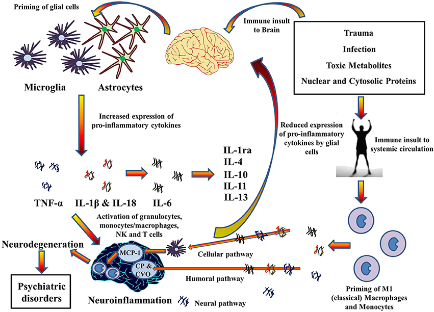
http://www.frontiersin.org/files/Articles/112233/fnins-08-00315-HTML/image_m/fnins-08-00315-g004.jpg

This post serves as a follow-up to yesterday's post on the relationship between trauma exposure and neuroinflammation. This review article comes from Frontiers in Neuroendocrine Science.

This is from the abstract:
Inflammasomes activate pro-inflammatory caspases 1 and 5, which then cleave the precursor forms of pro-inflammatory cytokines IL-1β, IL-18, and IL-33 into their active forms. These pro-inflammatory cytokines have been shown to promote a variety of innate immune processes associated with infection, inflammation, and autoimmunity, and thereby play an instrumental role in the instigation of neuroinflammation during old age and subsequent occurrence of neurodegenerative diseases, cognitive impairment, and dementia. In particular, NLRP inflammasomes may also have a role in the etiologies of depression, Alzheimer's disease (AD) and in metabolic disorders, such as Type II diabetes, obesity and cardiovascular diseases that have been shown to be co-morbid with psychiatric illnesses.
The researchers believe that NLRP inflammasomes might be able to decrease neuroinflammation and, therefore, become a possible treatment for neuroinflammation and its associated psychiatric diseases. The current review assesses the "complex inflammatory signaling pathways involved in the activation of NLRP inflammasomes and the role they play in promoting neuroinflammation and subsequent behavioral changes."

Full Citation: 
Singhal G, Jaehne EJ, Corrigan F, Toben C and Baune BT. (2014, Oct 7). Inflammasomes in neuroinflammation and changes in brain function: a focused review. Frontiers in Neuroscience: Neuroendocrine Science; 8:315. doi: 10.3389/fnins.2014.00315

Inflammasomes in neuroinflammation and changes in brain function: a focused review


Gaurav Singhal [1], Emily J. Jaehne [1], Frances Corrigan [2], Catherine Toben [1] and Bernhard T. Baune [1]
1. Psychiatric Neuroscience Lab, Discipline of Psychiatry, School of Medicine, University of Adelaide, Adelaide, SA, Australia
2. Discipline of Anatomy and Physiology, School of Medical Sciences, University of Adelaide, Adelaide, SA, Australia
Abstract

Recent literature has pointed to the existence of inflammasome-mediated inflammatory pathways in central nervous system (CNS) disorders and associated changes in behavior. Neuroinflammation, which is an innate immune response in the CNS against harmful and irritable stimuli such as pathogens and metabolic toxic waste, as well as to chronic mild stress, is mediated by protein complexes known as inflammasomes. Inflammasomes activate pro-inflammatory caspases 1 and 5, which then cleave the precursor forms of pro-inflammatory cytokines IL-1β, IL-18, and IL-33 into their active forms. These pro-inflammatory cytokines have been shown to promote a variety of innate immune processes associated with infection, inflammation, and autoimmunity, and thereby play an instrumental role in the instigation of neuroinflammation during old age and subsequent occurrence of neurodegenerative diseases, cognitive impairment, and dementia. In particular, NLRP inflammasomes may also have a role in the etiologies of depression, Alzheimer's disease (AD) and in metabolic disorders, such as Type II diabetes, obesity and cardiovascular diseases that have been shown to be co-morbid with psychiatric illnesses. It has been reported that while these inflammasomes may be activated through TNF-α dependent pathways, other cytokines, like IFN-γ, may assist in inhibiting their activation and thus delay disease progression. Furthermore, some other cytokines, including IL-6, may not have a direct role in inflammasome-mediated diseases. An array of recent research suggests that NLRP inflammasomes targeted therapies could be used for alleviating neuroinflammation and for treatment of associated psychiatric illnesses, although this still remains a challenge and necessitates further extensive research. This review examines the complex inflammatory signaling pathways involved in the activation of NLRP inflammasomes and the role they play in promoting neuroinflammation and subsequent behavioral changes.


Introduction


The discovery of inflammasomes by Martinon et al. (2002) has prompted considerable interest in the role that inflammasomes play in the mechanism of inflammation and associated disease patterns. Of late, an emerging body of literature points to the existence of inflammasome-mediated inflammatory pathways in central nervous system (CNS) disorders.


Neuroinflammation is a known factor in the pathogenesis of neurodegenerative diseases (Frank-Cannon et al., 2009), and psychiatric illnesses such as depression (Walker et al., 2014), Alzheimer's disease (AD) (Pimplikar, 2014), Parkinson's disease (PD) (Hirsch et al., 2012), Huntington's disease (Möller, 2010), and multiple sclerosis (Frohman et al., 2006). It has also been implicated in sickness behavior (Biesmans et al., 2013), diminished cognition (Ownby, 2010), and memory (Hein and O'Banion, 2009), as well as in age-related increased sensitization of the immune system to extrinsic and intrinsic stimuli (Godbout et al., 2005; Sparkman and Johnson, 2008). Pattern recognition receptors (PRRs) play an integral role in the innate immune response through recognition of pathogen specific proteins (PAMPs) and damage associated proteins (DAMPs). They are primarily expressed by glial cells, macrophages and oligodendrocytes within the brain and can be membrane bound (toll-like receptors) or within the cytoplasm [Nod-like receptors (NLRs)]. Activation of these NLRs leads to the assembly and activation of cytosolic protein complexes known as inflammasomes which then enable the activation of pro-inflammatory caspases, particularly caspase-1. This then leads to the activation of pro-inflammatory cytokines interleukin (IL)-1β, IL-18, and IL-33 (Arend et al., 2008; Chakraborty et al., 2010), which promote a number of innate immune processes associated with infection, inflammation and autoimmunity (Davis et al., 2011), thereby responsible for neuroinflammation and associated brain diseases (Tha et al., 2000; Cacquevel et al., 2004; Felderhoff-Mueser et al., 2005; Godbout and Johnson, 2009; Mawhinney et al., 2011; Zhang et al., 2014).


It has been known for some time that immunosenescence, in addition to neurodegenerative changes with age, predisposes the brain to higher risk of acquiring neuroinflammatory disorders. Considerable findings during the last decade have suggested an instrumental role of inflammasomes in the pathophysiology of neuroinflammation during neuronal ageing, and its associated neurodegenerative diseases such as dementia, leading to loss of memory and cognitive impairment (Simi et al., 2007; Chakraborty et al., 2010; Mawhinney et al., 2011; Liu and Chan, 2014). In particular, NLRP (NLR family, containing pyrin domain) inflammasomes have been shown to have a role in the etiologies of several neurological diseases such as depression (Zhang et al., 2014), AD (Tan et al., 2013), PD (Cedillos, 2013), and multiple sclerosis (Gris et al., 2010; Fischer et al., 2012). Systemically, NLRP inflammasome-driven inflammatory responses also play a role in the development of Type II diabetes (Grant and Dixit, 2013; Lee et al., 2013), obesity (Stienstra et al., 2011), and cardiovascular diseases (Garg, 2011), as well as cancer (Zitvogel et al., 2012). Given that metabolic disorders can predispose to the development of psychiatric disorders, it is possible that inflammasome-driven inflammatory pathways may be a potential mechanism driving this co-morbidity.


A number of studies have investigated the innate immune pathways associated with the activation of NLRP inflammasomes and the subsequent production of IL-1β, IL-18, and IL-33 from their precursors. The aim of this review is to examine these complex inflammatory signaling pathways associated with NLRP inflammasomes activation, and leading to neuroinflammation and behavioral changes that have commonly been observed during various psychiatric disorders and brain aging.


Materials and Methods


PRISMA criteria


The guidelines prescribed by PRISMA (Preferred reporting items for systematic reviews and meta-analyses) (Liberati et al., 2009; Moher et al., 2009) were followed while constructing this review. The checklist items from PRISMA as relevant to this review, for example those related to search and writing approaches, were included and the items not relevant, for example those related to meta-analyses, were excluded.

Search and Selection Process


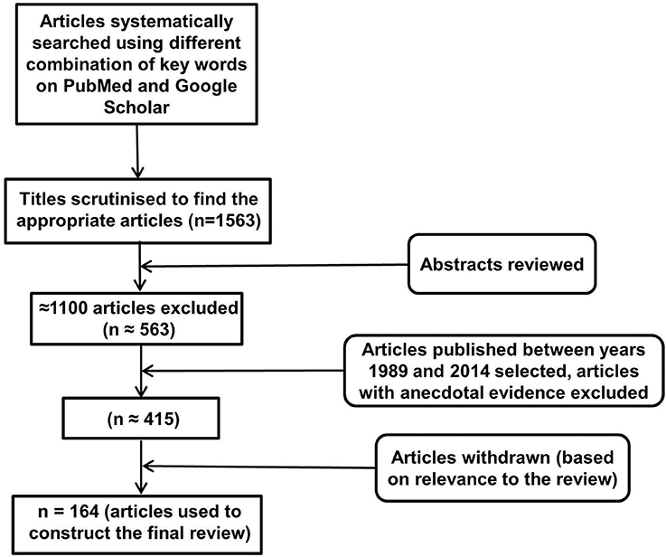
An electronic database search of PubMed and Google Scholar with several key terms in various permutations was performed. These included but were not limited to: inflammasomes, neuroinflammation, NLRP, NALP (NACHT, LRR, and PYD domains containing proteins), cytokines, IL-1, IL-18, IL-33, TNF, cellular, humoral, immune, aging, depression, AD, PD, Huntington's disease, multiple sclerosis, cognition, behavior, metabolic disorders, diabetes, obesity, cardiovascular disease, cancer, pathogen associated molecular patterns, damage associated molecular patterns, toll like receptors, and glial cells. At each stage of the search, titles and abstracts were scrutinized and the most appropriate organized into separate folders using End Note X6.0.1 software. In addition, articles relevant to our discussion were retrieved from the reference list of other online articles on each subtopic. This in total yielded 1563 papers. After placing all inclusion and exclusion criteria into our search (depicted in Figure 1), 164 articles closely related to the aims set forth for this review were selected and hence utilized.
FIGURE 1
Figure 1. Study inclusion flowchart. It depicts the methodology for search and collection of relevant articles for this review, following PRISMA guidelines (McGeer and McGeer, 2004; Heneka et al., 2010).
Inclusion and Exclusion Criteria


The emphasis of this review has been on inflammatory pathways associated with inflammasome activity in the brain, and as such articles investigating inflammasomes, in particular NLRP inflammasomes and their mechanism of actions in CNS disorders were selected for detailed analysis. In addition, articles addressing the effects of IL-1 family of cytokines in the brain were read thoroughly to understand and analyze the various mechanisms of actions of these cytokines, especially in the brain and their association with inflammasomes. Other immune factors related to inflammasomes and the role of inflammasomes in systemic diseases was also investigated while writing this review. All articles included in this review have been published between 1989 and 2014. Articles without the full text available and with anecdotal evidence were excluded from the review.


Structure of NLRP Inflammasomes


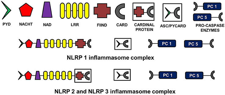
The NLR family, pyrin domain containing inflammasomes (NLRP) are the most studied and best characterized protein complex during inflammation (Stutz et al., 2009). NLRs are intracellular PRRs and function in association with Toll-Like Receptors to sense the presence of PAMPs which are found in a variety of microorganisms that enter cell through phagocytosis (infectious stimuli), and DAMPs such as nuclear and cytosolic protein characteristics of tissue injury/stress (non-infectious stimuli). In turn, this activates the innate and acquired immune response (Inohara et al., 2005; Kanneganti et al., 2007; Franchi et al., 2009). A key part of this process is the assembly of inflammasome complexes that generally have three main components: a cytosolic PRR (either from the NLR family or the pyrin and HIN domain containing family-PYHIN), caspase-1 and an adaptor protein ASC (apoptosis-associated speck like protein). The NLR family contains a leucine rich repeat domain (LRR), a central NACHT domain and a variable amino-terminal domain, which in the NLRP subfamily is an N-terminal pyrin domain (PYD). Activation of NLRPs leads to the recruitment of ASC which contains a caspase activation and recruitment domain (CARD). ASC then interacts with the CARD of pro-caspase-1. There are exceptions to this sequence, with for example NLRP1, directly interacting with pro-caspase 1, without necessarily needing ASC. Nonetheless the interaction with pro-capse-1 leads to its conversion to caspase 1, which then converts pro forms of IL-1β, IL-18, and IL-33 into their active forms, initiating an inflammatory response (Martinon et al., 2002; Petrilli et al., 2005). The NLRP3 is the largest and most studied inflammasome of all known at this stage (Stutz et al., 2009).


See Figure 2 for the schematic representation of the structures of different NLRP inflammasomes as described above.
FIGURE 2
Figure 2. Structure of NLRP inflammasomes. NLRP inflammasomes are intracellular protein complexes consisting of NLRP (NACHT, LRR, and PYD domains containing proteins 1, 2, or 3), the adapter protein ASC/Pycard, enzyme pro-caspases 1 and 5, and cardinal proteins. NLRP1 has Pyrin (PYD) domain on the amino (N)-terminal. This PYD domain is bonded to a NACHT domain followed by a NACHT-associated domain (NAD), several lucine-rich repeats (LRR), FIIND domain and the caspase recruitment domain (CARD) at the carboxy (C)-terminal. The molecular structures of NLRP2 and NLRP3 are similar to NLRP1, except that instead of being directly linked to FIIND domain, LRRs are linked to a cardinal protein which consists of FIIND domain on N-terminal and CARD domain on the C-terminal. NLRPs with the adapter protein ASC/Pycard and pro-caspase enzymes form the inflammasome complex responsible for converting pro-IL-1 cytokines into their active forms within the cytoplasm of glial cells primarily. ASC, apoptosis-associated speck-like protein containing a CARD.

The Role of IL-1 Family of Cytokines in Inflammation, Pathological States, and Homeostatic Response in the Brain


Pro-inflammatory cytokines function to attract leucocytes and enhance their proliferation at the site of inflammation. They stimulate cytotoxicity, release of proteolytic enzymes, synthesis of prostaglandins, and synthesis and secretion of secondary cytokines. This in turn promotes inflammation and increases thermoregulatory set point, generally associated with symptoms such as fever, tissue destruction, shock, and even death (Cannon, 2000). The IL-1 family of cytokines comprises 11 secreted factors, including IL-1α, IL-1β, IL-18, and IL-33, which are known for playing a role in host defense and immune system regulation in inflammatory diseases (Barksby et al., 2007; Arend et al., 2008; Dinarello, 2009; Sims and Smith, 2010). These cytokines have been shown to be involved in a variety of immune reactions as well as in the initiation, regulation, and maintenance of inflammation (Dinarello, 2000). In particular, cytokine mediated processes have been shown to result in long term neuropsychiatric disorders and were found to be related to major depression, dementia, and AD (Licastro et al., 2000; Cacquevel et al., 2004; McAfoose and Baune, 2009). The presence of IL-1β has been demonstrated in cerebrospinal fluid and plasma of patients with AD (Licastro et al., 2000; Tarkowski et al., 2003). Similarly, the roles of IL-18 and IL-33 in neuroinflammation and neurodegenerative diseases have also been well established (Felderhoff-Mueser et al., 2005; Arend et al., 2008; Liew et al., 2010).


However, it is important to note that although pro-inflammatory cytokines (IL-1 and TNF family of cytokines) have been shown to result in neuroinflammation and neurodegenerative diseases when expression is high, at constitutive levels they are required for normal physiological functioning, particularly in the molecular and cellular mechanisms responsible for learning, memory and cognition (McAfoose and Baune, 2009). They influence and maintain homeostasis in monoamine metabolism, neuronal genesis and survival, Hypothalamic-Pituitary-Adrenal (HPA) axis sensitivity to cortisol and certain cellular neuroimmune functions (Eyre and Baune, 2012). However, levels of both pro-inflammatory and anti-inflammatory cytokines have been shown to be elevated in many brain disorders, including AD, PD, and age related dementia, indicating their role in cognitive and memory deficits with age. When pro-inflammatory cytokines are overexpressed, anti-inflammatory cytokines potentially function to suppress the gene expression for pro-inflammatory cytokine production and control the pro-inflammatory response. For instance, gene knockout mice for anti-inflammatory cytokines, such as IL-1ra, IL-10, and TGF-β 1 showed enhanced inflammatory reactions (Dinarello, 2000). However, no study describes the effects of anti-inflammatory cytokines on activated inflammasomes. Activated microglia and astrocytes are the main source of cytokines in the brain (Rothwell et al., 1996; Hanisch, 2002).


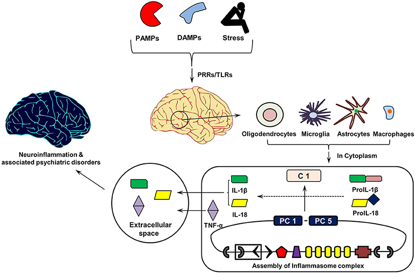
Figure 3 shows the cytokine cascade in brain following stimulation with an infectious agent, metabolic waste or foreign material. As this includes increase in the levels of IL-1β cytokine which can only be activated from its precursor in the presence of enzyme caspase 1, it indicates an active involvement of inflammasome action during this cytokine cascade.
FIGURE 3
Figure 3. Inflammasomes Cascade in Brain. On recognizing pathogen associated molecular patterns (PAMPs, found in phagocytized microorganisms) and damage associated molecular patters (DAMPs, e.g., nuclear and cytosolic proteins), intracellular pathogen recognition receptors (PRRs), and Toll-Like Receptors (TLRs) initiates assembly of cytosolic inflammasome complex. In turn, this activates the innate and acquired immune responses involving Interleukin (IL)-1 cytokines, which in addition to Tumor Necrosis Factor (TNF)-α initiate inflammatory reaction in the extracellular space. PC1 and PC5, Pro-caspases 1 and 5.
Link between Aging of Brain, IL-1 Cytokines, and Brain Disorders


Aging of the brain has been shown to be associated with many cognitive and memory deficit disorders and is believed to be regulated by extrinsic (e.g., environmental) and intrinsic (e.g., genotype) factors (van der Staay, 2002). Several brain disorders, such as AD and PD, are the products of chronic neuroinflammation and resultant neurodegeneration (Heneka et al., 2010; Hirsch et al., 2012), the symptoms which are also common to the aging brain (McGeer and McGeer, 2004). Dementia, decline in cognitive abilities and impairment of spatial memory are often seen during aging and are associated with neuroinflammatory changes within the brain accumulated over a period of time. Indeed, a significant association has been found between age related depression and level of pro-inflammatory cytokines in the brain (Godbout et al., 2008). Moreover, the risk of infections increases with age, mainly due to immunosenescence (Aw et al., 2007) and a rise in circulating autoantibodies and lymphoproliferative disorders, hence contributing toward greater morbidity and mortality in old age (Shinkai et al., 1998; Senchina and Kohut, 2007).


Structural and functional changes in the brain are coordinated by a range of intracellular signaling molecules. Consistent findings suggest that an increase in the expression of pro-inflammatory cytokines by astrocytes and microglia within the brain results in neuroinflammation followed by neurodegeneration, eventually resulting in cognitive and memory deficit and exacerbated sickness and depressive-like behavior (Mrak and Griffin, 2005; Huang et al., 2008). Microglia are primed with aging and upon secondary stimulation, these microglia release excessive quantities of pro-inflammatory cytokines, such as TNF-α, IL-1β, and IL-6 (Dilger and Johnson, 2008). The increase in the number of microglia, astrocytes and percentage of GFAP in the brain with age has been reported in rodents, which is subsequently found to be related to cognitive and memory impairment (Sugaya et al., 1996; Rozovsky et al., 1998), and to neurodegenerative diseases such as AD (Mrak and Griffin, 2005). The effects of aging on microglial functions in the brain has been reviewed in detail by Conde and Streit (2006). During aging, glial cells, particularly microglia show increased activation and expression of pro-inflammatory cytokines, such as IL-1, however, they also become increasingly dysfunctional and lose neuro-protective properties which predispose the brain to the neurodegenerative disorders in line with other genetic and acquired environmental risks (Mrak and Griffin, 2005; Streit, 2005). Similarly, astrocytes are the immune effector cells, which express cytokines (IL-1, IL-6, IL-10, interferons α and β, TNF α and β) and chemokines, and mediate inflammation and immune reactivity in the brain. The under-expression or over-expression of astrocytes can also lead to neurodegenerative diseases (Dong and Benveniste, 2001).

Inflammasome Mediated Inflammatory Pathways in the Aging Brain


It was not known until recently whether inflammasomes play any role in causing or aggravating neuroinflammation during neuronal aging. Considerable recent findings have suggested an instrumental role of inflammasomes in the pathophysiology of neuroinflammation during neuronal ageing, and associated neurodegenerative diseases, cognitive impairment and dementia (Simi et al., 2007; Chakraborty et al., 2010; Mawhinney et al., 2011; Liu and Chan, 2014). There is upregulation in the expression of several genes that signal inflammasome assembly and activation of caspase 1 (e.g., thioredoxin-interacting protein, P2X7, and pannexins), as well as signaling of TLRs (e.g., CD14, TLR2, TLR4, TLR7, TOLLIP, MYD88) in different regions of the brain such as hippocampus, post-central gyrus, and superior frontal gyrus during aging (Cribbs et al., 2012). TLRs are evolutionary conserved microbe specific structural motifs (e.g., PAMPs) and endogenous molecule (e.g., DAMPs) recognition transmembrane or endosomic membrane proteins, primarily expressed in various sentinel cells such as dendritic cells, macrophages, and plasmatoid dendritic cells that form the first line of defense (Kumar et al., 2009). The interaction between TLRs and LRRs on cytosolic NLRs trigger the assembly and activation of inflammasomes culminating in caspase 1 catalyzing pro-IL-1 cytokines into their active forms (van de Veerdonk and Netea, 2011). The increased activity of both TLRs and NLRs in the aging brain can therefore act as a deterrent to the negative regulation of their expression and a sustained expression could result in chronic neuroinflammation and associated neurodegenerative diseases. Moreover, production of reactive oxygen species (ROS) from dysfunctional mitochondria and increased NF-κB signaling with aging could also potentiate the priming of NLRP3 inflammasomes in the brain resulting in an inflammatory response (Salminen et al., 2012). Formation of mutant α-synuclein and Aβ fibrils, seen during PD and AD respectively, further pose greater danger in old age as they act to enhance the activity of inflammasomes in the brain (Salminen et al., 2009; Tschopp and Schroder, 2010; Cedillos, 2013).


Enhanced inflammasome activity could manifest in the form of cognitive decline in an aging population, as shown in 18 month old male Fisher 344 rats, however spatial learning improved when rats were treated with an anti-inflammatory drug probenecid (Mawhinney et al., 2011) which reduced NLRP1 inflammasome activation. However the mechanism for this is not yet completely understood.


Association between Neuroinflammation and Major Depressive Disorder


Major depressive disorder (MDD) is characterized by a distinct change of mood accompanied by sadness, irritability, loss of interest in all activities and events, as well as psychophysiological changes (Belmaker and Agam, 2008). A number of studies, both experimental and meta-analytic, have revealed increased expression of pro-inflammatory cytokines, TNF-α, IL-1β, and IL-6, in the brain of MDD patients leading to neuroinflammation (Maes et al., 1997; Howren et al., 2009; Dowlati et al., 2010; Hannestad et al., 2011). Role of chronic mild stress in neuroinflammation and subsequent occurrence of depression has also been established (Farooq et al., 2012), although it is not clear if inflammasomes have any role in causing neuroinflammation in response to chronic mild stress.

Inflammasome Mediated Inflammatory Pathways Associated with Major Depressive Disorder


The increase in IL-1β levels and neuroinflammation in the brain of MDD patients potentially suggests a role for inflammasomes in MDD. Researchers have indeed recently shown the involvement of the NLRP3 inflammasome in lipopolysaccharide (LPS)-induced mouse depressive-like behavior (Zhang et al., 2014). Similar findings were seen in human participants when activated NLRP3 inflammasomes were detected in blood mononuclear cells from depressive patients (Alcocer-Gómez et al., 2014). To investigate the etiological role of inflammasomes in depression, a panel of researchers conducted a clinical trial in major depression and schizophrenic patients. They concluded from the study that inflammasome-related inflammation is an ongoing process in psychiatric patients during diseased states (Hohmann et al., 2014). Moreover, the recent finding that mice lacking caspase-1 are resistant to LPS-induced depressive-like behavior further supports the role of inflammasomes in depression (Moon et al., 2009). Some authors recently reviewed the role of inflammasomes in MDD and its comorbidity with systemic illnesses, and proposed a new inflammasome hypothesis of depression and related comorbid systemic illnesses (Iwata et al., 2013). The review highlights the central mediator role that inflammasomes play in the contribution of psychological and physical stressors to the development of depression and its association with systemic illnesses. The activation of inflammasomes, particularly NLRP3, therefore could be indirectly related to the pathophysiology of depression and its comorbidity with other systemic diseases through an inflammatory response in the brain.


Association between Neuroinflammation and Alzheimer's Disease


AD is characterized by a debilitating chronic and progressive neurodegeneration leading to major clinical hallmarks of loss of memory, cognitive deficit, dementia, and behavioral impairment. Although, the prevalence of AD is higher in people over 60 and it increases proportionally with every 10 years of age (Younger/Early Onset Alzheimer's and Dementia: Alzheimer's Association, 2014), early onset of AD has also been reported in people in their 40s and 50s (Kim et al., 2014). Several factors such as genetic predisposition (Bertram and Tanzi, 2009; Kamboh et al., 2012), reduced synthesis of excitatory neurotransmitter acetyl choline (Babic, 1999), extracellular deposition of amyloid beta (Aβ) in the brain (Palop and Mucke, 2010), abnormalities in tau protein forming neurofibrillary tangles leading to disintegration of microtubules (Ballatore et al., 2007), and oxidative stress and inflammatory cascades mediated by primed glia cells (Agostinho et al., 2010) have been proposed to cause AD. These different hypotheses have been established after years of independent research; however, recent efforts toward finding the common link between the causal factors for AD have pointed toward the inflammatory cascade linking them in the brain. Indeed, damaged neurons, highly insoluble Aβ deposits and neurofibrillary tangles could provide stimuli for neuroinflammation (Wenk, 2003). Similarly, neurotransmitter acetylcholine has been shown to be involved in inhibiting the release of pro-inflammatory cytokines from microglia and monocytes (Tabet, 2006), an anti-inflammatory mechanism that could be disturbed during acetylcholine deficiency. This suggests that all above etiologies for AD, when accompanied by chronic neuroinflammation, lead to progressive neurodegeneration and behavioral impairment with age, characteristics of symptoms of AD. In the absence of neuroinflammation, these etiologies may not provide sufficient pathology to cause AD. This is supported by the finding that significant amyloid deposition could be present in the brain of healthy elderly individuals without cognitive impairment (Aizenstein et al., 2008). Likewise, while higher quantities of tau proteins have been reported in the brain of AD patients than unaffected individuals (Avila et al., 2004), some authors have challenged the tau protein hypothesis and proposed that tau phosphorylation is a compensatory mechanism to protect neurons against oxidative stress (Lee et al., 2005). Nonetheless, this suggests overall that a single factor alone may not be sufficient to cause AD, and irrespective of the causative factor, neuroinflammation essentially provides a central pathway to the onset of AD which is mediated by various pro-inflammatory cytokines and chemokines, including IL-1 family of cytokines that are activated by inflammasomes.


Indeed, IL-1β and IL-18 over-expression has been shown to initiate inflammatory process in the brain of AD patients (Rubio-Perez and Morillas-Ruiz, 2012; Liu and Chan, 2014). This over-expression has been detected in microglia, astrocytes as well as neurons, and found to be co-localized with both Aβ plaques and tau. Interestingly, it has also been suggested that chronic inflammation could be the cause for increase in Aβ and tau phosphorylation in the brain (Meraz-Ríos et al., 2013). In support of this, studies on transgenic mice with LPS-induced neuroinflammation have shown enhanced intracellular deposition of Aβ (Sheng et al., 2003; Lee et al., 2008) and tau phosphorylation (Kitazawa et al., 2005) in the brain of mice. Overall, this suggests a chain of continuous adverse events in the brain of AD patients, mediated by IL-1 family of pro-inflammatory cytokines.


Inflammasome Mediated Inflammatory Pathways Associated with Alzheimer's Disease


Recently, the role of inflammasomes, particularly NLRP3, in oxidative stress-induced neuroinflammation and impaired amyloid metabolism seen in AD brains has been evaluated and recognized (Halle et al., 2008; Marchesi, 2011; Tan et al., 2013). Neuronal injury caused by insoluble Aβ oligomers and fibrils releases DAMPs which are sensed by PRRs (NLR domain) on NALP inflammasomes initiating a chain of events leading to the maturation of proIL-1β and proIL-18 cytokines and release of their active forms as the final event (Halle et al., 2008; Salminen et al., 2009). Moreover, Aβ can interact with neuronal membranes to create ion channels that allow potassium ion (K+) efflux mediated by ATPase enzyme, activating inflammasomes and in turn secretion of the active IL-1 family of cytokines (Salminen et al., 2009; Tschopp and Schroder, 2010). Reduction in intracellular K+ to 90 mM though has been found to be a requirement for the activation of NLRP3 inflammasomes (higher intracellular concentration of K+ inhibits activation of inflammasomes) (Petrilli et al., 2007). Some authors however, found impaired activity of Na+/K+ ATPase in AD patients (Hattori et al., 1998) that is required for the active efflux of K+ across the cell membranes, which therefore raises the question whether efflux of K+ is essential for the activation of inflammasomes. ATPase is required to catalyze ATP into ADP and a phosphate ion with the release of energy that activates the purinergic P2X7 receptor. This receptor in turn decreases intracellular K+ levels (Perregaux and Gabel, 1994; Solle et al., 2001). Purinergic signaling has also been shown to control the cerebral vascular tone and this has been implicated in learning and memory, locomotor and feeding behavior and sleep (Burnstock, 2013). Nonetheless, diminished activity of Na+/K+ ATPase reduces the gradient of ions across the cell membranes causing an excitotoxic cellular response resulting in neuronal death (Hattori et al., 1998). This causes a release of DAMPs from dead neurons that potentially act as the activators of NLRP3 inflammasome dependent innate immune response (Rubartelli, 2014). It has also been shown that disease-associated extracellular amyloid and unique protein aggregates caused by inappropriate oligomerization or misfolding are sensed by NLRP3 inflammasomes (Masters and O'Neill, 2011), likely as DAMPs within the resident macrophages after engulfment in the brain. Research has also suggested that the brain in AD is under increased oxidative stress and Aβ peptides generates free radicals that together further enhance neuron degeneration and death (Markesbery, 1997). Mitochondrial ROS released during tissue injury/death could enhance oxidative damage and signal inflammasome activation up-regulating pro-inflammatory cytokine levels in brain (Martinon, 2010; Tschopp and Schroder, 2010; Naik and Dixit, 2011), potentially resulting in neuroinflammation.


Significant pathology and behavioral deficits characteristics of chronic neuroinflammation do not manifest until advanced age. This has been attributed to the capacity of the brain to compensate for the presence of chronic neuroinflammation by regulating the glutamatergic system (Brothers et al., 2013). This suggests that neuroinflammation in itself does not cause AD; however it acts as an initiator, enhancer and sustainer of AD disease during old age which is reinforced by various other etiologies. Since IL-1 cytokines are key contributors of chronic neuroinflammation and associated neurodegenerative diseases, including AD, inflammasomes provide a possible answer for the mechanism of IL-1 action and the ways in which IL-1 activity is regulated during chronic neuroinflammation in old age (Allan et al., 2005).


Immune Factors Associated with the Activity of Inflammasomes


It has been well established that both TNF-α and IL-1β stimulate each other's secretion and exhibit overlapping and synergistic effects (Akira et al., 1990; Ikejima et al., 1990; Knofler et al., 1997). For instance, while TNF-α enhances migration of leucocytes in inflamed tissue and promotes apoptosis, IL-1β acts as a potent pyrogen and decreases the threshold of pain by inducing the transcription of cyclooxygenase 2 enzyme, thereby enhancing production of prostaglandins E2, which is responsible for pain and fever. This raises a possibility that inflammasomes, which catalyze IL-1β precursors, may stimulate TNF-α secretion through an indirect pathway. Contrary to this, it has recently been reported that inflammasomes may also be activated independently of PRRs, through TNF-α dependent pathways. TNF-α has been shown to trigger the activation of caspase 1 and in turn secretion of IL-1β (Alvarez and Munoz-Fernandez, 2013), suggesting a possible bidirectional cause-effect relationship between inflammasomes and TNF-α. Although the precise mechanism for this has not yet been elucidated, this study indicated that TNF-α may potentially substitute for a TLR mediated stimulus required for inflammasome activation. Moreover, recent findings also suggest that TNF-α induces production of IL-33 in keratinocytes (Taniguchi et al., 2013) and regulates expression of IL-18 in dendritic precursor-like cell line KG-1 and cardiomyocytes (Chandrasekar et al., 2003; Koutoulaki et al., 2010), further supporting the hypothesis that TNF-α has a role in inflammasome activation, although this direct relation between TNF-α, and IL-18 and IL-33 is yet to be established in the brain. Furthermore, other cytokines such as Type I interferon (IFN) gamma have been shown to inhibit caspase-1 cleavage and reduce IL-1β secretion in rodents (Guarda et al., 2011). Conversely, although IFN-gamma does not cause inflammation by activating inflammasomes directly, it has been shown to augment TNF activity (Dinarello, 2000). Although IL6 is a reliable inflammatory marker it may not always be directly involved in inflammasome mediated inflammation as seen in an IL-6 knock in mouse model (McGeough et al., 2012). Overall, this suggests a complex and intricate immune pathway mediated by various cytokines that may be involved in the activation of inflammasomes in the brain, and resultant neuroinflammation and changes in brain function; however this requires validation through extensive research.


Inflammasome-Independent Neuroinflammation-Mediated Brain Pathologies


TNF-α is another pro-inflammatory cytokine, in addition to IL-1 cytokines, that has been primarily implicated in neuroinflammation. Elevated levels of TNF-α in particular have been shown to cause a reduction in hippocampal volumes through the neurodegenerative TNFR1 pathway (Baune et al., 2012) and can lead to the development of depressive-like behavior (Eyre et al., 2013). Glial cells, microglia, and astrocytes, are the primary immune effector cells and express various cytokines in the CNS (Rothwell et al., 1996; Hanisch, 2002). Though glial cells are neuroprotective, their over expression or sustained stimulation can result in enhanced production of cytokines (e.g., IL-1β and TNF-α) (Sawada et al., 1989; Dong and Benveniste, 2001; Hanisch, 2002) resulting in severe neuroinflammation, neurodegeneration and subsequent cognitive dysfunction and psychiatric diseases, such as AD.


While levels of both pro-inflammatory and anti-inflammatory cytokines in the peripheral circulation and CNS have been reported to rise during several brain disorders such as depression, schizophrenia and AD (Schwarz et al., 2001), other humoral immune factors, such as mitogen-activated protein kinases (MAPK), C reactive protein (CRP), the complement system and chemokines have also been reported to modify brain anatomy and functions. MAPKs are specific protein kinases (serine-threonine specific) that elicit pro-inflammatory and immunomodulatory functions (Lee et al., 1994; Dong et al., 2002). Similarly, CRP is an acute phase reactant protein which enhances inflammation and tissue damage by promoting phagocytosis by opsonization (Du Clos, 2000) and activating the complement system (Padilla and Perez, 2003). High levels of CRP in the brain have been linked to neuroinflammation and associated cognitive impairment and dementia (Kuo et al., 2005), and AD (McGeer et al., 2000). Researchers have observed upregulation of the complement system in human brain during AD and other neurodegenerative diseases (McGeer and McGeer, 1995; Yasojima et al., 1999). The complement system consists of distinct plasma proteins that act as opsonins and initiate a series of inflammatory responses (Janeway et al., 2001). Chemokines promote neuroinflammation by attracting leucocytes to the point of inflammation (Proost et al., 1996; Mélik-Parsadaniantz and Rostène, 2008). Neuroinflammation in turn has been implicated for the impairment of brain function (Campbell, 2004; Ownby, 2010; Tansey and Goldberg, 2010).


Further to the role of inflammasomes and humoral immune factors, several cellular immune factors such as granulocytes, monocytes/macrophages, NK cells, and T lymphocytes have also been shown to have a role in the pathophysiology of neuroinflammation (Petersen and Pedersen, 2005, 2006). The role of NK cells in various brain disorders such as depression, AD and PD has been reviewed and validated by some researchers (Poli et al., 2013). Likewise, the exchange of B cells across the BBB has been reported in patients with multiple sclerosis and associated with the development of autoimmunity in the CNS (von Büdingen et al., 2012).


Discussion


Neuroinflammation and Cytokines


Neuroinflammation is an innate mechanism to ward off any stimuli that may be harmful to the host and has been shown to be mediated by various immune factors, particularly cytokines (Cacquevel et al., 2004) and chemokines (Ubogu et al., 2006). In particular, pro-inflammatory cytokines, such as TNF-α and the IL-1 family of cytokines, are credited for initiating the inflammatory reactions in the brain in response to an adverse stimulus, for continuation of neuroinflammation by attracting leucocytes at the site of inflammation and activating other pro-inflammatory factors, as well as for the anti-inflammatory pathway by enhancing IL-6 production that in turn stimulates production and expression of anti-inflammatory cytokines and immune factors (Cannon, 2000). However, what is more important here is the mechanism for the activation of these pro-inflammatory cytokines in response to an adverse stimulus in the first instance. While the concept of an increase in concentration of pro-inflammatory cytokines within the brain during aging and infection is now established, less is known about the mechanisms of cell signaling that result in the pro-inflammatory cytokine gradients within the brain.

NLRP Inflammasomes in Neuroinflammation


While several theories have been postulated to explain this mechanism, recent findings suggest the role of inflammasomes to be important in particular for the IL-1 family of cytokines (Martinon and Tschopp, 2006; Stutz et al., 2009; Schroder and Tschopp, 2010). In response to PAMPs and DAMPs, trans-membranous TLRs that are present in semantic cells such as macrophages and dendritic cells, interact with NLRs on inflammasomes to recognize the stimulus, initiating an inflammasome cascade leading to the release of caspase 1 enzyme in the cytoplasm. Caspase 1 cleaves the pro-forms of the IL-1 family of cytokines to form their active forms (Inohara et al., 2005; Kanneganti et al., 2007; Franchi et al., 2009) that may result in neuroinflammation as a result of increased pro-inflammatory cytokine gradients. Although the acute neuroinflammatory response includes activation of resident tissue macrophages in the CNS and subsequent release of various cytokines and chemokines, this may also cause oxidative and nitrosative stress, which is a first line preventative mechanism against pathogenic extrinsic and intrinsic proteins and is less likely to cause long term damage to neurons (Frank-Cannon et al., 2009). However, it could still result in neurodegenerative changes, as well as in short-term cognitive impairment and exacerbated sickness behavior, as seen in rodent trials after LPS-induced acute neuroinflammation characterized by heightened pro-inflammatory cytokine response (Morimoto et al., 2002; Huang et al., 2008). Nevertheless, a bigger danger is posed by chronic neuroinflammation that is generally seen during old age (Sparkman and Johnson, 2008) and responsible for some brain pathologies such as depression (Wager-Smith and Markou, 2011), AD (Hauss-Wegrzyniak et al., 1998), PD (Tansey and Goldberg, 2010), and multiple sclerosis (Frischer et al., 2009).


For a chronic neuroinflammation to be sustained, the stimuli need to be continuous, potent and self-replicating. This could be explained from the findings that AD patients in old age suffer from a persistent degenerative condition that involves consistent increases in the various proposed etiologies, be it Aβ oligomerization or Tau phosphorylation, in the presence of neuroinflammation (Meraz-Ríos et al., 2013). A similar scenario could be plausible in the case of PD where formation of α-synuclein fibril aggregate increases in the presence of neuroinflammation. Accelerated formation of the mutant α-synuclein fibrils has been linked with the onset of PD (Conway et al., 1998, 2000). Although it has been shown that aggregated α-synuclein in microglia-like cells potentially activate the assembly of NLRP3 inflammasomes by inducing vesicle rupture in THP-1 cells that are sensed as danger signals (Cedillos, 2013), the opposite scenario still need to be studied (Cedillos, 2013). Consistent findings have also established the link between various etiologies of depression and neuroinflammation (Maes et al., 1997; Howren et al., 2009; Dowlati et al., 2010; Hannestad et al., 2011).

A Short Note on the Role of Inflammasome Mediated Neuroinflammatory Pathways in the Comorbidity of Systemic Illnesses and Psychiatric Disorders


High incidences of chronic inflammatory diseases such as cancer (Il'yasova et al., 2005), diabetes (De Rekeneire et al., 2006), osteoarthritis (Stannus et al., 2013), and cardiovascular disease (Volpato et al., 2001) have been demonstrated by prospective and correlative studies in aged cohorts. Investigation at the molecular level suggests increased levels of systemic pro-inflammatory cytokine IL-1β, in addition to TNF-α and IL-6, and acute phase proteins (e.g., CRP). Moreover, significant findings have confirmed an association between age related depression and level of pro-inflammatory cytokines in the brain (Godbout et al., 2008). This suggests a mechanism whereby pro-inflammatory cytokines migrate from systemic circulation to the brain and vice versa, especially during old age. Indeed, the pathways for the transport of pro-inflammatory cytokines to brain from systemic circulation have been described in a review by Capuron and Miller (2011) (See Figure 4). Rodent studies have shown increased production and expression of IL-1β in the brain after LPS-induced systemic inflammation (Cunningham et al., 2005) and changes in mood and behavior similar to depression after systemic administration of pro-inflammatory cytokines (Pollak and Yirmiya, 2002). This transport of pro-inflammatory cytokines into the brain and increase in their expression could be the reason for the comorbidity of systemic illnesses with psychiatric disorders in old age. Comorbid conditions, such as Type II diabetes (Grant and Dixit, 2013; Lee et al., 2013), obesity (Stienstra et al., 2011), cardiovascular diseases (Connat, 2011), and cancer (Fallowfield et al., 2001) with psychiatric illnesses therefore supports the hypothesis that inflammasomes play a large role in immunosenescence associated with aging and formation of psychiatric and systemic illnesses with age, the top-most reasons for deaths worldwide as mentioned by World Health Organization (2014). However, future research into the role of inflammasomes in these pathways during aging could possibly explain the link between age-related psychiatric and systemic illnesses.
FIGURE 4
Figure 4. Cytokines hypothesis of neuroinflammation: Implications in comorbidity of systemic illnesses with psychiatric disorders. Pro-inflammatory cytokines can migrate between systemic circulation and brain in both directions which could explain the comorbidity of systemic illnesses with psychiatric disorders. There are three pathways for the transport of pro-inflammatory cytokines from systemic circulation to brain as described by Capuron and Miller (Dong et al., 2002): Cellular, Humoral, and Neural. Moreover, PAMPs and DAMPs from trauma, infection and metabolic waste can prime glial cells to express pro-inflammatory cytokines TNF-α, IL-1β, and IL-6. When expressed, these cytokines activates granulocytes, monocytes/macrophages, Natural Killer, and T cells and together contribute to the pathophysiology of neuroinflammation. Chronic neuroinflammation could result in neurodegeneration and associated psychiatric disorders. These pro-inflammatory cytokines also stimulate production and expression of anti-inflammatory cytokine by glial cells that function as negative feedback to reduce the expression of pro-inflammatory cytokines, subsiding the neuroinflammation. MCP-1, Monocyte chemoattractant protein-1; CP, Choroid plexus; CVO, Circumventricular organ.
The above mentioned link between systemic inflammatory conditions and CNS neurological disorders via activation of inflammasomes provides a molecular platform on which to develop therapies to prevent the initiation of those pro-inflammatory chronic cascades which are detrimental to the CNS. However, although hypothesized a number of times, it is yet to be seen if these therapies can be used to treat diseases such as cancer, diabetes, CVD, and auto-inflammatory disorders (Wilson and Cassel, 2010) that are major killers worldwide and comorbid with brain disorders. Inflammasomes, a molecular platform, could therefore be regarded as an advent to the innovation of therapies in the near future.


Concluding Remarks


Taken together, it is clear that the discovery of the role of inflammasomes in neuroinflammation has opened an array of research opportunities to investigate the inflammasome targeted therapies for age-related and pathological changes in the brain. It is also clear from the above discussion that the inflammasome activation pathway is complex and may involve the role of other immune factors such as cytokines, as well as mutant protein aggregates such as Aβ and α-synuclein fibrils. However, further research into these mechanisms and inflammasome-targeted therapies is advisable for constructing and assessing the complete profile of inflammasome-driven inflammatory pathways in brain.

Conflict of Interest Statement


The presented work is supported by the National Health and Medical Research Council Australia (APP 1043771 to Bernhard T. Baune). The funders had no role in study design, data collection and analysis, decision to publish, or preparation of the manuscript. The authors declare that the research was conducted in the absence of any commercial or financial relationships that could be construed as a potential conflict of interest.


References at the Frontiers site