Despite her apt framing of the adaptation hypothesis, [Cecilia] Heyes actually argues against it. If she is right, then we’re all simply born with one set of visual neurons that becomes activated when observing an action, and a second set of motor neurons that activates when executing an action. Sometimes the activity of those neurons becomes correlated—perhaps because the two events occurred closely in time, or because one regularly precedes the other—and those motor neurons become mirror neurons as a result.The activity of visual neurons and motor neurons are correlated because that is the basic foundation of learning, and they appear to be synchronized because the brain is fully capable of parallel processing. This brief summary from Sigman & Dehaene (The Journal of Neuroscience, 2008; 28(30):7585–7598) should suffice:
According to a prominent theory, which emerged from numerous behavioral experiments, perceptual and response operations occur in parallel, and only a central decision stage, involved in coordinating sensory and motor operations, is delayed (Pashler, 1994).There you go - mystery solved. For a more in-depth explanation, I refer interested readers to "Eight Problems for the Mirror Neuron Theory of Action Understanding in Monkeys and Humans," by Gregory Hickok (J Cogn Neurosci. Jul 2009; 21(7): 1229–1243).
Anyway, this is an entertaining article from Nautilus on how mirror neuron research is refuting many of the initial claims.
Mirror Neurons Are Essential, but Not in the Way You Think
Posted By Jason G. Goldman on Jun 04, 2014
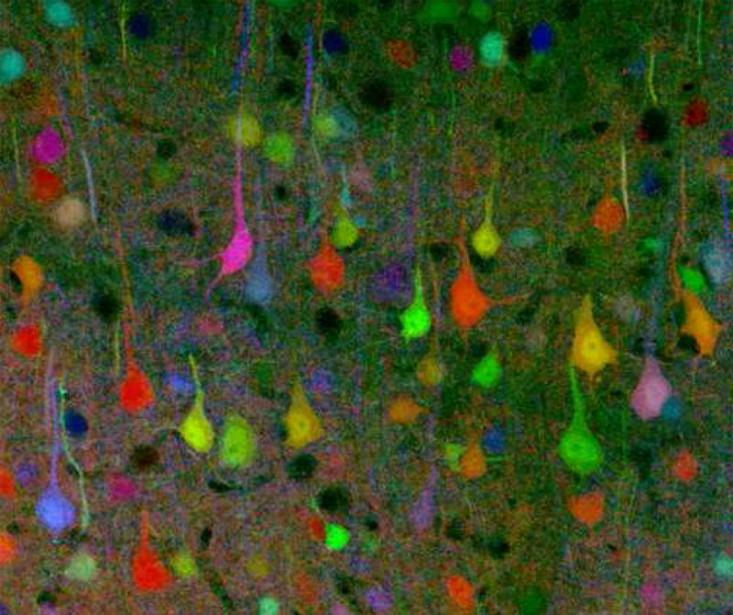
A “brainbow”: neurons labels with fluorescent tags, in this case, from a mouse. Stephen J. Smith via Wikipedia
In his 2011 book, The Tell-Tale Brain: A Neuroscientist's Quest for What Makes Us Human, neuroscientist V. S. Ramachandran says that some of the cells in your brain are of a special variety. He calls them the “neurons that built civilization,” but you might know them as mirror neurons. They’ve been implicated in just about everything from the development of empathy in earlier primates, millions of years ago, to the emergence of complex culture in our species.
Ramachandran says that mirror neurons help explain the things that make us so apparently unique: tool use, cooking with fire, using complex linguistics to communicate.
It’s an inherently seductive idea: that one small tweak to a particular set of brain cells could have transformed an early primate into something that was somehow more. Indeed, experimental psychologist Cecilia Hayes wrote in 2010 (pdf), “[mirror neurons] intrigue both specialists and non-specialists, celebrated as a ‘revolution’ in understanding social behaviour and ‘the driving force’ behind ‘the great leap forward’ in human evolution.”
The story of mirror neurons begins in the 1990s at the University of Parma in Italy. A group of neuroscientists were studying rhesus monkeys by implanting small electrodes in their brains, and they found that some cells exhibited a curious kind of behavior. They fired both when the monkey executed a movement, such as grasping a banana, and also when the monkey watched the experimenter execute that very same movement.
It was immediately an exciting find. These neurons were located in a part of the brain thought solely responsible for sending motor commands out from the brain, through the brainstem to the spine, and out to the nerves that control the body’s muscles. This finding suggested that they’re not just used for executing actions, but are somehow involved in understanding the observed actions of others.
After that came a flood of research connecting mirror neurons to the development of empathy, autism, language, tool use, fire, and more. Psychologist and science writer Christian Jarrett has twice referred to mirror neurons as “the most hyped concept in neuroscience.” Is he right? Where does empirical evidence end and overheated speculation begin?
The strongest claim about mirror neurons is that they are responsible for human uniqueness, because they turned us into a singularly social primate. “It is widely believed that hyper-sociality is what makes humans ‘special,’ the key to understanding why it is we, and not the members of any other species, who dominate the world with our language, artefacts and institutions,” wrote Heyes in her 2010 commentary. “Therefore, in the light of this ‘adaptation hypothesis,’ mirror neurons emerge as an evolutionary foundation of human uniqueness…If mirror neurons are an adaptation, and more ‘advanced’ in humans than in monkeys, they may well play a major role in explaining the evolutionary origins and online control of human social cognition,” she wrote. Indeed, that’s the same claim that Ramachandran makes.
But recent research casts doubt on the adaptation hypothesis. Increasing evidence indicates that the so-called “mirror effect” in brain cells can be enhanced, abolished, or even reversed due to the effects of learning. The mirror-neuron systems of dancers and musicians, for example, have different properties than those of others, and those tool-sensitive mirror neurons in monkeys only come about as a result of experience with tools.
Despite her apt framing of the adaptation hypothesis, Heyes actually argues against it. If she is right, then we’re all simply born with one set of visual neurons that becomes activated when observing an action, and a second set of motor neurons that activates when executing an action. Sometimes the activity of those neurons becomes correlated—perhaps because the two events occurred closely in time, or because one regularly precedes the other—and those motor neurons become mirror neurons as a result.
That means that mirror neurons didn’t evolve, per se. What evolved is the mechanism that produces mirror neurons: associative learning, our ability to identify statistical patterns in the world, to associate one event with another, like the ringing of a bell with a tasty dinner. And associative learning is present in a wide variety of species, meaning that its mere presence can’t be the “evolutionary foundation of human uniqueness.”
If mirror neurons are formed routinely by learning, that may explain the fact that since their initial discovery, researchers have spotted a wide variety of mirror neurons, in different parts of the brain. Some are highly tuned to one particular kind of action, firing for only precision grips (grasping an object between two fingers) rather than whole-hand grips. Others reliably fire for both kinds of grasping actions, but not for other sorts of behaviors. Some mirror neurons are willing to fire when an object is being grasped with a tool, like a pair of pliers, rather than with a hand. Others demand a biological hand in order to activate.
One study found that mirror neurons would still fire even if the action was hidden behind an occluder, revealing that they work when the monkey must imagine the action in their mind’s eye, but others do not. Some studies have found that some mirror neurons will fire if a certain action is viewed from the front, side, or back of the performer, while others fire only when an action is witnessed from a particular viewpoint.
Some mirror neurons are perfectly happy to fire when the performer is on a video screen, but others require a live, in-the-flesh performer. There are also, according to some reports, so-called “auditory mirror neurons,” discharging both when the monkey hears itself do an action, and when the monkey hears someone else do the same action.
Perhaps most interestingly, some mirror neurons appear to “care” about the goal or reward value of the performer’s actions, rather than the action itself. For example, in one experiment, monkeys viewed an actor reaching for and grasping an object, and then either eating it or placing it into a container. Some mirror neurons fired for the eating action, others only for the placing action, and others fired in both cases.
Since mirror neurons seem to be involved with so much that our brains do, it is less likely that they suddenly appeared and made one momentous change to one of our ancestors. Instead, it seems like they’re a widespread part of how neurons become organized as we learn.
Even if they’re the result of experience, even if they’re a byproduct of the evolution of associative learning rather than adaptations themselves, mirror neurons could still play an important role in complex social phenomena. Heyes compares mirror neurons to the neural systems used in reading. “The neural mechanisms involved in reading did not evolve for that ‘purpose,’ but through explicit training they are made to fulfill an important function,” she says. Mirror-neuron systems can be “recruited” in the development of social-cognitive skills like empathy, Heyes suggests, just as visual pattern-recognition systems are recruited in the development of reading skills. But mirror neurons are not “for” social cognition any more than pattern-recognition neurons are “for” reading, which has only been practiced in a very recent slice of human existence.
In reflecting on mirror neurons, it is perhaps easy to see how the reasonable, evidence-based claims about some brain cells made in the mid-1990s snowballed into the media-driven frenzy that dominates the discourse of mirror neurons now, some 20 years later. It’s a compelling idea, that a tiny group of brain cells could have a hand in all that we think of as unique to our species. Mirror neurons are indeed amazing and fascinating and worthy of awe themselves. But the difficult problem of how to understand our own species will not be solved by as simple an answer as the mirror neuron. Perhaps its time to smash the mirror neuron’s hype and steel ourselves against the inevitable seven years of bad luck.
~ Jason G. Goldman received his Ph.D. in developmental psychology at the University of Southern California in Los Angeles and writes a blog called The Thoughtful Animal, hosted at Scientific American. His doctoral research focused on the evolution and architecture of the mind, and how different early experiences might affect innate knowledge systems.