Showing posts with label psychedelic mushrooms. Show all posts

Thursday, October 30, 2014

This Is Your Brain on Psychedelic Drugs (via Discover)

Dr. David Nutt and a team of researchers have published a study on the psychoactive substance in mushrooms, psilocybin, and how it impacts the brain circuits. As you can see in the picture below, the effect of psilocybin is a much more interconnected brain (which suggests some other circuits that limit activity are inactive under the influence of psilocybin).

Interesting stuff.

The summary below is from Discover, then the whole article, which is open access, is also included below.

This Is Your Brain on Psychedelic Drugs


By Ben Thomas | October 29, 2014 4:16 pm


Left, the stable brain activity in a normal brain. Right, under the influence of psilocybin, diverse brain regions not normally in communication become strongly linked.

Psychedelic substances can change a user’s mindset in profound ways — a fact that’s relevant even to those who’ve never touched the stuff, because such altered states of consciousness give scientists a window into how our brains give rise to our normal mental states. But neuroscientists are only beginning to understand how and why those mental changes occur.

Now some mathematicians have jumped into the fray, using a new mathematical technique to analyze the brains of people on magic mushrooms.

Psychedelic Puzzles

Scientists have known for decades that many of psychedelic drugs’ most famous effects — visual hallucinations, heightened sensory and emotional sensitivity, etc. — are linked to elevated levels of the neurotransmitter serotonin.

But increasingly neuroscience researchers are interested not just in single chemicals but also in overall brain activity, because the most complicated brain functions arise from lots of different regions working together. Over the last several years, a branch of mathematics known as network theory has been applied to study this phenomenon.

Paul Expert, a complexity researcher at the Imperial College London, and his team took this approach to analyzing fMRI data from people who’d taken psilocybin, the psychedelic chemical in magic mushrooms. The team had recently been working on a new technique for network modeling — one designed to highlight small but unusual patterns in network connectivity.

Brains on Drugs

The team used fMRI data from a previous study, in which 15 healthy people rested inside an fMRI scanner for 12 minutes on two separate occasions. The volunteers received a placebo in one of those sessions, and a mild dose of psilocybin during the other, but they weren’t told which was which.

The investigators crunched the data, specifically studying the brain’s functional connectivity — the amount of active communication among different brain areas.

They found two main effects of the psilocybin. First, most brain connections were fleeting. New connectivity patterns tended to disperse more quickly under the influence of psilocybin than under placebo. But, intriguingly, the second effect was in the opposite direction: a few select connectivity patterns were surprisingly stable, and very different from the normal brain’s stable connections.

This indicates “that the brain does not simply become a random system after psilocybin injection, but instead retains some organizational features, albeit different from the normal state,” the authors write in their paper in the Journal of the Royal Society Interface.

Far Out
The findings seem to explain some of the psychological experiences of a psilocybin trip. Linear thinking and planning become extremely difficult, but nonlinear “out of the box” thinking explodes in all directions. By the same token, it can become difficult to tell fantasy apart from reality during a psilocybin trip; but focusing on a certain thought or image — real or imagined — often greatly amplifies that thought’s intensity and vividness.

The authors suggest that effects like these may be rooted in the two connectivity traits they spotted, since the connectivity patterns that rapidly disperse may reflect unorganized thinking, while the stable inter-regional connections may reflect information from one sensory domain “bleeding” into other areas of sensory experience. In fact, the researchers also suggest that synesthesia — the sensory blurring that causes users of psychedelics to experience sounds as colors, for example — may be a result of these connectivity changes too.

The researchers hope that the patterns they’ve found will provide neuroscientists with new approaches for studying the brain on psychedelic drugs, and therefore better understand the strange psychological effects their users report.
* * * * *

Full Citation:
Petri, G, Expert, P,  Turkheimer, F, Carhart-Harris, R, Nutt, D, Hellyer, PJ, and Vaccarino, F. (2014, Oct 29). Homological scaffolds of brain functional networks. J. R. Soc. Interface; 11(101). doi: 10.1098/​rsif.2014.0873 [Print: 6 December 2014] 

Homological scaffolds of brain functional networks


G. Petri [1], P. Expert [2], F. Turkheimer [2], R. Carhart-Harris [3], D. Nutt [3], P. J. Hellyer [4] and
F. Vaccarino [1,5]
1. ISI Foundation, Via Alassio 11/c, 10126 Torino, Italy
2. Centre for Neuroimaging Sciences, Institute of Psychiatry, Kings College London, De Crespigny Park, London SE5 8AF, UK
3. Centre for Neuropsychopharmacology, Imperial College London, London W12 0NN, UK
4. Computational, Cognitive and Clinical Neuroimaging Laboratory, Division of Brain Sciences, Imperial College London, London W12 0NN, UK
5. Dipartimento di Scienze Matematiche, Politecnico di Torino, C.so Duca degli Abruzzi no 24, Torino 10129, Italy
Abstract

Networks, as efficient representations of complex systems, have appealed to scientists for a long time and now permeate many areas of science, including neuroimaging (Bullmore and Sporns 2009 Nat. Rev. Neurosci. 10, 186–198. (doi:10.1038/nrn2618)). Traditionally, the structure of complex networks has been studied through their statistical properties and metrics concerned with node and link properties, e.g. degree-distribution, node centrality and modularity. Here, we study the characteristics of functional brain networks at the mesoscopic level from a novel perspective that highlights the role of inhomogeneities in the fabric of functional connections. This can be done by focusing on the features of a set of topological objects—homological cycles—associated with the weighted functional network. We leverage the detected topological information to define the homological scaffolds, a new set of objects designed to represent compactly the homological features of the correlation network and simultaneously make their homological properties amenable to networks theoretical methods. As a proof of principle, we apply these tools to compare resting-state functional brain activity in 15 healthy volunteers after intravenous infusion of placebo and psilocybin—the main psychoactive component of magic mushrooms. The results show that the homological structure of the brain's functional patterns undergoes a dramatic change post-psilocybin, characterized by the appearance of many transient structures of low stability and of a small number of persistent ones that are not observed in the case of placebo.

1. Motivation

The understanding of global brain organization and its large-scale integration remains a challenge for modern neurosciences. Network theory is an elegant framework to approach these questions, thanks to its simplicity and versatility [1]. Indeed, in recent years, networks have become a prominent tool to analyse and understand neuroimaging data coming from very diverse sources, such as functional magnetic resonance imaging (fMRI), electroencephalography and magnetoencephalography [2,3], also showing potential for clinical applications [4,5]. 

A natural way of approaching these datasets is to devise a measure of dynamical similarity between the microscopic constituents and interpret it as the strength of the link between those elements. In the case of brain functional activity, this often implies the use of similarity measures such as (partial) correlations or coherence [6–8], which generally yield fully connected, weighted and possibly signed adjacency matrices. Despite the fact that most network metrics can be extended to the weighted case [9–13], the combined effect of complete connectedness and edge weights makes the interpretation of functional networks significantly harder and motivates the widespread use of ad hoc thresholding methods [7,14–18]. However, neglecting weak links incurs the dangers of a trade-off between information completeness and clarity. In fact, it risks overlooking the role that weak links might have, as shown for example in the cases of resting-state dynamics [19,20], cognitive control [21] and correlated network states [22]. 

In order to overcome these limits, Rubinov & Sporn [13,23,24] recently introduced a set of generalized network and community metrics for functional networks that among others were used to uncover the contrasting dynamics underlying recollection [25] and the physiology of functional hubs [26]. 

In this paper, we present an alternative route to the analysis of brain functional networks. We focus on the combined structure of connections and weights as captured by the homology of the network. A summary of all the keywords and concepts introduced in this paper can be found in table 1.
View this table:
Table 1. List of notations.
2. From networks to topological spaces and homology

Homology is a topological invariant that characterizes a topological space X by counting its holes and their dimensions. By hole, we mean a hollow region bounded by the parts of that space. The dimension of a hole is directly related to the dimension of its boundary. The boundary of a two-dimensional hole is a one-dimensional loop; the three-dimensional inner part of a doughnut, where the filling goes, is bounded by two-dimensional surface; for dimensions higher than 2, it becomes difficult to have a mental representation of a hole, but k-dimensional holes are still bounded by (k − 1) dimensional faces. In our work, we start with a network and from it construct a topological space. We now use figure 1 to show how we proceed and make rigorous what we mean by boundaries and holes. 

Figure 1.
Figure 1. Panels (a,b) display an unweighted network and its clique complex, obtained by promoting cliques to simplices. Simplices can be intuitively thought as higher-dimensional interactions between vertices, e.g. as a simplex the clique (b,c,i) corresponds to a filled triangle and not just its sides. The same principle applies to cliques—thus simplices—of higher order. (Online version in colour.) 
In a network like that of figure 1a, we want the ring of nodes (a,b,c,d) to be a good candidate for a one-dimensional boundary, whereas the other rings of three nodes should not constitute interesting holes. The reason for this choice comes from the formalization of the notion of hole. One way to formalize this is by opposition that is we define what we mean by a dense subnetwork in order to highlight regions of reduced connectivity, i.e. holes. The most natural and conservative definition we can adopt for a dense subnetwork is that of clique, a completely connected subgraph [27]. Moreover, cliques have the crucial property, which will be important later, of being nested, i.e. a clique of dimension k (k-clique) contains all the m-cliques defined by its nodes with m < k. Using this definition and filling in all the maximal cliques, the network in figure 1a can be represented as in figure 1b: 3-cliques are filled in, becoming tiles, and the only interesting structure left is the square (a,b,c,d). It is important at this point to note that a k-clique can be seen as a k − 1 simplex, i.e. as the convex hull of k-points. Our representation of a network can thus be seen as a topological space formed by a finite set of simplices that by construction satisfy the condition that defines the type of topological spaces called abstract simplicial complexes [28]: each element of the space is a simplex, and each of its faces (or subset in the case of cliques) is also a simplex. 

This condition is satisfied, because each clique is a simplex, and subsets of cliques are cliques themselves, and the intersection of two cliques is still a clique. 

The situation with weighted networks becomes more complicated. In the context of a weighted network, the holes can be thought of as representing regions of reduced connectivity with respect to the surrounding structure. 

Consider, for example, the case depicted in figure 2a: the network is almost the same as figure 1 with the two exceptions that it now has weighted edges and has an additional very weak edge between nodes a and c. The edges in the cycle [a,b,c,d] are all much stronger than the link (a,c) that closes the hole by making (a,b,d) and (b,c,d) cliques and therefore fills them. The loop (e,f,g,h,i) has a similar situation, but the difference in edge weights between the links along the cycle and those crossing, is not as large as in the previous case. It would be therefore useful to be able to generalize the approach exposed earlier for binary networks to the case of weighted networks in such a way as to be able to measure the difference between the two cases (a,b,c,d) and (e,f,g,h,i). As shown by figure 2b, this problem can be intuitively thought of as a stratigraphy in the link-weight fabric of the network, where the aim is to detect the holes, measure their depth and when they appear as we scan across the weights' range. 

Figure 2.
Figure 2. Panels (a­–c) display a weighted network (a), its intuitive representation in terms of a stratigraphy in the weight structure according the weight filtration described in the main text (b) and the persistence diagram for H1 associated with the network shown (c). By promoting cliques to simplices, we identify network connectivity with relations between the vertices defining the simplicial complex. By producing a sequence of networks through the filtration, we can study the emergence and relative significance of specific features along the filtration. In this example, the hole defined by (a,b,c,d) has a longer persistence (vertical solid green bars) implying that the boundary of the cycle are much heavier than the internal links that eventually close it. The other hole instead has a much shorter persistence, surviving only for one step and is therefore considered less important in the description of the network homological properties. Note that the births and deaths are defined along the sequence of descending edge weights in the network, not in time. (Online version in colour.) 
From figure 2b, it becomes clear that the added value of this method over conventional network techniques lies in its capability to describe mesoscopic patterns that coexist over different intensity scales, and hence to complement the information about the community structure of brain functional networks. A way to quantify the relevance of holes is given by persistent homology. We describe it and its application to the case of weighted networks in full detail in §3.
3. A persistent homology of weighted networks

The method that we adopt was introduced in references [29,30] and relies on an extension of the metrical persistent homology theory originally introduced by references [31,32]. Technical details about the theory of persistent homology and how the computation is performed can be found in the works of Carlsson, Zomorodian and Edelsbrunner [28,31–35]. Persistent homology is a recent technique in computational topology developed for shape recognition and the analysis of high dimensional datasets [36,37]. It has been used in very diverse fields, ranging from biology [38,39] and sensor network coverage [40] to cosmology [41]. Similar approaches to brain data [42,43], collaboration data [44] and network structure [45] also exist. The central idea is the construction of a sequence of successive approximations of the original dataset seen as a topological space X. This sequence of topological spaces X0, X1, … , XN = X is such that Graphic whenever i < j and is called the filtration. Choosing how to construct a filtration from the data is equivalent to choosing the type of goggles one wears to analyse the data. 

In our case, we sort the edge weights in descending order and use the ranks as indices for the subspaces. More specifically, denote by Graphic the functional network with vertices V, edges E and weights Graphic. We then consider the family of binary graphs Gω = (V, Eω), where an edge e ∈ E is also included in Gω if its weight ωe is larger than ω (Graphic). 

To each of the Gω, we associate its clique, or flag complex Kω, that is the simplicial complex that contains the k-simplex [n0, n1, n2, … nk − 1] whenever the nodes n0, n1, n2, … nk −1 define a clique in Gω [27]. As subsets of cliques and intersections of cliques are cliques themselves, as we pointed out in §2, our clique complex is thus a particular case of a simplicial complex. 

The family of complexes {Kω} defines a filtration, because we have Graphic for ω > ω′. At each step, the simplices in Kω inherit their configuration from the underlying network structure and, because the filtration swipes across all weight scales in descending order, the holes among these units constitute mesoscopic regions of reduced functional connectivity.

Moreover, this approach also highlights how network properties evolve along the filtration, providing insights about where and when lower connectivity regions emerge. This information is available, because it is possible to keep track of each k-dimensional cycle in the homology group Hk. A generator uniquely identifies a hole by its constituting elements at each step of the filtration process. The importance of a hole is encoded in the form of ‘time-stamps' recording its birth βg and death δg along the filtration {Kω} [31]. These two time-stamps can be combined to define the persistence πg = δg − βg of a hole, which gives a notion of its importance in terms of it lifespan. Continuing the analogy with stratigraphy, βg and δg correspond, respectively, to the top and the bottom of a hole and πg would be its depth. As we said above, a generator Graphic, or hole, of the kth homology group Hk is identified by its birth and death along the filtration. Therefore, Graphic is described by the point Graphic. A standard way to summarize the information about the whole kth persistent homology group is then to consider the diagram obtained plotting the points corresponding to the set of generators. The (multi)set {(βg,δg} is called the persistence diagram of Hk. In figure 2c, we show the persistence diagram for the network shown in figure 2a for H1. Axes are labelled by weights in decreasing order. It is easy to check that the coordinates correspond exactly to the appearance and disappearance of generators. The green vertical bars highlight the persistence of a generator along the filtration. The further a point is from the diagonal (vertically), the more persistent the generator is. In §4, we introduce two objects, the persistence and the frequency homological scaffolds, designed to summarize the topological information about the system. 

4. Homological scaffolds

Once one has calculated the generators Graphic of the kth persistent homology group Hk, the corresponding persistence diagram contains a wealth of information that can be used, for example, to highlight differences between two datasets. It would be instructive to obtain a synthetic description of the uncovered topological features in order to interpret the observed differences in terms of the microscopic components, at least for low dimensions k. Here, we present a scheme to obtain such a description by using the information associated with the generators during the filtration process. As each generator, Graphic is associated with a whole equivalence class, rather than to a single chain of simplices, we need to choose a representative for each class, we use the representative that is returned by the javaplex implementation [46] of the persistent homology algorithm [47]. For the sake of simplicity in the following, we use the same symbol Graphic to refer to a generator and its representative cycle. 

We exploit this to define two new objects, the persistence and the frequency homological scaffolds Graphic and Graphic of a graph G. The persistence homological scaffold is the network composed of all the cycle paths corresponding to generators weighted by their persistence. If an edge e belongs to multiple cycles g0,g1, … ,gs, its weight is defined as the sum of the generators' persistence:

Formula 4.1

Similarly, we define the frequency homological scaffold Graphic as the network composed of all the cycle paths corresponding to generators, where this time, an edge e is weighted by the number of different cycles it belongs to

Formula 4.2

where Graphic is the indicator function for the set of edges composing gi. By definition, the two scaffolds have the same edge set, although differently weighted. 

The construction of these two scaffolds therefore highlights the role of links which are part of many and/or long persistence cycles, isolating the different roles of edges within the functional connectivity network. The persistence scaffolds encodes the overall persistence of a link through the filtration process: the weight in the persistence scaffold of a link belonging to a certain set of generators is equal to the sum of the persistence of those cycles. The frequency scaffold instead highlights the number of cycles to which a link belongs, thus giving another measure of the importance of that edge during the filtration. The combined information given by the two scaffolds then enables us to decipher the nature of the role different links have regarding the homological properties of the system. A large total persistence for a link in the persistence scaffold implies that the local structure around that link is very weak when compared with the weight of the link, highlighting the link as a locally strong bridge. We remark that the definition of scaffolds we gave depends on the choice of a specific basis of the homology group, and the choice of a consistent basis is an open problem in itself, therefore the scaffolds are not topological invariants. Moreover, it is possible for an edge to be added to a cycle shortly after the cycle's birth in such a way that it creates a triangle with the two edges composing the cycle. In this way, the new edge would be part of the shortest cycle, but the scaffold persistence value would be misattributed to the two other edges. This can be checked, for example, by monitoring the clustering coefficient of the cycle's subgraph as edges are added to it. We checked for this effect and found that in over 80% of the cases the edges do not create triangles that would imply the error, but instead new cycles are created, whose contribution to the scaffold is then accounted for by the new cycle. Finally, we note also that, when a new triangle inside the cycle is created, the two choices of generator differ for a path through a third strongly connected node, owing to the properties of boundary operators. Despite this ambiguity, we show in the following that they can be useful to gain an understanding of what the topological differences detected by the persistent homology actually mean in terms of the system under study.
5. Results from fMRI networks

We start from the processed fMRI time series (see Methods for details). The linear correlations between regional time series were calculated after covarying out the variance owing to all other regions and the residual motion variance represented by the 24 rigid motion parameters obtained from the pre-processing, yielding a partial-correlation matrix χα for each subject. The matrices χα were then analysed with the algorithm described in the previous sections. We calculated the generators Graphic of the first homological group H1 along the filtration. As mentioned before, each of these generators identifies a lack of mesoscopic connectivity in the form of a one-dimensional cycle and can be represented in a persistence diagram. We aggregate together the persistence diagrams of subjects belonging to each group and compute an associated persistence probability density (figure 3). These probability density functions constitute the statistical signature of the groups' H1 features. 

Figure 3.
Figure 3. Probability densities for the H1 generators. Panel (a) reports the (log-)probability density for the placebo group, whereas panel (b) refers to the psilocybin group. The placebo displays a uniform broad distribution of values for the births–deaths of H1 generators, whereas the plot for the psilocybin condition is very peaked at small values with a fatter tail. These heterogeneities are evident also in the persistence distribution and find explanation in the different functional integration schemes in placebo and drugged brains. (Online version in colour.) 
We find that, although the number of cycles in the groups are comparable, the two probability densities strongly differ (Kolmogorov–Smirnov statistics: 0.22, p-value less than 10−10). 

The placebo group displays generators appearing and persisting over a limited interval of the filtration. On the contrary, most of the generators for the psilocybin group are situated in a well-defined peak at small birth indices, indicating a shorter average cycle persistence. However, the psilocybin distribution is also endowed with a longer tail implying the existence of a few cycles that are longer-lived compared with the placebo condition and that influences the weight distribution of the psilocybin persistence scaffold. The difference in behaviour of the two groups is made explicit when looking at the probability distribution functions for the persistence and the birth of generators (figure 4), which are both found to be significantly different (Kolmogorov–Smirnov statistics: 0.13, p-value < 10−30 for persistence and Kolmogorov–Smirnov statistics: 0.14, p-value < 10−35 for births). In order to better interpret and understand the differences between the two groups, we use the two secondary networks described in §4, Graphic and Graphic for the placebo group and Graphic and Graphic for the psilocybin group. The weight of the edges in these secondary networks is proportional to the total number of cycles an edge is part of, and the total persistence of those cycles, respectively. They complement the information given by the persistence density distribution, where the focus is on the entire cycle's behaviour, with information on single links. In fact, individual edges belonging to many and long persistence cycles represent functionally stable ‘hub’ links. As with the persistence density distribution, the scaffolds are obtained at a group level by aggregating the information about all subjects in each group. These networks are slightly sparser than the original complete χα networks

Formula 5.1 

and Formula 5.2 

and have comparable densities. A first difference between the two groups becomes evident when we look at the distributions for the edge weights (figure 5a). In particular, the weights of Graphic display a cut-off for large weights, whereas the weights of Graphic have a broader tail (Kolmogorov–Smirnov statistics: 0.06, p-value < 10−20; figure 5a). Interestingly, the frequency scaffold weights probability density functions cannot be distinguished from each other figure 5a (inset) (Kolmogorov–Smirnov statistics: 0.008, p-value = 0.72). Taken together, these two results imply that while edges statistically belong to the same number of cycles, in the psilocybin scaffold, there exist very strong, persistent links. 

Figure 4.
Figure 4. Comparison of persistence π and birth β distributions. Panel (a) reports the H1 generators' persistence distributions for the placebo group (blue line) and psilocybin group (red line). Panel (b) reports the distributions of births with the same colour scheme. It is very easy to see that the generators in the psilocybin condition have persistence peaked at shorter values and a wider range of birth times when compared with the placebo condition. (Online version in colour.) 

Figure 5.
Figure 5. Statistical features of group homological scaffolds. Panel (a) reports the (log-binned) probability distributions for the edge weights in the persistence homological scaffolds (main plot) and the frequency homological scaffolds (inset). While the weights in the frequency scaffold are not significantly different, the weight distributions for the persistence scaffold display clearly a broader tail. Panel (b) shows instead the scatter plot of the edge frequency versus total persistence. In both cases, there is a clear linear relationship between the two, with a large slope in the psilocybin case. Moreover, the psilocybin scaffold has a larger spread in the frequency and total persistence of individual edges, hinting to a different local functional structure within the functional network of the drugged brains. (Online version in colour.) 
The difference between the two sets of homological scaffolds for the two groups becomes even more evident when one compares the weights between the frequency and persistence scaffolds of the same group. Figure 5b is a scatter plot of between the weights of edges from both scaffolds for the two groups. The placebo group has a linear relationship between the two quantities meaning that edges that are persistent also belongs to many cycles (R2 = 0.95, slope = 0.23). Although the linear relationship is still a reasonable fit for the psilocybin group (R2 = 0.9, slope = 0.3), the data in this case display a larger dispersion. In particular, it shows that edges in Graphic can be much more persistent/longer-lived than in Graphic but still appear in the same number of cycles, i.e. the frequency of a link is not predictive of its persistence or simply put: some connections are much more persistent in the psychedelic state. Moreover, the slopes of linear fits of the two clouds are statistically different (p-value < 10−20, npla = 13 200 and npsi = 13 275 [48]) pointing to a starkly different local functional structure in the two conditions. 

The results from the persistent homology analysis and the insights provided by the homological scaffolds imply that although the mesoscopic structures, i.e. cycles, in the psilocybin condition are less stable than in the placebo group, their constituent edges are more stable.
6. Discussion

In this paper, we first described a variation of persistent homology that allows us to deal with weighted and signed networks. We then introduced two new objects, the homological scaffolds, to go beyond the picture given by persistent homology to represent and summarize information about individual links. The homological scaffolds represent a new measure of topological importance of edges in the original system in terms of how frequently they are part of the generators of the persistent homology groups and how persistent are the generators to which they belong to. We applied this method to an fMRI dataset comprising a group of subjects injected with a placebo and another injected with psilocybin. 

By focusing on the second homology group H1, we found that the stability of mesoscopic association cycles is reduced by the action of psilocybin, as shown by the difference in the probability density function of the generators of H1 (figure 3). 

It is here that the importance of the insight given by the homological scaffolds in the persistent homology procedure becomes apparent. A simple reading of this result would be that the effect of psilocybin is to relax the constraints on brain function, ascribing cognition a more flexible quality, but when looking at the edge level, the picture becomes more complex. The analysis of the homological scaffolds reveals the existence of a set of edges that are predominant in terms of their persistence although they are statistically part of the same number of cycles in the two conditions (figure 5). In other words, these functional connections support cycles that are especially stable and are only present in the psychedelic state. This further implies that the brain does not simply become a random system after psilocybin injection, but instead retains some organizational features, albeit different from the normal state, as suggested by the first part of the analysis. Further work is required to identify the exact functional significance of these edges. Nonetheless, it is interesting to look at the community structure of the persistence homological scaffolds in figure 6. The two pictures are simplified cartoons of the placebo (figure 6a) and psilocybin (figure 6b) scaffolds. In figure 6a,b, the nodes are organized and coloured according to their community membership in the placebo scaffold (obtained with the Louvain algorithm for maximal modularity and resolution 1 [50]). This is done in order to highlight the striking difference in connectivity structure in the two cases. When considering the edges in the tail of the distribution, weight greater than or equal to 80, in figure 5a, only 29 of the 374 edges present in the truncated psilocybin scaffold are shared with the truncated placebo scaffold (165 edges). Of these 374 edges, 217 are between placebo communities and are observed to mostly connect cortical regions. This supports our idea that psilocybin disrupts the normal organization of the brain with the emergence of strong, topologically long-range functional connections that are not present in a normal state. 


Figure 6. Simplified visualization of the persistence homological scaffolds. The persistence homological scaffolds (a) and (b) are shown for comparison. For ease of visualization, only the links heavier than 80 (the weight at which the distributions in figure 5a bifurcate) are shown. This value is slightly smaller than the bifurcation point of the weights distributions in figure 5a. In both networks, colours represent communities obtained by modularity [49] optimization on the placebo persistence scaffold using the Louvain method [50] and are used to show the departure of the psilocybin connectivity structure from the placebo baseline. The width of the links is proportional to their weight and the size of the nodes is proportional to their strength. Note that the proportion of heavy links between communities is much higher (and very different) in the psilocybin group, suggesting greater integration. A labelled version of the two scaffolds is available as GEXF graph files as the electronic supplementary material. (Online version in colour.)
The two key results of the analysis of the homological scaffolds can therefore be summarized as follows (i) there is an increased integration between cortical regions in the psilocybin state and (ii) this integration is supported by a persistent scaffold of a set of edges that support cross modular connectivity probably as a result of the stimulation of the 5HT2A receptors in the cortex [51]. 

We can speculate on the implications of such an organization. One possible by-product of this greater communication across the whole brain is the phenomenon of synaesthesia which is often reported in conjunction with the psychedelic state. Synaesthesia is described as an inducer-concurrent pairing, where the inducer could be a grapheme or a visual stimulus that generates a secondary sensory output—like a colour for example. Drug-induced synaesthesia often leads to chain of associations, pointing to dynamic causes rather than fixed structural ones as may be the case for acquired synaesthesia [52]. Broadly consistent with this, it has been reported that subjects under the influence of psilocybin have objectively worse colour perception performance despite subjectively intensified colour experience [53]. 

To summarize, we presented a new method to analyse fully connected, weighted and signed networks and applied it to a unique fMRI dataset of subjects under the influence of mushrooms. We find that the psychedelic state is associated with a less constrained and more intercommunicative mode of brain function, which is consistent with descriptions of the nature of consciousness in the psychedelic state.
7. Methods

7.1. Dataset


A pharmacological MRI dataset of 15 healthy controls was used for a proof-of-principle test of the methodology [54]. Each subject was scanned on two separate occasions, 14 days apart. Each scan consisted of a structural MRI image (T1-weighted), followed by a 12 min eyes-close resting-state blood oxygen-level-dependent (BOLD) fMRI scan which lasted for 12 min. Placebo (10 ml saline, intravenous injection) was given on one occasion and psilocybin (2 mg dissolved in 10 ml saline) on the other. Injections were given manually by a study doctor situated within the scanning suite. Injections began exactly 6 min after the start of the 12-min scans, and continued for 60 s. 

7.1.1. Scanning parameters

The BOLD fMRI data were acquired using standard gradient-echo EPI sequences, reported in detail in reference [54]. The volume repetition time was 3000 ms, resulting in a total of 240 volumes acquired during each 12 min resting-state scan (120 pre- and 120 post-injection of placebo/psilocybin). 

7.1.2. Image pre-processing

fMRI images were corrected for subject motion within individual resting-state acquisitions, by registering all volumes of the functional data to the middle volume of the acquisition using the FMRIB linear registration motion correction tool, generating a six-dimension parameter time course [55]. Recent work demonstrates that the six parameter motion model is insufficient to correct for motion-induced artefact within functional data, instead a Volterra expansion of these parameters to form a 24 parameter model is favoured as a trade-off between artefact correction and lost degrees of freedom as a result of regressing motion away from functional time courses [56]. fMRI data were pre-processed according to standard protocols using a high-pass filter with a cut-off of 300 s.
Structural MRI images were segmented into n = 194 cortical and subcortical regions, including white matter cerebrospinal fluid (CSF) compartments, using Freesurfer (http://surfer.nmr.mgh.harvard.edu/), according to the Destrieux anatomical atlas [57]. In order to extract mean-functional time courses from the BOLD fMRI, segmented T1 images were registered to the middle volume of the motion-corrected fMRI data, using boundary-based registration [58], once in functional space mean time-courses were extracted for each of the n = 194 regions in native fMRI space. 

7.1.3. Functional connectivity

For each of the 194 regions, alongside the 24 parameter motion model time courses, partial correlations were calculated between all couples of time courses (i,j), non-neural time courses (CSF, white matter and motion) were discarded from the resulting functional connectivity matrices, resulting in a 169 region cortical/subcortical functional connectivity corrected for motion and additional non-neural signals (white matter/CSF). 

7.2. Persistent homology computation


For each subject in the two groups, we have a set of persistence diagrams relative to the persistent homology groups Hn. In this paper, we use the H1 persistence diagrams of each group to construct the corresponding persistence probability densities for H1 cycles. 

Filtrations were obtained from the raw partial-correlation matrices through the Python package Holes and fed to javaplex [46] via a Jython subroutine in order to extract the persistence intervals and the representative cycles. The details of the implementation can be found in reference [30], and the software is available at Holes [59].
Funding statement

G.P. and F.V. are supported by the TOPDRIM project supported by the Future and Emerging Technologies programme of the European Commission under Contract IST-318121. I.D. P.E. and F.T. are supported by a PET methodology programme grant from the Medical Research Council UK (ref no. G1100809/1). The authors acknowledge support of Amanda Feilding and the Beckley Foundation and the anonymous referees for their critical and constructive contribution to this paper.

© 2014 The Authors. Published by the Royal Society under the terms of the Creative Commons Attribution License http://creativecommons.org/licenses/by/4.0/, which permits unrestricted use, provided the original author and source are credited.
References at the Journal of the Royal Society Interface site

Saturday, July 05, 2014

How Psilocybin Mushrooms Induce a Dream-Like State


Researchers in England have identified a possible mechanism for how psilocybin (the natural, hallucinogenic tryptamine found in various mushroom species) affects its actions in the brain.

Turns out that the state of consciousness while "tripping" is quite similar to the state of consciousness while dreaming. And now everyone who has ever used hallucinogens is releasing a collective, "Duh!"

Using brain imaging technology, the researchers identified "increased activity in the hippocampus and anterior cingulate cortex, areas involved in emotions and the formation of memories," areas considered to be more primitive in that they develop earlier. Meanwhile, there was "decreased activity was seen in 'less primitive' regions of the brain associated with self-control and higher thinking, such as the thalamus, posterior cingulate and medial prefrontal cortex."

How magic mushrooms induce a dream-like state


The chemical psilocybin causes the same brain activation as dreaming does <i>(Image: Ryan Wendler/Corbis)</i>
The chemical psilocybin causes the same brain activation as dreaming does  
(Image: Ryan Wendler/Corbis)

Anyone who has enjoyed a magical mystery tour thanks to the psychedelic powers of magic mushrooms knows the experience is surreally dreamlike. Now neuroscientists have uncovered a reason why: the active ingredient, psilocybin, induces changes in the brain that are eerily akin to what goes on when we're off in the land of nod.

For the first time, we have a physical representation of what taking magic mushrooms does to the brain, says Robin Carhart-Harris of Imperial College London, who was part of the team who carried out the research.

Researchers from Imperial and Goethe University in Frankfurt, Germany, injected 15 people with liquid psilocybin while they were lying in an fMRI scanner. The scans show the flow of blood through different regions of the brain, giving a measure of how active the different areas are.

The images taken while the volunteers were under the influence of the drug were compared with those taken when the same people were injected with an inert placebo. This revealed that during the psilocybin trip, there was increased activity in the hippocampus and anterior cingulate cortex, areas involved in emotions and the formation of memories. These are often referred to as primitive areas of the brain because they were some of the first parts to evolve.
 

Primal depths

At the same time, decreased activity was seen in "less primitive" regions of the brain associated with self-control and higher thinking, such as the thalamus, posterior cingulate and medial prefrontal cortex.

This activation pattern is similar to what is seen when someone is dreaming.

"This was neat because it fits the idea that psychedelics increase your emotional range," says Carhart-Harris.

Neuroscientific nuts and bolts aside, the findings could have genuine practical applications, says psychiatrist Adam Winstock at the Maudsley Hospital and Lewisham Drug and Alcohol Service in London. Psilocybin – along with other psychedelics – could be used therapeutically because of its capacity to probe deep into the primal corners of the brain.

"Dreaming appears to be an essential vehicle for unconscious emotional processing and learning," says Winstock. By using psilocybin to enter a dreamlike state, people could deal with stresses of trauma or depression, he says. "It could help suppress all the self-deceiving noise that impedes our ability to change and grow."

Next, the team wants to explore the potential use of magic mushrooms, LSD and other psychedelics to treat depression.

Journal reference: Human Brain Mapping, DOI: 10.1002/hbm.22562

* * * * *

Here is the abstract and introduction to the full article, which is freely available online.

Full Citation:
Tagliazucchi, E., Carhart-Harris, R., Leech, R., Nutt, D. and Chialvo, D. R. (2014, Jul 2). Enhanced repertoire of brain dynamical states during the psychedelic experience. Human Brain Mapping; ePub ahead of print. doi: 10.1002/hbm.22562

Enhanced repertoire of brain dynamical states during the psychedelic experience

Enzo Tagliazucchi, Robin Carhart-Harris, Robert Leech, David Nutt, and Dante R. Chialvo

Article first published online: 2 JUL 2014
Abstract

The study of rapid changes in brain dynamics and functional connectivity (FC) is of increasing interest in neuroimaging. Brain states departing from normal waking consciousness are expected to be accompanied by alterations in the aforementioned dynamics. In particular, the psychedelic experience produced by psilocybin (a substance found in “magic mushrooms”) is characterized by unconstrained cognition and profound alterations in the perception of time, space and selfhood. Considering the spontaneous and subjective manifestation of these effects, we hypothesize that neural correlates of the psychedelic experience can be found in the dynamics and variability of spontaneous brain activity fluctuations and connectivity, measurable with functional Magnetic Resonance Imaging (fMRI). Fifteen healthy subjects were scanned before, during and after intravenous infusion of psilocybin and an inert placebo. Blood-Oxygen Level Dependent (BOLD) temporal variability was assessed computing the variance and total spectral power, resulting in increased signal variability bilaterally in the hippocampi and anterior cingulate cortex. Changes in BOLD signal spectral behavior (including spectral scaling exponents) affected exclusively higher brain systems such as the default mode, executive control, and dorsal attention networks. A novel framework enabled us to track different connectivity states explored by the brain during rest. This approach revealed a wider repertoire of connectivity states post-psilocybin than during control conditions. Together, the present results provide a comprehensive account of the effects of psilocybin on dynamical behavior in the human brain at a macroscopic level and may have implications for our understanding of the unconstrained, hyper-associative quality of consciousness in the psychedelic state.

INTRODUCTION

Psilocybin (phophoryl-4-hydroxy-dimethyltryptamine) is the phosphorylated ester of the main psychoactive compound found in magic mushrooms. Pharmacologically related to the prototypical psychedelic LSD, psilocybin has a long history of ceremonial use via mushroom ingestion and, in modern times, psychedelics have been assessed as tools to enhance the psychotherapeutic process [Grob et al., 2011; Krebs et al., 2012; Moreno et al., 2006]. The subjective effects of psychedelics include (but are not limited to) unconstrained, hyperassociative cognition, distorted sensory perception (including synesthesia and visions of dynamic geometric patterns) and alterations in one's sense of self, time and space. There is recent preliminary evidence that psychedelics may be effective in the treatment of anxiety related to dying [Grob et al., 2011] and obsessive compulsive disorder [Moreno et al., 2006] and there are neurobiological reasons to consider their potential as antidepressants [Carhart-Harris et al., 2012, 2013]. Similar to ketamine (another novel candidate antidepressant) psychedelics may also mimic certain psychotic states such as the altered quality of consciousness that is sometimes seen in the onset-phase of a first psychotic episode [Carhart-Harris et al., 2014]. There is also evidence to consider similarities between the psychology and neurobiology of the psychedelic state and Rapid Eye Movement (REM) sleep [Carhart-Harris, 2007; Carhart-Harris and Nutt, 2014], the sleep stage associated with vivid dreaming [Aserinsky and Kleitman, 1953].

The potential therapeutic use of psychedelics, as well as their capacity to modulate the quality of conscious experience in a relatively unique and profound manner, emphasizes the importance of studying these drugs and how they act on the brain to produce their novel effects. One potentially powerful way to approach this problem is to exploit human neuroimaging to measure changes in brain activity during the induction of the psychedelic state. The neural correlates of the psychedelic experience induced by psilocybin have been recently assessed using Arterial Spin Labeling (ASL) and BOLD fMRI [Carhart-Harris et al., 2012]. This work found that psilocybin results in a reduction of both CBF and BOLD signal in major subcortical and cortical hub structures such as the thalamus, posterior cingulate (PCC) and medial prefrontal cortex (mPFC) and in decreased resting state functional connectivity (RSFC) between the normally highly coupled mPFC and PCC. Furthermore, our most recent study used magnetoencephalography (MEG) to more directly measure altered neural activity post-psilocybin and here we found decreased oscillatory power in the same cortical hub structures [Muthukumaraswamy et al., 2013, see also Carhart-Harris et al., 2014 for a review on this work).

These results establish that psilocybin markedly affects BOLD, CBF, RSFC, and oscillatory electrophysiological measures in strategically important brain structures, presumably involved in information integration and routing [Carhart-Harris et al., 2014; de Pasquale et al., 2012; Hagmann et al., 2008; Leech et al., 2012]. However, the effects of psilocybin on the variance of brain activity parameters across time has been relatively understudied and this line of enquiry may be particularly informative in terms of shedding new light on the mechanisms by which psychedelics elicit their characteristic psychological effects. Thus, the main objective of this article is to examine how psilocybin modulates the dynamics and temporal variability of resting state BOLD activity. Once regarded as physiological noise, a large body of research has now established that resting state fluctuations in brain activity have enormous neurophysiological and functional relevance [Fox and Raichle, 2007]. Spontaneous fluctuations self-organize into internally coherent spatiotemporal patterns of activity that reflect neural systems engaged during distinct cognitive states (termed “intrinsic” or “resting state networks”—RSNs) [Fox and Raichle, 2005; Raichle, 2011; Smith et al., 2009]. It has been suggested that the variety of spontaneous activity patterns that the brain enters during task-free conditions reflects the naturally itinerant and variegated quality of normal consciousness [Raichle, 2011]. However, spatio-temporal patterns of resting state activity are globally well preserved in states such as sleep [Boly et al., 2008, 2012; Brodbeck et al., 2012; Larson-Prior et al., 2009; Tagliazucchi et al., a,b,c] in which there is a reduced level of awareness—although very specific changes in connectivity occur across NREM sleep, allowing the decoding of the sleep stage from fMRI data [Tagliazucchi et al., 2012c; Tagliazucchi and Laufs, 2014]. Thus, if the subjective quality of consciousness is markedly different in deep sleep relative to the normal wakeful state (for example) yet FC measures remain largely preserved, this would suggest that these measures provide limited information about the biological mechanisms underlying different conscious states. Similarly, intra-RSN FC is decreased under psilocybin [Carhart-Harris et al., 2013] yet subjective reports of unconstrained or even “expanded” consciousness are common among users (see Carhart-Harris et al. [2014] for a discussion). Thus, the present analyses are motivated by the view that more sensitive and specific indices are required to develop our understanding of the neurobiology of conscious states, and that measures which factor in variance over time may be particularly informative.

A key feature of spontaneous brain activity is its dynamical nature. In analogy to other self-organized systems in nature, the brain has been described as a system residing in (or at least near to) a critical point or transition zone between states of order and disorder [Chialvo, 2010; Haimovici et al., 2013; Tagliazucchi and Chialvo, 2011; Tagliazucchi et al., 2012a]. In this critical zone, it is hypothesized that the brain can explore a maximal repertoire of its possible dynamical states, a feature which could confer obvious evolutionary advantages in terms of cognitive and behavioral flexibility. It has even been proposed that this cognitive flexibility and range may be a key property of adult human consciousness itself [Tononi, 2012]. An interesting research question therefore is whether changes in spontaneous brain activity produced by psilocybin are consistent with a displacement from this critical point—perhaps towards a more entropic or super-critical state (i.e. one closer to the extreme of disorder than normal waking consciousness) [Carhart-Harris et al., 2014]. Further motivating this hypothesis are subjective reports of hyper-associative cognition under psychedelics, indicative of unconstrained brain dynamics. Thus, in order to test this hypothesis, it makes conceptual sense to focus on variability in activity and FC parameters over time, instead of the default procedure of averaging these over a prolonged period. In what follows, we present empirical data that tests the hypothesis that brain activity becomes less ordered in the psychedelic state and that the repertoire of possible states is enhanced. After the relevant findings have been presented, we engage in a discussion to suggest possible strategies that may further characterize quantitatively where the “psychedelic brain” resides in state space relative to the dynamical position occupied by normal waking consciousness.

Sunday, June 22, 2014

The Effects of Psilocybin and MDMA on Between-Network Resting State Functional Connectivity in Healthy Volunteers


I've been binge watching a new series called Black Box, kind of like House, MD, but specific to neuroscience diagnostic mysteries (oh, and the main character is bipolar rather than narcissistic).

In the 5th episode (there's only 7 so far), one of the recurring characters has a brain tumor metastasize to his liver, which leaves him less than 6 months to live. He freaks out because the glioblastoma (one of the most common forms of brain tumor) had responded to treatment and he had finally reconnected with his estranged son.

The main character, a neuroscientist, suggests that maybe psilocybin will help ease his death anxiety. She repeats statistics of a study by her friend that showed 72% of terminal subjects in the study who received psilocybin had profound spiritual experiences that eased their anxiety.

And that was on ABC, not HBO or some other cable network.

A 2011 study out of UCLA showed that 11 of 12 subjects in the psilocybin study experienced reductions in Profile of Mood States (POMS) scores following administration of psilocybin. At 5 and 6 months post trial, the scores remained lower (this despite a rebound in the scores during the 2nd, 3rd, and 4th months).

You can read the study (Pilot Study of Psilocybin Treatment for Anxiety in Patients With Advanced-Stage Cancer) in JAMA Psychiatry. I posted back in 2012 on some of the research into psilocybin for cancer patients.

There are studies underway in the use of psilocybin in treating:
  • addiction in treatment-resistant nicotine addicts
  • anxiety in terminal cancer patients
  • recall of remote autobiographical memories
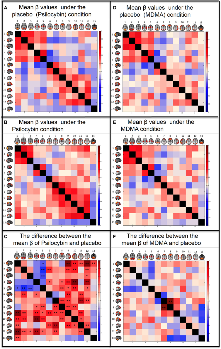
All of this is background to a new study in Frontiers in Human Neuroscience that looks at both psilocybin and MDMA and how they generate changes in resting-state functional connectivity (RSFC). Between-network RSFC increased under psilocybin, suggesting reduced differentiation between networks in the altered state of consciousness. There was also decreased RSFC between visual and sensorimotor resting state networks (RSN). MDMA had a much smaller effect on between-network RSFC. The suggestion is that classic psychedelics are more likely to produce profound effects on perception.

Full Citation: 
Roseman, L, Leech, R, Feilding, A, Nutt, DJ, and Carhart-Harris, RL. (2014, May 27). The effects of psilocybin and MDMA on between-network resting state functional connectivity in healthy volunteers. Frontiers in Human Neuroscience; 8:204. doi: 10.3389/fnhum.2014.00204

The effects of psilocybin and MDMA on between-network resting state functional connectivity in healthy volunteers

Leor Roseman [1,2], Robert Leech [2], Amanda Feilding [3], David J. Nutt [1], and Robin L. Carhart-Harris [1]
1. Centre for Neuropsychopharmacology, Division of Brain Sciences, Department of Medicine, Imperial College London, London, UK
2. Computational, Cognitive and Clinical Neuroscience Laboratory, Division of Brain Sciences, Department of Medicine, Imperial College London, London, UK
3. The Beckley Foundation, Oxford, UK

Abstract


Perturbing a system and observing the consequences is a classic scientific strategy for understanding a phenomenon. Psychedelic drugs perturb consciousness in a marked and novel way and thus are powerful tools for studying its mechanisms. In the present analysis, we measured changes in resting-state functional connectivity (RSFC) between a standard template of different independent components analysis (ICA)-derived resting state networks (RSNs) under the influence of two different psychoactive drugs, the stimulant/psychedelic hybrid, MDMA, and the classic psychedelic, psilocybin. Both were given in placebo-controlled designs and produced marked subjective effects, although reports of more profound changes in consciousness were given after psilocybin. Between-network RSFC was generally increased under psilocybin, implying that networks become less differentiated from each other in the psychedelic state. Decreased RSFC between visual and sensorimotor RSNs was also observed. MDMA had a notably less marked effect on between-network RSFC, implying that the extensive changes observed under psilocybin may be exclusive to classic psychedelic drugs and related to their especially profound effects on consciousness. The novel analytical approach applied here may be applied to other altered states of consciousness to improve our characterization of different conscious states and ultimately advance our understanding of the brain mechanisms underlying them.


Introduction


Psychedelic drugs have been used throughout history by different cultures as a means of altering consciousness. They are powerful tools for understanding the neurobiology of consciousness yet they have been underutilized by modern science, arguably due to political rather than scientific circumstances (Nutt et al., 2013). The majority of consciousness research has focused on states of reduced consciousness such as coma and sleep (Laureys, 2005). Indeed, consciousness has been defined as that which is lost during dreamless sleep (Tononi, 2004) but consciousness can also be studied in terms of changes in the mode or style of waking consciousness, such as is seen in the psychedelic state. Another popular model of consciousness describes it using two parameters: (1) wakefulness or arousal and (2) awareness (Laureys et al., 2009). It is recognized that these parameters have a mostly linear relationship; however, REM sleep and the vegetative state are considered anomalies, since the former involves greater awareness than would be predicted by wakefulness and the latter displays less (Laureys et al., 2009). The position of the psychedelic state in this model has never been considered before and it presents another interesting anomaly. There is no evidence of reduced wakefulness in the psychedelic state and although awareness is altered, it would be misleading to say that it is reduced. Indeed, the psychedelic state has been referred to as an “expansive” state of consciousness (Huxley, 1959). Thus, it is important to investigate what the neurobiological basis of this putative broadening of consciousness is.

One of the most popular theories of consciousness is the “information integration” theory of Tononi (2012). This proposes that consciousness depends on the presence of two key parameters: (1) information and (2) integration. Information is derived from information theory (Shannon and Weaver, 1949) and in the context of consciousness, refers to the potential size of the repertoire of different metastable states (Tognoli and Kelso, 2014) (or “sub-states”) the mind/brain can enter over time. Integration refers to the capacity of the mind/brain to integrate processes into a collective whole. The parameter of awareness is likely to be related to the property of information, since the greater the repertoire of sub-states the mind can enter, and the easier it can move between these, the broader consciousness will be.

In recent years, there has been an increasing interest in human fMRI measures of resting state functional connectivity (RSFC) (Damoiseaux and Greicius, 2009). Resting state networks (RSN) can be identified using seed-based approaches (Biswal et al., 1995) and independent component analysis (ICA) (Beckmann et al., 2005). These RSNs resemble stimulus-evoked networks (Smith et al., 2009) and may be thought of as metastable sub-states making-up a particular (macro) state of consciousness (Carhart-Harris et al., 2014a). Thus, one way to describe the quality of a macro-state of consciousness may be to investigate the integrity and dynamics of its sub-states and how they interact with each other. One way this can be done is by looking at the internal stability (integrity) of an RSN, i.e., reflected in the strength of the coupling between its constituent nodes. For example, we have found decreased intra-RSN connectivity post-psilocybin with both fMRI (Carhart-Harris et al., 2012a) and magnetoencephalography (MEG) (Muthukumaraswamy et al., 2013), implying a general breakdown of the integrity or internal stability of RSNs under psilocybin.

Another way to address the behavior of a system's sub-states is to look at their relationship with each other, e.g., by measuring between-RSN functional connectivity or coupling. A frequently investigated RSN is the default mode network (DMN) (Raichle et al., 2001). The DMN is known to be more active during rest than during goal-directed cognition and its activity has been found to be “anti-correlated” or at least uncorrelated or orthogonal with activity in networks that are engaged during goal-directed cognition - referred to generically as “task positive networks” or TPNs. This anticorrelation is preserved under task free conditions (Fox et al., 2005), implying that it is an important feature of normal consciousness, perhaps accounting for the distinction between externally focused cognition and introspection (Carhart-Harris et al., 2012b). We recently found that the classic psychedelic drug psilocybin reduces the anticorrelation between DMN and a number of TPNs during resting conditions, and this was interpreted as a decrease in the natural distinction between externally-focused attention and introspection (Carhart-Harris et al., 2012b), which is relevant to the notion of “ego-boundaries,” i.e., an agent's sense of being apart from or separate to its environment. It would be a natural extension of the above analysis to address the full gamut of between-RSN FC identified by ICA rather than just focusing on just the DMN-RSN RSFC. This was the aim of the present study.

The primary focus of the present paper is the classic psychedelic state and determining its underlying neurodynamics as measured with fMRI. However, in order to understand the psychedelic state, it is useful to compare it with other states of consciousness to see how it relates to these. Thus, the present analysis focuses on the brain effects of a classic psychedelic drug, psilocybin (the active component of magic mushrooms) and compares this with the effects of the pro-serotonergic stimulant, 3–4 methylenedioxymethamphetanine, MDMA. MDMA is a potent monoamine releaser that produces an acute euphoria in most individuals but it is not considered a classic psychedelic, as psilocybin is. Direct 5-HT2AR stimulation is the defining pharmacological property of classic serotonergic psychedelics, but relative to classic psychedelics, MDMA has a far weaker affinity for the 5-HT2A receptor (Green et al., 2003). Instead, MDMA produces a more generalized, non-selective activation of monoamine receptors by increasing the concentration of their endogenous ligands in the synapse via transporter-mediated release (Green et al., 2003). The primary subjective effects of MDMA include increased positive mood, heightened sensations and prosocial sentiments and although it can produce mild visual hallucinatory phenomena, it does not alter consciousness in the same fundamental manner as classic psychedelics (Gouzoulis-Mayfrank et al., 1996).

Thus, comparing changes in RSFC under psilocybin and MDMA can enable us to isolate and identify effects that are unique to the psychedelic-induced altered state of consciousness produced by classic psychedelics such as psilocybin. Considering the previous findings of decreased intra-RSN FC and DMN-TPN anti-correlation under psilocybin (Carhart-Harris et al., 2012a,b; Muthukumaraswamy et al., 2013), we hypothesized that the normal differentiation between RSNs would be affected by psilocybin such that RSNs whose activity is usually highly correlated would show reduced RSFC under psilocybin (but not MDMA) and that networks that are normally anti-correlated would show reduced anti-correlation under psilocybin (but not MDMA). If the hypothesized effects are present under psilocybin but absent under MDMA, this will strengthen the inference that they are specifically related to psilocybin more profound effects on consciousness.


Materials and Methods


Design

Psilocybin

This is an entirely new analysis on a previously published data set (Carhart-Harris et al., 2012a,b). This was a within-subjects placebo-controlled study that was approved by a local NHS Research Ethics Committee and Research and Development department, and conducted in accordance with Good Clinical Practice guidelines. A Home Office License was obtained for storage and handling of a Schedule 1 drug. The University of Bristol sponsored the research. The research was carried out at CUBRIC, University of Cardiff.

MDMA

This is also an entirely new analysis on a previously published dataset (Carhart-Harris et al., 2014b). This was a within-subjects, double-blind, randomized, placebo-controlled study. Participants were scanned twice, 7 days apart, once after MDMA and once after placebo. The study was approved by NRES West London Research Ethics Committee, Imperial College London's Joint Compliance and Research Office (JCRO), Imperial College's Research Ethics Committee (ICREC), the Head of Imperial College's Department of Medicine, Imanova Center for Imaging Science and Imperial College London's Faculty of Medicine, and was conducted in accordance with Good Clinical Practice guidelines. A Home Office License was obtained for the storage and handling of a Schedule 1 drug and Imperial College London sponsored the research.

Participants

Psilocybin

Fifteen healthy subjects took part: 13 males and 2 females (mean age = 32, SD = 8.9). Recruitment was via word of mouth. All subjects were required to give informed consent and undergo health screens prior to enrolment. Entry criteria were: at least 21 years of age, no personal or immediate family history of a major psychiatric disorder, substance dependence, cardiovascular disease, and no history of a significant adverse response to a hallucinogenic drug. All of the subjects had used psilocybin at least once before (mean number of uses per subject = 16.4, SD = 27.2) but not within 6 weeks of the study.

MDMA

The original study sample comprised of 25 healthy participants (mean age = 34, SD = 11, 7 females) with at least 1 previous experience with MDMA. None of the participants had used MDMA for at least 7 days and other drugs for at least 48 h, and this was confirmed by a urine screen. As a conservative step to control for between-study differences in the global intensity of the subjective effects produced by the different drugs, 11 subjects who gave ratings of <50% for the intensity of MDMA's effects were excluded from the analysis. This step meant that ratings of drug effects intensity were comparable across the two studies (i.e., the mean intensity of psilocybin's subjective effects was 67 ± 19 at peak and MDMA's was 69 ± 15). An additional subject was excluded because of significant head movements (mean head motion > one voxel width). Thus, a total of 13 subjects were included in the analysis (i.e., 12 excluded). An alcohol Breathalyzer test confirmed that none of the participants had recently consumed alcohol. For the sample of 13, participants had used MDMA an average of 29 (±35) times before (range = 1–100) and the mean time since last use was 983 (±1998) days (range = 7–6570 days). Participants were screened for general health, MR-compatibility and present mental health. Screening involved routine blood tests, electrocardiogram, heart rate, blood pressure and a brief neurological exam. All subjects were deemed physically and mentally healthy at the time of study entry and none had any history of drug or alcohol dependence.

Anatomical Scans

Psilocybin

Imaging was performed on a 3T GE HDx system. Anatomical scans were performed before each functional scan. These were 3D fast spoiled gradient echo scans in an axial orientation, (1 mm isotropic voxels).

MDMA

Imaging was performed on a 3T Siemens Tim Trio (Siemens Healthcare, Erlangen, Germany) using a 32-channel phased array head coil. Anatomical reference images were acquired using the ADNI-GO recommended MPRAGE parameters (1 mm isotropic voxels).

Drug and Scanning Parameters

Psilocybin

All subjects underwent two 12-min eyes-closed resting-state blood oxygen–level dependent (BOLD) fMRI scans on 2 separate occasions at least 7 days apart: placebo (10 ml saline, 60-s intravenous injection) was given on 1 occasion and psilocybin (2 mg dissolved in 10 ml saline) on the other. Seven of the subjects received psilocybin in scan 1, and 8 received it in scan 2. Injections were given manually by a study doctor situated within the scanning suite. The 60-s infusions began exactly 6 min after the start of the 12-min scans. Subjective ratings were given post-scan using visual analog scales (VAS). The subjective effects of psilocybin were felt almost immediately after injection and were sustained for the duration of the scan.

MDMA

Two BOLD resting-state scans were performed during each functional scanning session (duration of functioning scanning = 60 min). The first resting-state BOLD scan took place 60 min after capsule ingestion and the second resting-state BOLD scan occurred 113 min after capsule ingestion. Peak subjective effects were reported 100 min post administration of MDMA, generally consistent with the plasma t-max of MDMA (Kolbrich et al., 2008). The order of MDMA and placebo administration was counterbalanced.

fMRI Data Acquisition

Psilocybin

BOLD-weighted fMRI data were acquired using a gradient echo planar imaging sequence, 3 mm isotropic voxels, TR = 3000 ms, TE = 35 ms, field-of-view = 192 mm, 90° flip angle, 53 axial slices in each TR, parallel acceleration factor = 2, 64 × 64 acquisition matrix. The psilocybin and placebo scans for this analysis were of 5 min (1 min post infusion).

MDMA

BOLD-weighted fMRI data were acquired using a gradient echo planar imaging sequence, 3 mm isotropic voxels, TR = 2000 ms, TE = 31 ms, field-of-view = 192 mm, 80° flip angle, 36 axial slices in each TR, GRAPPA acceleration = 2, bandwidth = 2298 Hz/pixel. For each condition, MDMA and placebo, two scans were used for the analysis, each one of 6 min (performed 60 min and 113 min post-capsule ingestion)

Resting State Networks (RSN)

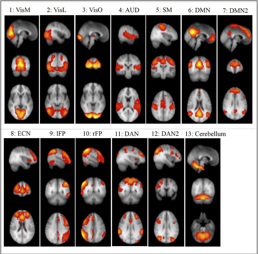
We used RSNs that were identified in Smith et al. (2009) using ICA (Figure 1). Ten of these components were given functional labels based on their correspondence to the BrainMap database of functional imaging studies, involving task-evoked FMRI data from nearly 30,000 human subjects. These networks were: Visual-Medial Network (VisM), Visual-Lateral Network (VisL), Visual-Occipital pole Network (VisO), Auditory Network (AUD), Sensorimotor Network (SM), Default Mode Network (DMN), Executive Control Network (ECN), Left frontoparietal Network(lFP), Right frontoparietal Network (rFP) and Cerebellar network. In addition, we used three more components from Smith et al, that we named DMN2 (an anterior DMN and ECN hybrid), Dorsal Attention Network 1 and 2 (DAN1 and DAN2). Another 6 components were identified as non-neural noise (likely generated by head motion and non-neural physiological fluctuations).
FIGURE 1  
http://www.frontiersin.org/files/Articles/78277/fnhum-08-00204-HTML/image_m/fnhum-08-00204-g001.jpg

Figure 1. Non-noise resting State Networks (RSN) from Smith et al., 2009: (1) Visual–Medial (VisM), (2) Visual–Lateral (VisL), (3) Visual–Occipital pole (VisO), (4) Auditory (AUD), (5) Sensorimotor (SM), (6) Default Mode Network (DMN), (7) DMN2–A hybrid of anterior DMN and Executive Control Network, (8) Executive Control Network (ECN), (9) left Frontoparietal Network (lFP), (10) right Frontoparietal Network (rFP), (11) Dorsal Attention Network (DAN), (12) DAN2, (13) Cerebellum. Ten of these components were given functional labels based on their correspondence to the BrainMap database of functional imaging studies. (RSNs 1, 2, 3, 4, 5, 6, 8, 9, 10, 13), additional networks (7, 11, 12) were labeled by the experimenters in the current study based on the regional distribution of activity.

Preprocessing

All analyses were performed using the Functional Magnetic Resonance Imaging of the Brain (FMRIB) Software Library (FSL, www.fmrib.ox.ac.uk/fsl) (Smith et al., 2004). We used the standard imaging preprocessing FSL pipeline that involved brain extraction (Smith, 2002), motion correction using MCFLIRT (Jenkinson et al., 2002), spatial smoothing (FWHM) of 5 mm (Smith and Brady, 1997) and a high-pass filter of 100 s. The scans were registered to the subjects' T1-weighted high-resolution (2 × 2 × 2 mm) anatomical scans and were then registered to the Montreal Neurological Institute standard brain (2 × 2 × 2 mm) (Jenkinson et al., 2002). The data was resampled into 4 mm space as part of the default processing pipeline for Melodic and was done to make the analysis more computational efficient.

Between Networks Functional Connectivity (FC)

Psilocybin

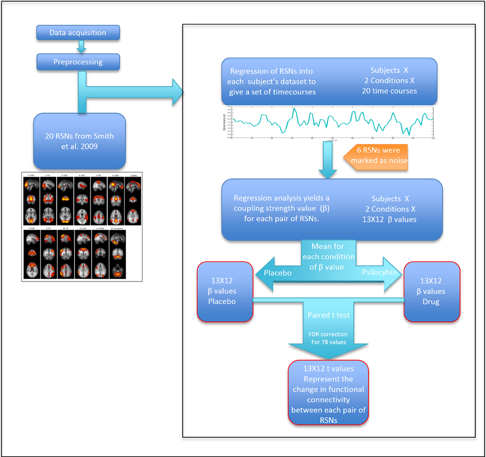
To extract time courses for each subject for each RSN and for each condition, we back-projected the components from Smith et al. (2009) into each 4D fMRI dataset using a general linear model. Specifically, we took the 20 components ICA map from Smith et al. as the set of template ICAs for the dual regression pipeline. The first step of the “dual regression” pipeline was then applied to each 4D dataset, resulting in a specific timecourse for each component for each dataset (Beckmann et al., 2009). Between-RSN coupling was presented graphically using a 13 × 13 correlation (or more strictly, regression) matrix in which the color in each square represents a beta weight or coupling strength for the corresponding RSN-RSN pair. Specifically, these weights were calculated by entering the time course for a specific RSN as a dependent variable in a general linear model, with the time course of another RSN entered as an independent variable—with this procedure repeated for each RSN pair. The mean head motion under psilocybin and its placebo condition were 0.1 ± 0.05 mm and 0.06 ± 0.015 mm, respectively (p < 0.01). Therefore, to further partial out non-neural noise confounds, six motion time courses (estimated from the motion correction) and motion outliers (estimated using the “fsl_motionoutlier” command implemented in FSL), as well as the time courses for 6 non-neural noise components were entered as confounds (some of this noise is driven by head motion). The model resulted in a parameter estimate or unstandardized beta weight (β) representing the strength of functional coupling between each RSN pair. The general linear model was estimated twice for each RSN pair: with each RSN as dependent variable in one model and as an independent variable in the second model. Since we were not looking at effective or directed connectivity (Friston et al., 2003), we created a symmetrical connectivity matrix by averaging together each subject's two β values for each RSN pair. For each RSN pair, three results were calculated: (a) group mean β value for the placebo condition; (b) group mean β value for the psilocybin condition; (c) Paired t-test (2-tail) for the difference between the mean β values of each condition (Figure 2). To correct for multiple comparisons, a false discovery rate (FDR) threshold was calculated using q = 0.05 and q = 0.1 (N = 78).
FIGURE 2  
http://www.frontiersin.org/files/Articles/78277/fnhum-08-00204-HTML/image_m/fnhum-08-00204-g002.jpg

Figure 2. Scheme of the analysis by steps. Calculating t-values for each RSN pair that represent the change in coupling strength between placebo and drug.

MDMA

The MDMA RSFC was analyzed using the same procedure described above (Figure 2). The only difference was that there were two resting state scans in the MDMA study, so β values from the two scans (performed 60 min and 113 min post-capsule ingestion) were averaged together before comparing between the placebo and drug conditions. The mean head motion under MDMA and its placebo condition were 0.083 ± 0.036 mm and 0.061 ± 0.019 mm, respectively (p = 0.047). The same procedure to control for motion in the psilocybin analysis was used for MDMA.


Results


Subjective Effects

Psilocybin

The subjective effects of psilocybin have been documented elsewhere (Carhart-Harris et al., 2011, 2012a). Briefly, the subjective effects of 2 mg psilocybin given as an intravenous injection over 60 s begin at the end of the injection period, reach a sustained peak after approximately 5 min, and subside completely after 45–60 min. Primary subjective effects include altered visual perception (e.g., hallucinated motion and geometric patterns), an altered sense of space and time, and vivified imagination. The intensity of psilocybin's global subjective effects was rated using a VAS format. The mean intensity at peak effects (5 min post-infusion) was 67% ±19.

MDMA

The subjective effects of MDMA are reported in a separate paper (Carhart-Harris et al., 2014b). At their peak, the average intensity of MDMA's global subjective effects was 69% ±15 (n = 13). There was no significant difference between intensity ratings under the two different drugs.

Between Networks FC

Psilocybin

The coupling strengths (β) for each condition can be seen graphically in the correlation matrixes in Figure 3 and numerically in the Supplementary material. For the placebo condition, see Figure 3A and Supplementary Table 1A and for the psilocybin condition see Figure 3B and Supplementary Table 1B. A paired t-test (2-tail) was done across subjects to compare the β values for each RSN pair in the drug and placebo (Figure 3C and Supplementary Table 1C). The results were corrected for multiple comparisons using FDR with q = 0.05 (resulting in a threshold of p < 0.0167) and q = 0.1 (resulting a threshold of p < 0.042). The RSN pairs that showed a significant decrease in coupling under psilocybin were: SM-VisM (p = 0.0265), SM-VisL (p = 0.0051) and SM-VisO (p = 0.0151). The RSN pairs that showed a significant increase in coupling were: VisM-lFP (p = 0.0001), VisM-DAN (p = 0.0156), VisM-rFP (p = 0.0023), VisM-DAN2 (p = 0.0002), VisM-Cerebellum (p = 0.0108), VisL-DMN (p = 0.0046), VisL-lFP (p = 0.0056), VisL-rFP (p = 0.0031), VisL-DAN2 (p = 0.0142), VisO-DAN2 (p = 0.0256), AUD-DMN (p = 0.028), AUD-ECN (p = 0.0323), AUD-lFP (p = 0.0029), AUD-rFP (p = 0.0001), AUD-DAN2 (p = 0.0005), SM-ECN (p = 0.0105), SM-lFP (p = 0.022), SM-rFP (p = 0.0026), SM-DAN2 (p = 0.034), DMN-lFP (p = 0.0029), DMN-DAN (p = 0.0058), DMN2-ECN (p = 0.0071) DMN2-lFP (p = 0.0101), DMN2-DAN (p = 0.0005), DMN2-DAN2 (p = 0.0091), ECN-lFP (p = 0.0077), ECN-rFP (p = 0.0098), lFP-DAN (p = 0.0026), rFP-DAN (p = 0.0187), and DAN-DAN2 (p = 0.0161).
FIGURE 3  
http://www.frontiersin.org/files/Articles/78277/fnhum-08-00204-HTML/image_m/fnhum-08-00204-g003.jpg

Figure 3. Between networks resting state functional connectivity results. Within each matrix, each colored square represents coupling between corresponding RSN pairs with the color of the square denoting the coupling strength (A,B,D,E) or change in coupling strength (C,F) between the RSN pairs (blue, negative coupling or a decrease in coupling; red, positive coupling or an increase in coupling). The six images are: (A) Group mean of β values for the placebo of psilocybin condition. (B) Group mean of β values for the psilocybin condition. (C) Paired t-test (2-tail) for the difference between the mean β values of psilocybin and placebo. (D) Group mean of β values for the placebo of MDMA condition. (E) Group mean of β values for the MDMA condition. (F) Paired t-test (2-tail) for the difference between the mean β values of MDMA and placebo. The networks from Smith et al., (2009) are: (1) Visual—Medial (VisM), (2) Visual—Lateral (VisL), (3) Visual—Occipital pole (VisO), (4) Auditory (AUD), (5) Sensorimotor (SM), (6) Default Mode Network (DMN), (7) DMN2—A hybrid of anterior DMN and Executive Control Network, (8) Executive Control Network (ECN), (9) left Frontoparietal Network (lFP), (10) right Frontoparietal Network (rFP), (11) Dorsal Attention Network (DAN), (12) DAN2, (13) Cerebellum. FDR correction for multiple comparison (N = 78) was applied on the t-tests: *0.05 < q < 0.1. **q < 0.05.

MDMA

The same analysis as above was repeated for the MDMA condition using a q of 0.05, resulting in a threshold of p < 0.0006 and q = 0.1, resulting in a threshold of p < 0.0012. Only one RSN pair showed a significant change in coupling under MDMA, i.e., increased coupling between the DMN2-ECN (p = 0.0001).

Differences in Movement

Both drugs showed significant, yet relatively modest, increased head motion between conditions. The mean head motion under psilocybin and its placebo condition were 0.1 ± 0.05 mm and 0.06 ± 0.015 mm, respectively (p < 0.01). The mean head motion under MDMA and its placebo condition were 0.083 ± 0.036 mm and 0.061 ± 0.019 mm, respectively (p = 0.047). Power et al. (2012) suggest that head motion can change the results of RSFC, therefore, in the regression analysis, we added several motion confounds: six motion time courses, motion outliers [similar to the procedure of scrubbing within regression (spike regression) mentioned by Yan et al. (2013) and Satterthwaite et al. (2013)] and time courses of RSNs that were driven by motion. However, it still remains possible that the increased movement under the drugs may have caused the changes in RSFC. Hence, we investigated if there was a relationship between the change in estimated motion (mean framewise displacement) between placebo and drug and the change in coupling strength (for pairs of RSNs that showed significant differences in coupling). For most of the RSN pairs no relationship was found (p < 0.05). However, under psilocybin, there were significant correlations with motion in the following RSN pairs: VisM-SM (p = 0.002), VisL-SM (p = 0.001), VisO-SM (p = 0.02), VisL-DMN (p = 0.03), VisM-rFN (p = 0.048), VisL-rFN (p = 0.01), VisO-DAN2, DMN-lFN (p = 0.001), DMN-DAN (p = 0.01). For that reason, the significant results of these RSN pairs should be approached with caution.


Discussion


To our knowledge, this is the first analysis to test the effects of different pharmacological agents using a standard ICA-derived template of RSNs to construct between-network functional connectivity matrixes for different drug states. This approach may have wider application, enabling researchers to determine connectivity “fingerprints” for characterizing different states of consciousness, i.e., not only those induced by pharmacological agents but sleep states and even pathological states. This will enable informed comparisons to be made between different states, potentially allowing us to categorize different states based on their connectivity profiles. Functional connectivity matrixes have been used before to differentiate between pathology states such as schizophrenia and bipolar disorder (Mamah et al., 2013) and here we suggest that they could be used more broadly to characterize states of consciousness, including those induced by psychoactive drugs.

Probably the most striking result of the present study was the marked increases in between-network RSFC under psilocybin. These increases were evident for heteromodal networks, both in terms of increased unimodal-heteromodal (e.g., AUD-rFP) and heteromodal-heteromodal network RSFC (e.g., lFP-ECN). Based on previous analyses (Carhart-Harris et al., 2012b), we had predicted that RSN pairs with weak or negative RSFC at baseline would show increased coupling post-psilocybin, and this was found (e.g., DMN-VisL). However, the increases in between-network RSFC were more fundamental than this, being evident for RSN pairs that were already positively coupled at baseline (e.g., DMN2-ECN). The increase in correlated brain activity across normally distinct brain networks was particularly true for heteromodal RSNs, where the distribution of 5-HT2A receptors is known to be highest (Erritzoe et al., 2010) and 5-HT2A receptor stimulation is linked to desynchronous cortical activity (Riba et al., 2002; Wood et al., 2012; Muthukumaraswamy et al., 2013) and network disintegration (Muthukumaraswamy et al., 2013; Carhart-Harris et al., 2014a).

The pattern of increased between-network RSFC under psilocybin did not apply universally for the whole of the brain. Decreased RSFC was observed between the three visual RSNs and the sensorimotor network [these networks are known to be highly connected (Wise et al., 1997; Van Den Heuvel et al., 2008)], and there was a general trend toward decreased unimodal-unimodal network RSFC (e.g., VisM-AUD and SM-AUD showed decreased RSFC under psilocybin but this failed to survive FDR correction, see Supplementary Table 1). However these decreases can also be explained by the changes in head motion between conditions and further work is required to test whether these decreases in sensory RSN RSFC under psilocybin relate to the drug's characteristic perceptual/hallucinatory effects.

Previous neuroimaging studies with psychedelics have so far failed to reveal a simple and compelling explanation for their characteristic hallucinogenic effects (Vollenweider et al., 1997; Carhart-Harris et al., 2012a; Muthukumaraswamy et al., 2013) (but see De Araujo et al., 2012) and so drug-induced visual hallucinations remain poorly understood. Under normal conditions, activity in the visual cortex is driven by and thus anchored to visual input. Moreover, activity in other networks (e.g., the DMN), concerned with other distinct functions (e.g., introspection), is often weakly or inversely coupled to visual activity (e.g., see the pale and blue colored squares for the visual-RSN pairs in Figures 3A,D). Thus, increased communication between the visual system and systems that are usually reserved for distinct functions may lead to erroneous perceptual associations. For example, increased DMN-visual network RSFC, may relate to an increased influence of imagination (mediated by the DMN) on visual perception (mediated by the visual networks). A similar process may occur in situations of sensory deprivation where sensory processing becomes decoupled from sensory stimulation, allowing the system to “free-wheel” with the potential for the spontaneous emergence of internally-generated percepts. Decreased cross-modality RSFC and increased unimodal to heteromodal network RSFC may be a common characteristic of such states but future studies are required to test this. For example, comparisons between the present results and changes in RSFC in the meditative state could inform these speculations.

Given reports of synesthesia-like experiences under psychedelics (e.g., participants reported that the noise of the MR scanner influenced the rate and content of their closed eye visual hallucinations Carhart-Harris et al., 2012a and see also Luke and Terhune, 2013) one may have predicted increased cross-modality communication under psilocybin rather than the decreased coupling that was observed here. However, it has yet to be determined whether synesthesia-like experiences in drug-induced altered states of consciousness are qualitatively and mechanistically related to synesthesia experienced outside of this context and it is also worth noting that increased visual to heteromodal cortical functional connectivity has been found in color-grapheme synesthesia (Dovern et al., 2012; Sinke et al., 2012) as well as in the present study.

Taking a dynamical systems theory approach to the present results, RSNs can be conceived of as “attractors,” i.e., patterns of activity into which the brain tends to gravitate for short periods of time (Deco et al., 2009; Hellyer et al., 2014). A macro-state of consciousness (such as normal waking, deep sleep or the psychedelic state) may, therefore, be graphically represented as an “attractor landscape” in which the depth of “basins of attraction” (valleys in an otherwise flat 2D-plane) reflect the stability of particular RSNs or metastable “sub-states,” i.e., more long lasting sub-states will have deep basins of attraction and unstable sub-states will have shallow ones. A recent paper (Kanamaru et al., 2013) has described brain function in these terms, suggesting that the shape of attractors depends on selective attention. In this particular model, high levels of acetylcholine activating muscarinic receptors were found to produce an attractor landscape with more stable sub-states. Relating this to the present results, the increased RSFC observed between different RSNs could be interpreted as a flattening of the attractor landscape, in which the basins of attraction are shallower, implying that the global system will move more easily between different metastable sub-states. A flattened (but not flat) attractor landscape would be consistent with increased “information” in the sense of the “information-integration” theory of consciousness (Tononi, 2012) since greater movement between metastable sub-states would imply that a larger number of these sub-states (or a broader “repertoire”) can be entered over a given time. At a critical flatness, the size of the repertoire of metastable states will be maximal but if the landscape is too flat, information will be reduced because attractors will become too unstable. This scenario is referred to as “super-criticality” (Chialvo, 2010), and if taken to the extreme, an entirely flat landscape would imply that the system has no metastable states, or just one entirely disordered one. Future studies are required to determine whether the psychedelic state is “critical” or “super-critical” in this sense (Tagliazucchi et al., 2012; Carhart-Harris et al., 2014a). Another way these results could be perceived however, is that increased between-RSN RSFC under psilocybin is representative of a “sub-critical” system, i.e., one that is more globally synchronous and therefore ordered; however, that there were also decreases in between-RSN RSFC under psilocybin, does not support this view. We intend to follow-up this matter in order to test our hypothesis that it is specifically the ease of transition (or transition probability) between RSNs/metastable sub-states that is facilitated under the drug.

In contrast to the marked changes in between-network RSFC observed with psilocybin, only one RSN-pair showed a significant change in RSFC under MDMA, i.e., increased ECN-DMN2 RSFC (Figure 3F). This result is difficult to interpret in isolation; however, it is worth noting that ECN-DMN2 RSFC was also significantly increased under psilocybin (Figure 3C). MDMA is not considered a classic psychedelic, although like psilocybin, its subjective effects are known to be significantly mediated by serotonergic mechanisms (Liechti and Vollenweider, 2001; Van Wel et al., 2011). Thus, increased ECN-DMN2 RSFC may relate to a shared aspect of these drugs' subjective effects, such as their propensity to alter mood and cognition (Carhart-Harris et al., 2014b). Pre-treatment studies with selective receptor antagonists would help to inform these matters.

There is an important caveat to be addressed about the present analysis. It should be noted that the two studies from which the data was derived employed quite different methodologies (e.g., intravenous administration of psilocybin vs. oral administration of MDMA, different MR scanners and different study samples). Thus, it would be problematic to attempt to make inferences based entirely on a comparison of their relative RSFC profiles. This analysis was not intended to be a formal comparison of the brain effects of MDMA and psilocybin and if this was the intention, then a standardized methodology would need to be employed. Rather, the present analysis has focused on understanding the neural correlates of the psychedelic state as produced by the classic psychedelic, psilocybin, and the finding that MDMA had a less marked effects on between-network RSFC has merely served to emphasize that the psychedelic state rests on a particularly profound disturbance of brain function. This does not imply that MDMA's own subjective effects are unimportant or that they do not involve some (albeit more subtle) changes in between-network RSFC.

The significant change in head movement under psilocybin implies that some of the results should be interpreted with caution, in particular the decreases in coupling strength. We have used multiple ways to model motion as a possible confound but for a subset of the RSN pairs, the changes with drug correlate with the differences in mean motion. These significant correlations do not necessarily mean that motion is responsible for these changes, since intensity of drug is likely to be associated with increased movement, meaning that disambiguating the two effects is problematic for some RSN pairs. In support of this, we found a marginally significant correlation between changes in motion and changes in the subjective intensity rating (r = 0.382, p = 0.08). Future work restricting head motion in the scanner and with larger samples is necessary to be able to demonstrate that changes in these RSN pairs that correlate with motion reflect genuine brain activity or not.

In conclusion, this new analysis has used between-network functional connectivity to investigate the effects of two distinct serotonergic compounds on spontaneous brain function. It was found that psilocybin produced marked changes in between-network RSFC, generally in the direction of increased coupling between RSNs, with an additional decrease in coupling between visual and sensorimotor networks. MDMA had a notably less marked effect on between-network RSFC implying that psilocybin's more profound effects on global brain function (at least as determined by this measure) may explain its more profound effects on consciousness. The analytic methods used in this study, i.e., using ICA templates to determine functional connectivity matrixes for different drug states, may have wider application, enabling researchers to more objectively describe and potentially categorize different states of consciousness.

Conflict of Interest Statement

The authors declare that the research was conducted in the absence of any commercial or financial relationships that could be construed as a potential conflict of interest.

Acknowledgments

These studies received financial and intellectual support from the Beckley Foundation. We would like to thank the reviewers for their useful comments on previous versions of this manuscript.

Supplementary Material

The Supplementary Material for this article can be found online at: http://www.frontiersin.org/journal/10.3389/fnhum.2014.00204/abstract

References available at the Frontiers site.