Showing posts with label adverse childhood experiences. Show all posts

Wednesday, September 24, 2014

Epigenomic Profiling of Men Exposed to Early-Life Stress Reveals DNA Methylation Differences in Association with Current Mental State

http://upload.wikimedia.org/wikipedia/commons/d/dd/Epigenetic_mechanisms.jpg

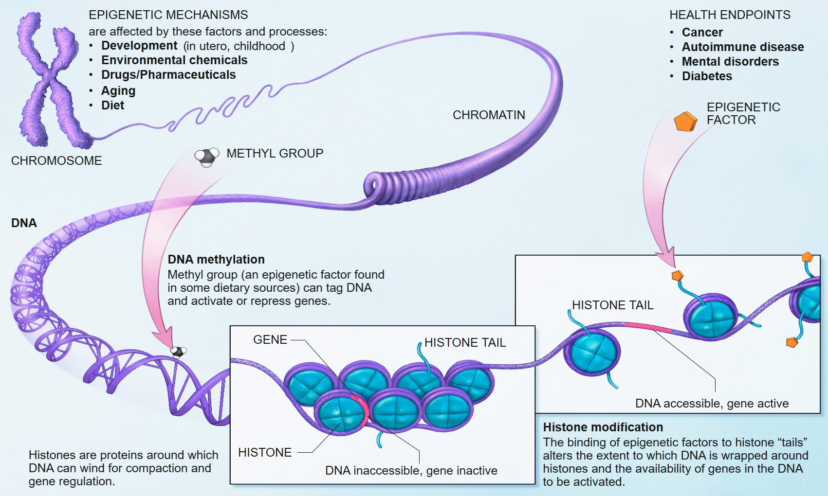
We know that early life adversity for children can cause long-lasting psychological issues, and one of the mechanisms of this outcome is inhibited DNA methylation. In biological systems, methylation is catalyzed by enzymes; such methylation can be involved in regulation of gene expression, regulation of protein function, and RNA processing, among other processes.
Research in humans has shown that repeated high level activation of the body's stress system, especially in early childhood, can alter methylation processes and lead to changes in the chemistry of the individual's DNA. The chemical changes can disable genes and prevent the brain from properly regulating its response to stress. Researchers and clinicians have drawn a link between this neurochemical dysregulation and the development of chronic health problems such as depression, obesity, diabetes, hypertension, and coronary artery disease.[9][10][11][12][13]
This new study adds to the evidence of how stress can derail normal developmental processes.

Full Citation:
Khulan, B, Manning, JR, Dunbar, DR, Seckl, JR, Raikkonen, K, Eriksson, JG, and Drake, AJ. (2014, Sep 23). Epigenomic profiling of men exposed to early-life stress reveals DNA methylation differences in association with current mental state. Translational Psychiatry; 4, e448; doi:10.1038/tp.2014.94


Epigenomic profiling of men exposed to early-life stress reveals DNA methylation differences in association with current mental state


B Khulan [1], J R Manning [1], D R Dunbar [1], J R Seckl [1], K Raikkonen [2], J G Eriksson [3,4,5,6,7] and A J Drake [1]
1. Endocrinology Unit, University/BHF Centre for Cardiovascular Science, Queen's Medical Research Institute, University of Edinburgh, Edinburgh, UK
2. Institute of Behavioural Sciences, University of Helsinki, Helsinki, Finland
3. Department of Chronic Disease Prevention, National Institute for Health and Welfare, Helsinki, Finland
4. Department of General Practice and Primary Health Care, University of Helsinki, Helsinki, Finland
5. Vasa Central Hospital, Vasa, Finland
6. Folkhälsan Research Center, Helsinki, Finland
7. Unit of General Practice, Helsinki University Central Hospital, Helsinki, Finland

Abstract


Early-life stress (ELS) is known to be associated with an increased risk of neuropsychiatric and cardiometabolic disease in later life. One of the potential mechanisms underpinning this is through effects on the epigenome, particularly changes in DNA methylation. Using a well-phenotyped cohort of 83 men from the Helsinki Birth Cohort Study, who experienced ELS in the form of separation from their parents during childhood, and a group of 83 matched controls, we performed a genome-wide analysis of DNA methylation in peripheral blood. We found no differences in DNA methylation between men who were separated from their families and non-separated men; however, we did identify differences in DNA methylation in association with the development of at least mild depressive symptoms over the subsequent 5–10 years. Notably, hypomethylation was identified at a number of genes with roles in brain development and/or function in association with depressive symptoms. Pathway analysis revealed an enrichment of DNA methylation changes in pathways associated with development and morphogenesis, DNA and transcription factor binding and programmed cell death. Our results support the concept that DNA methylation differences may be important in the pathogenesis of psychiatric disease.


Introduction


Early-life stress (ELS) is recognised as a risk factor for later mental health disorders. Findings from a number of studies have linked events in childhood, such as physical or sexual abuse, parental separation, neglect or adoption, with an increased risk of subsequent mental health disorders.1, 2, 3 The long-term adverse effects of ELS are not limited to neuropsychiatric problems and childhood exposure to socioeconomic disadvantage, maltreatment or social isolation is also associated with an increased risk of an adverse cardiometabolic disease risk profile in adulthood.3

Longitudinal cohort studies are an invaluable resource for the study of factors impacting on health, particularly in addressing the long-term impact of ELS. The Helsinki Birth Cohort Study (HBCS), comprising 13 345 individuals born in Helsinki between 1934 and 1944, is one such resource.4 During the Second World War, almost 70 000 Finnish children of varying socioeconomic status were evacuated from their homes to temporary foster families, mainly in the nearby countries of Sweden and Denmark. The evacuations, which occurred between 1939 and 1944, were arranged by the Finnish government or independently by families. Evacuations were voluntary, but heavily promoted by the government and particularly targeted at children living in cities. Documents on the evacuations and their timing and length have been retained in the Finnish National Archives’ Register. Linking evacuation data with the HBCS has allowed the identification of 11 028 individuals within the HBCS who were not separated from their parents as children, and 1719 individuals who were temporarily separated from their parents. The average age at separation was 4.6 years (s.d.=2.4, range=0.17–10.6) and the average length of separation 1.7 years (s.d.=1.6, range=0.05–8.1).

Previous studies in this cohort have identified a number of long-term consequences of early-life separation. Separated individuals have a higher prevalence of mental health disorders including depressive symptoms and personality disorders5 and an increased risk of substance abuse.6 The risk of any mental and substance use disorder was highest among those with an upper childhood socioeconomic background, perhaps suggesting increased vulnerability.6 Separation is also associated with effects on stress biology; separated individuals have higher average salivary cortisol and plasma ACTH concentrations and higher salivary cortisol reactivity to a Trier Social Stress Test.7 In men, separation associates with poorer cognitive performance8 and with negative effects on physical and psychosocial functioning.9 The consequences of separation were not limited to effects on mental health and the hypothalamic–pituitary–adrenal (HPA) axis, as cohort members who were separated from their parents in childhood also had a higher cardiovascular morbidity, including coronary artery disease, hypertension and type 2 diabetes,10,11 with the highest prevalence of cardiovascular disease in those who were evacuated for the longest period.11 Finally, early-life separation also associated with differences in reproductive and marital traits in both sexes.12

One of the potential mechanisms by which the environment in early life might have a lasting impact on the phenotype of an individual is through effects on the epigenome, particularly changes in DNA methylation13 and consistent with this, a few studies have shown alterations in DNA methylation in association with exposure to ELS.14,15 In this study of a unique cohort of men in their 70s from the HBCS we have investigated the long-term impact of early-life separation on genome-wide DNA methylation and whether this associated with a number of clinical and psychological variables.


Materials and Methods

Cohort

The HBCS comprises 13 345 individuals (6370 women and 6975 men), born as singletons between 1934 and 1944 in one of the two main maternity hospitals in Helsinki and who were living in Finland in 1971 when a unique personal identification number was allocated to each member of the Finnish population. The HBCS, which has been described in detail elsewhere,4 has been approved by the Ethics Committee of the National Public Health Institute. Register data were linked with permission from the Finnish Ministry of Social Affairs and Health and the Finnish National Archives.

In 2001–2004 at an average age of 61.5 years (s.d.=2.9 and range=56.7–69.8 years), a randomly selected subsample of the cohort comprising 2003 individuals (1075 women and 928 men) was invited to a clinical examination including collection of a blood sample for (epi)genetic and biochemical studies and a psychological survey including a measure of depressive symptoms. For 283 participants, extraction of DNA was not successful, or DNA showed gender discrepancy or close relatedness. The excluded and the included participants did not differ from each other in any of the study variables (P-values>0.13). From the remaining sample of 1720 individuals, 115 women and 97 men had been evacuated according to the Finnish National Archives’ register. Of them nine women and 12 men had missing data on age at and length of evacuation, respectively, and one man had missing data on father’s occupational status in childhood. In this study, the analyses are based on 83 evacuated men and 83 non-evacuated controls matched for sex, birth year and father’s occupational status in childhood. For this group the mean age was 64.0 years (s.d.=2.9) for separated and 62.9 (s.d.=2.5) for non-separated individuals. The mean difference in birth year is 1.21 years (P-value=0.01) such that evacuated individuals were born on average earlier. This reflects the fact that ‘matching’ in terms of birth year was not ‘perfect’ as older children were evacuated more frequently than younger children.

In 2009–2010 at an average age of 70.2 years (s.d.=2.8 and range=65.0–76.0 years), the evacuated cases and non-evacuated controls who were still traceable (n=65, 78.3% and n=63, 75.9%, respectively) were invited for a psychological follow-up, including a re-test on depressive symptoms. Of the evacuated cases and controls, 20 and 20 had died, their addresses were not traceable or they had refused participation in further follow-ups, respectively. Of them, 45 and 52 had data available on depressive symptoms.

The clinical variables available for the cohort included age at separation, length of separation, socioeconomic status and education level, history of mental health disorders and a number of biological measures including glucose, insulin, IL6, TNFα and CRP. The Beck Depression Inventory16 (BDI; performed at the 2001–2004 clinical assessment) and the BDI II17 (performed at the 2009–2010 clinical assessment) were used to measure the frequency of depressive symptoms. The BDI and BDI II consist of 21 items assessing symptoms of depression during the past two weeks. Each item contains four statements reflecting varying degrees of symptom severity. Respondents are instructed to circle the number that corresponds with the statement that best describes them. Ratings are summed to calculate a total score which can range from 0 to 63. Although the BDI and the BDI II are designed to screen but not diagnose major depression, BDI and BDI II cutoff scores of 10 and 14 or more, respectively, are suggestive of mild-to-severe depressive symptoms.17, 18, 19, 20, 21, 22

DNA extraction

Genomic DNA was extracted from EDTA-anti-coagulated whole peripheral blood collected at the first clinical assessment (2001–2004) using the QIAamp DNA Blood Maxi Kit (Qiagen, Crawley, UK). The samples were stored at −20 °C before and after DNA extraction.

DNA methylation analysis

DNA methylation analysis was performed at the Genetics Core of the Wellcome Trust Clinical Research Facility (Edinburgh, UK). Bisulphite conversion of 500 ng input DNA was carried out using the EZ DNA Methylation Kit (Zymo Research, Freiburg, Germany). Four microlitres of bisulphite-converted DNA was processed using the Infinium HD Assay for Methylation. This was performed using the Illumina Methylation 450 k beadchip and Infinium chemistry (Illumina, Inc., San Diego, CA, USA). Each sample was interrogated on the arrays against 485 000 methylation sites. The arrays were imaged on the Illumina HiScan platform and genotypes were called automatically using GenomeStudio Analysis software version 2011.1.


Data analysis

Data processing

Data were processed with the Lumi23 package of Bioconductor,24 with Infinium-centric routines. CpG loci were annotated with the gene of the nearest transcription start site, as defined in UCSC hg19 and retrieved with the ‘Genomic Features’25 Bioconductor package. Variables associated with each individual were examined independently for association with M values of methylation using the ‘phenotest’ Bioconductor package. P-values were corrected for probe-wise multiple testing with the Benjamini–Hochberg (BH) method. To remove any variable associations indicated by chance, a variable was only considered further if at least one probe passed a variable-wise Bonferroni correction. Having removed such spurious associations, downstream analyses were applied to values without this second correction. Individual pairwise contrasts were then constructed from the pairs of values for all categorical variables and examined for differential methylation using (a) linear modelling using Limma,26 including adjustment for multiple testing and (b) the methyAnalysis package to smooth processed data and identify differentially methylated regions by use of t-tests applied between groups (also BH-corrected for multiple testing).

Gene set enrichment

Gene set enrichment was analysed using annotations from the Kyoto Encyclopedia of Genes and Genomes (KEGG)27 and Gene Ontology.28 Only probes with methylation above a background of 0.2 in at least 17 (~10%) of samples were used in the enrichment background set. Enrichment was assessed by the use of hypergeometric statistic as implemented in the GO stats package of Bioconductor.

Genome location

To examine the genomic location of significantly associated loci independent of gene annotations, the genome was binned into overlapping bins of 100 kb in size and bins with three or more significant loci were reported.

Validation by pyrosequencing

Pyrosequencing was used to validate DNA methylation at five CpG sites within 200 bp of the TSS of the leucine-rich, glioma inactivated 1 (LGI1) gene and additionally at a number of CpG sites with a spectrum of high, low and intermediate methylation levels on the array (IL17C, SAGE1, MIR4493, MIR548M, CLDN9 and TACC3). Bisulphite conversion was performed on 1 μg of genomic DNA with the EZ DNA methylation kit (Zymo Research). The converted DNA was amplified using the AmpliTaq Gold 360 kit (Applied Biosystems, Warrington, UK) with primers mapping to target regions containing CpGs assayed within the array. PCR primers (Supplementary Table 1) were designed using PyroMark Assay Design Software 2.0 (Qiagen). Pyrosequencing was performed using PyroMark Q24Gold reagents on a PyroMark Q24 Pyrosequencer (Qiagen) according to the manufacturer’s instructions. Data were extracted and analysed using PyroMark Q24 1.0.10 software (Qiagen). Background non-conversion levels were <3%.


Results

Association of methylation with clinical variables

Principal components analysis showed no clear clusters and no obvious differences between the way separated and non-separated individuals or different socioeconomic groupings clustered. Linear modelling between clinical variables and methylation showed no associations between DNA methylation and most clinical variables including separation status and socioeconomic status. There were significant associations between DNA methylation in peripheral blood taken at the first clinical assessment (2001–2004) and categorical scores on the BDI II performed at the second assessment some years later (Table 1). When specific contrasts in categorical variables were queried using linear modelling, differential methylation was identified at 474 probes representing 445 genes, all of which associated with the clinical cutoffs indicative of at least mild depressive symptoms on the BDI II (representing a score of 14–25; Table 1 and Supplementary Table 2). This contrast involves a relatively small number of eight individuals, compared to 88 individuals with minimal depressive symptomatology (a score of 1–13). Running methyAnalysis with its associated smoothing produced a larger number of significant probes (491 probes representing 351 genes; Table 1), again predominantly in association with BDI II categorical score indicative of at least mild depressive symptoms. Of these, 80 genes showed differential methylation at more than one probe. There were 170 of the significant probes and 175 associated genes present in the lists from both methods. MethyAnalysis results were used in subsequent analyses.
Table 1 - Significant associations between methylation and BDI II score.

  
Full table
For genes, which had methylation changes observed in the same direction for more than one CpG site, DNA methylation was decreased in ~2/3 and increased in ~1/3 in association with a BDI II score indicative of at least mild symptomatology (Table 1). Differential methylation in association with a BDI II score indicative of mild depressive symptoms was identified at a number of genes with possible roles in brain development and/or function (Table 2), all of which showed hypomethylation (Figure 1). Among the most well-supported genes in terms of multiple significant probes were LGI1 and LGI2 which are thought to have important roles in brain development and function.29,30 Hypomethylation was present at five probes within the promoter of LGI1,31 and at three probes in a CpG island shore close to LGI2. Overall, significant loci were more likely to be identified in CpG island shores rather than in CpG islands or at the TSS (χ2 P-value=6 × 10⁻⁸; Supplementary Table 3), in agreement with the data from a study in postmortem brains.32 A number of 100 Kb bins in the genome contained three or more significant loci in the output from methyAnalysis associated with Beck Depression Questionnaire (Table 3).


Unfortunately we are unable to provide accessible alternative text for this. If you require assistance to access this image, please contact help@nature.com or the author

Figure 1. - Differential methylation in association with Beck Depression Inventory II (BDI II) score. Hypomethylation was present at multiple probes corresponding to genes with possible roles in brain development and/or function. BDI II=1 indicates a score of 1–13, that is, minimal symptomatology; BDI II=2 indicates a score of 14–25, that is, at least mild symptomatology. Full figure and legend (106K)
Table 2 - Genes with differential methylation in relation to depression scores which may be involved in the development and functioning of the brain.

 
Full table
Table 3 - 100 Kb bins in the genome containing three or more significant loci in the output from methyAnalysis associated with Beck Depression Questionnaire.

 
Full table

Pathway analysis

Analysis of gene ontology for the genes associated with altered DNA methylation and BDI II score using the molecular function, cellular component and biological processes categories of the Gene Ontology database28 revealed enrichment of a number of gene ontology terms, particularly within the molecular function and biological processes categories (Table 4). Mean methylation was decreased for all the genes within enriched pathways. The set of overrepresented categories included a significant number associated with development and morphogenesis, and DNA and transcription factor binding. In addition, consistent with the findings of a recent study in postmortem brain from individuals with depression compared with individuals without, a number of the identified gene sets are involved in programmed cell death;32 notably, hypomethylation was also found in association with depression in this study.
Table 4 - Gene enrichment: gene ontology overrepresented categories following assessment of DNA methylation differences in association with mild depression on the BDI II scale.

 
Full table

Array validation

To validate the array findings, pyrosequencing was performed to confirm the findings at LGI1 and additionally for a number of genes which had low, high or intermediate methylation levels. DNA methylation levels at the five CpG sites within the promoter of LGI1 were highly correlated with each other (r=0.68–0.98; P<0.01) and highly significant correlations between array and pyrosequencing data were confirmed for all five CpGs (r=0.86–0.97; all P<0.0001). Significant correlations were also confirmed for probes located in IL17C, SAGE1, MIR4493 and MIR548M (r ranging from 0.28–0.97, all P<0.05. Combining all loci: r=0.9770; P<0.0001). Although no correlations were observed between the array and pyrosequencing for CLDN9 and TACC3, this is not surprising as pyrosequencing analysis revealed that DNA methylation levels were <5% at these loci, which is below the reliable limit of detection.

Discussion

Studies in animal models have shown a significant influence of early-life adversity on behaviour and stress axis responsiveness (reviewed in ref. 44). ELS in rodents is associated with effects on DNA methylation in the brain in both candidate gene45, 46, 47 and genome-wide studies.48 In humans, genome-wide methylation profiling of hippocampal tissue from suicide victims has revealed that experience of abuse during childhood is associated with altered DNA methylation at multiple gene promoters.49 Importantly, and perhaps not surprisingly given that the long-term effects of ELS are seen in multiple systems, changes in DNA methylation have also been reported in cells which are more accessible for large studies in human populations, for example, lymphocytes and buccal cells. Altered DNA methylation has been found in DNA from buccal cells from adolescents whose parents reported experiencing high levels of stress during their children’s early years50 and a recent small study in children showed differences in DNA methylation in peripheral blood between children raised by their parents and children who had been institutionalized.51 Furthermore, candidate gene studies in peripheral blood DNA have identified associations between ELS and DNA methylation at the glucocorticoid receptor52, 53, 54, 55 and the serotonin transporter.56, 57, 58 Nevertheless, not all studies report differences in DNA methylation as a consequence of ELS, for example, Smith et al.59 found no association between child abuse and global or gene-specific DNA methylation in peripheral blood from African-American adults.

Although the deleterious effects of separation in infancy and childhood persist throughout life, so that more than 60 years later, those who were evacuated in infancy/childhood, when compared with non-evacuated individuals, display over 20% higher levels of depressive symptoms,5 a more than twofold higher risk of cardiovascular morbidity and a 1.4-fold in type 2 diabetes risk,11 we did not identify differences in DNA methylation between men who were separated from their families compared with men who remained with their families. In addition, although several studies have reported associations between early-life socioeconomic status and DNA methylation in adulthood,60,61 we found no evidence for this in these men from the HBCS. This is one of the largest studies of genome-wide methylation in the context of ELS and the individuals participating in the study have been extremely well phenotyped. The men studied here were not selected on the basis of current mental health status and although other studies have reported small differences in DNA methylation at candidate genes in otherwise healthy adults with a reported history of ELS,56,57 others have reported effects of ELS on DNA methylation specifically in the context of ongoing mental health disorders,52,62 so that this may be one explanation for the lack of any association between ELS and DNA methylation in the HBCS.

We did identify differences in DNA methylation at a number of genes in association with the development of at least mild depressive symptomatology over the subsequent 5–10 years. Differential methylation in association with a BDI II score indicative of mild depressive symptoms was identified at a number of genes with possible roles in brain development and function, all of which showed hypomethylation, consistent with recent studies in brain tissue from individuals with depression, although the mechanisms accounting for this loss of methylation is not clear.32 Notably, differential methylation was noted at a number of CpGs within the promoter of LGI1, a gene associated with epilepsy and psychiatric disorders.30 LGI1 appears to function in the synapse and has important roles in brain development and function, including in the regulation of postsynaptic function during development, in dendritic pruning and in the maturation of pre- and postsynaptic membrane functions of glutamatergic synapses during postnatal development.29,30 Differential methylation was also noted near the gene encoding LGI2; intriguingly, differential methylation at LGI2 has previously been identified in association with major depressive disorder (MDD).36

Consistent with studies showing alterations in DNA methylation in postmortem brain or blood of individuals with MDD, major psychosis and posttraumatic stress disorder, the observed differences in DNA methylation in our study were small (generally <10%).36,62, 63, 64 Three recent studies have analysed DNA methylation in individuals with MDD compared with a control population.32,36,65 Using DNA from peripheral blood Byrne et al.65 found no significant methylation differences between twins discordant for MDD although the twins with MDD showed increased variation in methylation across the genome. Another slightly larger study identified DNA methylation differences at 224 candidate regions, which were highly enriched for neuronal growth and development genes in DNA from frontal cortex collected from postmortem brains.36 While the differentially methylated gene regions identified in brain in the latter study showed little overlap with those identified in the HBCS, analysis of gene ontology identified an overrepresentation of developmental pathways in both studies, suggesting potential common mechanisms in the pathogenesis of depression.

There are tissue-specific differences in DNA methylation profiles, which are likely to reflect cellular identity and tissue function,66,67 so that it is difficult to infer causality from studies performed on DNA from peripheral blood with respect to conditions primarily affecting other organs. For studies in the brain, this is further complicated by differences in DNA methylation across different brain regions, which may be one explanation for the disparities between studies.68 Detailed studies of different regions of the brain in specific disease states are needed to identify additional and/or more subtle epigenetic changes which may be particularly pertinent to particular phenotypes and which can be related to gene expression and function in the region of interest.49,63 Nevertheless, as data from an increasing number of studies suggest that some disease-relevant epigenetic changes are conserved across different tissues,64,69, 70, 71, 72 the findings from studies using accessible tissues such as blood may indeed give important insights into disease pathogenesis and lead to the development of new biomarkers which can be utilized in population studies.

In conclusion, although we were unable to identify differences in DNA methylation as a consequence of early-life separation in a large study of well-phenotyped men from the HBCS, methylation differences were identified in peripheral blood in association with the finding of depressive symptoms some 5–10 years later. Although the numbers of men with at least mild depressive symptoms was small compared with those without, the numbers are comparable to other studies of this nature in individuals with psychiatric disorders.32,64,65 Our study involved only a cohort of well-studied men, closely matched for a number of factors including age, for whom longitudinal data were available and such prospective cohort studies have been highlighted as being of particular importance in improving the understanding of the role of age-related epigenetic changes in the development of disease;73 further studies will be necessary to study any similar effects in women. The findings of hypomethylation in association with the subsequent development of depressive symptoms, and the identification of common pathways and candidate genes are in agreement with other studies. Our results support the concept that DNA methylation differences may be important in the pathogenesis of psychiatric disease and raise the intriguing possibility that changes in DNA methylation may be predictive of the development of depression.

Conflict of interest

The authors declare no conflict of interest.

References at the Nature Translational Psychiatry site.

Wednesday, September 17, 2014

Mindfulness Protects Adults' Health from the Impacts of Childhood Adversity

http://boldogsagtervezes.hu/wp-content/uploads/2014/05/mindfulness.jpg

A recently developed model (late 1990s) for assessing early childhood experience as it relates to adult physical and mental health is the Adverse Childhood Experiences (ACE) assessment (Felitti, et al., 1998; get your score here). The ACE scale identifies ten forms of adverse childhood experience (occurring before the age of 18): (1) verbal and emotional abuse, (2) physical abuse, (3) sexual abuse, (4) emotional neglect, (5) physical neglect, (6) battered parent (originally included as “battered mother”), (7) household substance abuse, (8) household psychological distress, (9) parental separation or divorce, and (10) criminal household member(s) (Dong, et al., 2003). The more ACEs that have been experienced, the greater the risk for later life health issues (Weiss & Wagner, 1998), including heart disease (Dong, et al., 2004), obesity (Williamson, et al., 2002), cancer (Brown, et al., 2010), drug use (Dube, et al., 2003), suicide risk (Dube, et al., 2001), smoking (Anda, et al., 1999), and psychological distress (Anda, et al., 2007; Chapman, et al., 2004; Chapman, et al., 2007; Edwards, et al., 2003), among many others (Felitti, et al., 1998, Felitti, 2002).

None of this deals with the intense mental distress (depression, anxiety, PTSD, psychosis) than can result from ACEs.

The new study below shows that the impact of ACEs can be mitigated by mindfulness meditation.  
References

1. Felitti, V.J., Anda, R.F., Nordenberg, D., Williamson, D.F., Spitz, A.M., Edwards, V., Koss, M.P. & Marks, J.S. (1998, May). Relationship of childhood abuse and household dysfunction to many of the leading causes of death in adults. The Adverse Childhood Experiences (ACE) Study. American Journal of Preventive Medicine; 14(4):245-58.
2. Maxia Dong, M., Anda, R.E., Dube, S.R., Giles, W.H., and Felitti, V.J. (2003). The relationship of exposure to childhood sexual abuse to other forms of abuse, neglect, and household dysfunction during childhood. Child Abuse & Neglect; 27: 625–639.
3. Weiss JS, Wagner SH. (1998). What explains the negative consequences of adverse childhood experiences on adult health? Insights from cognitive and neuroscience research (editorial). American Journal of Preventive Medicine; 14:356-360.
4. Dong M, Giles WH, Felitti VJ, Dube, SR, Williams JE, Chapman DP, Anda RF. (2004). Insights into causal pathways for ischemic heart disease: Adverse Childhood Experiences Study. Circulation; 110:1761-1766.
5. Williamson DF, Thompson TJ, Anda RF, Dietz WH, Felitti VJ. (2002). Adult Body Weight, Obesity, and Self-Reported Abuse in Childhood. International Journal of Obesity; 26: 1075–1082.
6. Brown DW, Anda RA, Felitti VJ, Edwards VJ, Malarcher AM, Croft JB, Giles WH. (2010). Adverse childhood experiences are associated with the risk of lung cancer: a prospective cohort study. BMC Public Health; 10: 20-32.
7. Dube SR, Anda RF, Felitti VJ, Chapman DP, Giles WH. (2003). Childhood Abuse, Neglect, and Household Dysfunction and the Risk of Illicit Drug Use: The Adverse Childhood Experiences Study. Pediatrics; 111:564-572.
8. Dube SR, Anda RF, Felitti VJ, Chapman D, Williamson DF, Giles WH. (2001). Childhood abuse, household dysfunction and the risk of attempted suicide throughout the life span: Findings from the Adverse Childhood Experiences Study. Journal of the American Medical Association; 286:3089-3096.
9. Anda RF, Croft JB, Felitti VJ, Nordenberg D, Giles WH, Williamson DF, Giovino GA. (1999). Adverse childhood experiences and smoking during adolescence and adulthood. Journal of the American Medical Association; 282:1652-1658.
10. Felitti VJ. (2002). The relationship between adverse childhood experiences and adult health: Turning gold into lead. The Permanente Journal; 6:44-47.
Here, then, is the article (a summary press release of the original article, which is behind a paywall).
 

Mindfulness protects adults' health from the impacts of childhood adversity

Date: September 13, 2014
Source: Temple University
Summary:
Adults who were abused or neglected as children are known to have poorer health, but adults who tend to focus on and accept their reactions to the present moment—or are mindful—report having better health, regardless of their childhood adversity, researchers report.
Adults who were abused or neglected as children are known to have poorer health, but adults who tend to focus on and accept their reactions to the present moment -- or are mindful -- report having better health, regardless of their childhood adversity. These findings, to be published in the October issue of Preventive Medicine, are based on the first study ever conducted to examine the relationship between childhood adversity, mindfulness, and health.

Led by Robert Whitaker, professor of public health and pediatrics at Temple University, the researchers surveyed 2,160 adults working in Head Start, the nation's largest federally-funded early childhood education program.

Survey respondents, who worked in 66 Pennsylvania Head Start programs, were asked if they experienced any of eight types of childhood adversity, such as being abused or having a parent with alcoholism or drug addiction. In addition, respondents were asked questions about their current health, as well their mindfulness, meaning their tendency in daily life to pay attention to what is happening in the moment and to be aware of and accepting of their thoughts and feelings.

Nearly one-fourth of those surveyed reported three or more types of adverse childhood experiences, and almost 30 percent reported having three or more stress-related health conditions like depression, headache, or back pain, noted the researchers. However, the risk of having multiple health conditions was nearly 50 percent lower among those with the highest level of mindfulness compared to those with the lowest. This was true even for those who had multiple types of childhood adversity.

Regardless of the amount of childhood adversity, those who were more mindful also reported significantly better health behaviors, like getting enough sleep, and better functioning, such as having fewer days per month when they felt poorly -- either mentally or physically, said Whitaker.

"Our results suggest that mindfulness may provide some resilience against the poor adult health outcomes that often result from childhood trauma," he said. "Mindfulness training may help adults, including those with a history of childhood trauma, to improve their own well-being and be more effective with children."

While many smaller studies have shown that learning mindfulness practices like meditation can improve psychological and physical symptoms such as depression and pain, more research is needed to see if interventions to increase mindfulness can improve the health and functioning of those who have had adverse childhood experiences, Whitaker said.

With nearly two-thirds of U.S. adults reporting one or more types of adverse childhood experiences, Whitaker noted that "mindfulness practices could be a promising way to reduce the high costs to our society that result from the trauma adults experienced during childhood."

The findings are a follow-up to the researchers' previous study which found that women working in Head Start programs reported higher than expected levels of physical and mental health problems. That study was published in 2013 in the journal Preventing Chronic Disease.


Story Source:
The above story is based on materials provided by Temple University. Note: Materials may be edited for content and length.

Journal Reference:
Robert C. Whitaker, Tracy Dearth-Wesley, Rachel A. Gooze, Brandon D. Becker, Kathleen C. Gallagher, Bruce S. McEwen. (2014). Adverse childhood experiences, dispositional mindfulness, and adult health. Preventive Medicine; 67: 147 DOI: 10.1016/j.ypmed.2014.07.029

Thursday, August 14, 2014

Thaddeus Pace | Mindfulness Training May Assuage Early-Life Trauma

Via Scientific American Mind.
Dr. Pace studies biological mechanisms linking psychological stress to illness, and novel ways to combat stress to promote optimal health. He is Assistant Professor in the Colleges of Nursing and Medicine (Department of Psychiatry) at the University of Arizona, and also the director of the Arizona Stress and Health Collaboratory (based in the College of Nursing at Arizona). Dr. Pace received his Ph.D. in Neuroscience and Psychology from the University of Colorado at Boulder for his studies on brain regulation of the cortisol response to psychological stress. His work at Arizona explores endocrine and immune system changes in people who suffer from stress-related psychiatric illness, including major depression and posttraumatic stress disorder. He has also studied endocrine and inflammatory immune alterations as a result of adverse early life experiences. Informed by this work, Dr. Pace investigates the effectiveness of novel contemplative interventions to optimize psychological, inflammatory immune, and endocrine responses to stress including Compassion Meditation (in collaboration with Dr. Chuck Raison and Emory's Dr. Lobsang Tenzin Negi). He is also interested in novel, natural anti-inflammatory compounds such as curcumin to promote health and wellness. Dr. Pace is the recipient of a NARSAD young investigator award, and his research is supported by grants from the National Institutes of Health. He is also a 2012 Pop!Tech Science Fellow.
This guy sounds like someone I would like to know!

Mindfulness Training May Assuage Early-Life Trauma

By Thaddeus Pace | August 11, 2014 

The views expressed are those of the author and are not necessarily those of Scientific American.

We live in an increasingly stressful world. There’s an aspirational sense things should improve with time, witness the U.S. War on Poverty or the U.N. Millennium Development Goals. But in the last 50 years, many risks, perceived and real, have grown worse: extreme weather, violent conflict, economic dislocation, poverty (especially for children), abuse and domestic violence. Traumatic and chronic stress affects millions. Many become sick and marginalized because of it; others manage to survive and thrive. What explains the difference?

“Resilience” is a popular answer these days. But it’s a buzzword in danger of losing its meaning through overuse. As the need for resilience grows, it’s important to be specific about the term. A new white paper, “The Human Dimensions of Resilience,” of which I’m a co-author, reviews relevant research and proposes evidence-based ways of defining and building resilience. Published by the Garrison Institute, a non-profit that promotes “contemplative” solutions to social and environmental concerns, the paper is intended to advance conversations about our wellbeing.


Intel employees participate in Awake@Intel in 2013, a program that teaches mindfulness techniques to improve performance and reduce stress at work. (Credit: Intel Free Press via Flickr)

Science views resilience as part of the response to stress. Not all stress is bad; short stressors can inspire outstanding performance. But extreme or acute stress can be traumatizing and damaging. When physiological responses to stress like cortisol, adrenaline and inflammation persist even after a stressor has ended, they can undermine mental and physical health. Unchecked behavioral responses to stress can lead to sleep and diet problems. Besides PTSD, exposure to chronic and/or traumatic stress can also lead to other serious conditions including heart disease, hypertension, type 2 diabetes, anxiety, depression and cognitive problems – maybe even DNA damage.

Traumatic stress can undermine and shorten peoples’ lives, especially if they’re exposed before age 18. They’re more likely to have lower achievement and wellness, and experience more illness. “Early life adversity”—experiencing abuse or household dysfunction during childhood—correlates not only with more psychological problems, but also with elevated inflammatory markers like C-reactive protein or higher insulin levels that persist into adulthood. Studies show a strong, graded relationship between early life adversity and risk factors for the leading causes of death in adults.

Resilience can mitigate those effects. Extraordinarily resilient people can thrive in adversity and use difficult experiences as opportunities for growth. But resilience isn’t an inscrutable, innate personality trait you’re either born with or not. It’s likely a spectrum of qualities that people possess in varying degrees that help them survive challenges, shut off aspects of stress response when they’re no longer needed, and return to a pre-stressor, baseline state. As such, resilience is something we should be able to analyze and teach, and anyone should be able to learn.


Buddha has left the building. (Credit: Mindfulness via Flickr)

Studies show contemplative practices such as mindfulness meditation, compassion training, yoga, etc. can reduce harmful impacts of stress, and they can be helpful in building resilience. However, recent media coverage gushing over how contemplative practices like mindfulness make you happier, healthier, sharper and richer spreads confusion about how those practices work.

Contemplative practices weren’t invented to fight cancer or boost performance, but rather to tackle big issues like living purposefully and facing death with equanimity. One fundamental skill they build is attention, the simple act of consciously choosing what to focus on instead of letting the mind wander. Having strong attention is an important component of resilience, because it develops a sense of agency and choice in directing one’s thoughts and influencing one’s inner landscape – a powerful counterweight to the sense of helplessness or passivity that traumatic stress can produce.

Colleagues and I recently studied teenagers in foster care in Georgia who were exposed to early life adversity. They were taught a form of meditation called Cognitively Based Compassion Training. After six weeks, the kids who really practiced not only reported feeling better and coping better with anger and stress (“At school, someone threw M&M’s at me and I ignored him. Normally I would have thrown things back and been negative.”). Pre- and post- saliva testing also showed their C-reactive protein levels dropped, which means they actually had less inflammation in their bodies. That suggests increased resilience, because it shows some better functioning and movement back toward baseline.

We recently launched a similar Cognitively Based Compassion Training program in Arizona. The next horizon for research is determining whether kids in such programs perform better in school and generally thrive. Failure to thrive—not taking advantage of the opportunities that arise in life and work—is a symptom of traumatization. Effective resilience building should be able to ameliorate it.

If contemplative practice can help accomplish that for these kids, imagine what it might do for people working in fields with high trauma exposure and burnout risk, like first responders or humanitarian aid and relief workers. For example the Garrison Institute’s Contemplative-Based Resilience Training program designs trainings for aid workers that incorporate meditation, yoga and other contemplative techniques to help them cope with stress, avoid burnout, and thrive in their work. It hypothesizes that more resilient individuals make for more resilient communities, but how and why that’s the case is a subject for another blog.


About the Author: 
Thaddeus Pace, PhD, is Assistant Professor in the College of Nursing at the University of Arizona. He is also Assistant Professor in the Department of Psychiatry in the College of Medicine at Arizona and Director of the Arizona Stress and Health Collaboratory. His research explores stress, health, wellness and nonpharmacological, contemplative-based ways to limit stress responses and improve health.

The views expressed are those of the author and are not necessarily those of Scientific American.

Sunday, August 10, 2014

Childhood Abuse and Neglect - The Objective Effects and the Subjective Experience


The two articles below are complimentary in their description of the impact of childhood maltreatment (CM: abuse and/or neglect). The first is only available as an abstract (paywall, of course) and the second comes from Psych Central, a nice resource for lay readers in psychology.

Together these articles show the impact of CM on the function and structure of the brain and the subjective suffering that can result from CM years later. This is the "conclusion" of the first article:
Maltreatment was associated with decreased centrality in regions involved in emotional regulation and ability to accurately attribute thoughts or intentions to others and with enhanced centrality in regions involved in internal emotional perception, self-referential thinking, and self-awareness. This may provide a potential mechanism for how maltreatment increases risk for psychopathology.
In the adults molested as children (AMAC) clients I work with, I see these two processes playing themselves out in their lives every week. The limited affect regulation and the strong tendency toward inaccurate attribution of intentions to others creates a near-constant state of hypervigilance and a general sense of being unsafe with anyone, anywhere.

Likewise, the accentuated interior focus creates a self-sustaining cycle of anxiety, depression, self-blame, and rumination on past wounding. This too can be very debilitating. 


Full Citation:
Teicher, MH, Anderson, CM, Ohashi, K, and Polcari, A. (2013, Aug 15). Childhood Maltreatment: Altered Network Centrality of Cingulate, Precuneus, Temporal Pole and Insula. Biological Psychiatry; 76(4): 297–305. DOI: http://dx.doi.org/10.1016/j.biopsych.2013.09.016

Childhood Maltreatment: Altered Network Centrality of Cingulate, Precuneus, Temporal Pole and Insula

Martin H. Teicher, Carl M. Anderson, Kyoko Ohashi, Ann Polcari

Background

Childhood abuse is a major risk factor for psychopathology. Previous studies have identified brain differences in maltreated individuals but have not focused on potential differences in network architecture.

Methods

High-resolution T1-weighted magnetic resonance imaging scans were obtained from 265 unmedicated, right-handed 18- to 25-year-olds who were classified as maltreated (n = 142, 55 men/87 women) or nonmaltreated (n = 123, 46 men/77 women) based on extensive interviews. Cortical thickness was assessed in 112 cortical regions (nodes) and interregional partial correlations across subjects were calculated to derive the lowest equivalent cost single-cluster group networks. Permutation tests were used to ascertain whether maltreatment was associated with significant alterations in key centrality measures of these regions and membership in the highly interconnected “rich club.”

Results

Marked differences in centrality (connectedness, “importance”) were observed in a handful of cortical regions. Left anterior cingulate had the second highest number of connections (degree centrality) and was a component of the “rich club” in the control network but ranked low in connectedness (106th of 112 nodes) in the network derived from maltreated-subjects (p < .01). Conversely, right precuneus and right anterior insula ranked first and 15th in degree centrality in the maltreated network versus 90th (p = .01) and 105th (p < .03) in the control network.

Conclusions

Maltreatment was associated with decreased centrality in regions involved in emotional regulation and ability to accurately attribute thoughts or intentions to others and with enhanced centrality in regions involved in internal emotional perception, self-referential thinking, and self-awareness. This may provide a potential mechanism for how maltreatment increases risk for psychopathology.
* * * * *

This article comes from Psych Central's World of Psychology blog.

Consequences of Emotional Abuse

By Archana Sankaran
August 1, 2014


I come from a family where abuse has had a generational continuity. My grandfather abused my grandmother. My grandmother abused her son, daughter-in-law and other people. (She threw food at me once.) My father bullies his wife and daughter. My mother is emotionally violent to me. I go crazy and can break stuff around my mother.

Overall it is a very disturbing home environment. No one knows how to get out of the situation and we continue to harm each other. At times it feels like a spiraling battle to death. My grandpa passed away recently, ending his part.

Abuse has many forms. Sometimes it involves power over decision-making, where some people’s opinions do not count in matters related to them. Sometimes the emotional reactions of one person are projected onto others, shifting responsibility. It also can be physically violent, involving breaking things, hitting or cutting. Gossip and social shaming was one of my grandmother’s favorite ways to get control over my father.

I think that abuse is basically a perverted mechanism for control when the healthy ways to influence people seem infeasible. Often with dysfunctional families there is a repetitive nature to these conflicts.

After a few weeks with my family, my body seems to be permanently ready for attack. My shoulder hunches up and there is constant fear in the pit of my stomach. It feels like every person around me who I let into my territory is out to harm me. And no one will choose to spend time with me if they know me fully.

For years the only places I could feel safe or relax in were ashrams and meditation halls. I spent a lot of time by myself in nature. That would eventually calm me down. I was greatly anxious in social interactions, even of a functional nature such as asking for a room to rent.

My father told me a few years ago that every man I am with would leave me. I could not believe that he had used those words on me, knowing that I hurt terribly on this topic. I had just come out of four dark years of matrimony-related sorrow. There was a sense of being boxed in and bashed up.

My father, in his anger, tuned into my wounds and stabbed me where it always hurt most. It took me a while to understand this. I reacted in shock, numbness, severe depression at times. At other times I screamed at him and he released more toxic words.

Always there was a need in me to go closer, to understand the abuse and resolve it. Not one situation resolved. I am being forced to see that there is no healthy closure available to these situations. It is wounded people reacting and damaging others from their woundedness.

Family dynamics harmed me even in less-dramatic situations. For example, I do not recall being able to relax at home with family as a child. Any time I sat down with people at home, I had to perform — an activity such as cleaning the table, or listening to a story or dreaming up projects to do.

That made me always tense when I sat down with people in social situations. How should I entertain them? Often in a group of friends this behavior of mine was not received as my insecurity but as my need to show off.

As a child, positive social stamping was extremely important to me. It was the one way to get attention from my father. I could get warmth and respect from my family and from society if I was a successful person. Social regard became a very important part of my psyche’s feel-good mechanism. I didn’t realize that they would turn completely against me if they perceived me as a failure, which happened later.

In India’s strictly traditional society, I remained unmarried. I was not able to dismiss the social rejection and shaming easily. It was a painful lesson — not only but my society is extreme. Arranged marriages still account for the majority of Indian marriages. Most of the population is married and there is little acceptance of any other choice of living.

I believe that life is a series of lessons that we have to learn and graduate from. Most of us remain broken, wounded individuals trying to cope with our ceaseless desires. May we awaken to an awareness of our wounds. May we find our path to wholeness.

~ Archana Sankaran is an artist and therapist who lives in south India. She writes on alternative health, psychology and gardening. Her blog is at http://energyclinic.wordpress.com

Friday, June 20, 2014

Effects of Early Life Adverse Experiences on the Brain: Implications from Maternal Separation Models in Rodents

Adverse Childhood Experiences (ACE) Study Pyramid

In the past, I have posted quite a few time about how adverse childhood experiences (ACE) can have a profound impact on physical and mental health (see here, here, here, here, here, and here, for example). This new study looks at how maternal separation in a mouse model impacts brain development.

It' easy to dismiss research like this because it involves mice, not humans. First of all, we could never do this research with humans - it's not ethical. More importantly, however, nearly all mammals hve similar parent/offspring bonding drives and needs, so for the first few months of life, we can model these behaviors in rodents with relative assurance that they will at least partially translate to humans.

In this particular mini-review, the researchers are seeking a molecular explanation of how early life stress impacts brain development and dysfunction (hint: stress hormones).

Full Citation: 
Nishi, M., Horii-Hayashi, N. and Sasagawa, T. (2014, Jun 17). Effects of early life adverse experiences on the brain: implications from maternal separation models in rodents. Frontiers in Neuroscience: Neuroendocrine Science; 8:166. doi: 10.3389/fnins.2014.00166

Effects of early life adverse experiences on the brain: implications from maternal separation models in rodents

Mayumi Nishi, Noriko Horii-Hayashi and Takayo Sasagawa
  • Department of Anatomy and Cell Biology, Nara Medical University, Kashihara, Japan
During postnatal development, adverse early life experiences affect the formation of neuronal networks and exert long-lasting effects on neural function. Many studies have shown that daily repeated maternal separation (MS), an animal model of early life stress, can regulate the hypothalamic-pituitary-adrenal axis (HPA axis) and affect subsequent brain function and behavior during adulthood. However, the molecular basis of the long-lasting effects of early life stress on brain function has not been fully elucidated. In this mini review, we present various cases of MS in rodents and illustrate the alterations in HPA axis activity by focusing on corticosterone (CORT). We then show a characterization of the brain regions affected by various patterns of MS, including repeated MS and single time MS at various stages before weaning, by investigating c-Fos expression. These CORT and c-Fos studies suggest that repeated early life stress may affect neuronal function in region- and temporal-specific manners, indicating a critical period for habituation to early life stress. Next, we introduce how early life stress can impact behavior, namely by inducing depression, anxiety or eating disorders, and alterations in gene expression in adult mice subjected to MS.

Introduction


As our contemporary society changes rapidly, changes in family structure can have a large influence on the mother–child relationship, as well as on other social environmental factors. In adult patients with various neuropsychiatric disorders, childhood abuse including sexual and/or physical abuse and neglect, is one of the most serious causes (Bremne and Vermetten, 2001; Heim and Nemeroff, 2001; Teicher et al., 2006). Adverse experiences occurring during critical periods of development, such as perinatal life, harmfully influence behavior, and physiological functions, including growth, metabolism, reproduction, and immune responses. Stressful environments in early life may induce permanent rather than transient consequences in animals. Previous studies have indicated that early unfavorable events augment the risk of behavioral disorders in adulthood, including neuropsychiatric disorders, such as depression (Kendler et al., 2002) and psychosis (Morgan et al., 2007). In rodent and primate models, adverse environments during the neonatal periods seem to play a critical role in developing the brain systems important to regulate behavior and stress responsiveness. In particular, the responsiveness of the hypothalamic-pituitary-adrenal (HPA) axis can be deteriorated by interrupting usual mother-pup interactions, which may induce persistent changes in the neurobiology, physiology, and emotional behavior in adult animals (Ellenbroek et al., 1998; Lyons et al., 1998; Pryce et al., 2005; Enthoven et al., 2008; Nishi et al., 2013).

In this mini review, we will focus on the response of corticosterone (CORT), an end product of the HPA axis in rodents, and c-Fos expression for examining the activated brain regions induced by maternal separation (MS), a model of rodent early life stress. Furthermore, we will also present alterations of behavioral aspects and alterations in gene expression.

Early MS


The inventive studies of Levine and colleagues, and consequently of Meaney, Plotsky, and their collaborators have demonstrated that changes in rodents' early postnatal experiences can induce profound long-lasting effects on emotionality and stress response (Levine, 1967; Meaney, 2001; Plotsky et al., 2005), which have spurred the employment of the rodent MS for investigating early life stress. This early life stress model is based on the evidence that unfavorable events in early life cause the vulnerability for developing various kinds of diseases in later life. In this type of study, MS should be carefully discussed in comparison to the appropriate control group, which may or may not be undisturbed from mother.

The procedure of MS showed a variety of the duration (e.g., 60 min–24 h) and the number of days (e.g., 1–14 days, 15–21 days) for the separation experiences among laboratories (Biagini et al., 1998; Caldji et al., 2000; Barreau et al., 2004; Arborelius and Eklund, 2007; Carrera et al., 2009; Tjong et al., 2010). In MS paradigm, many experiments, but certainly not all, have demonstrated that separation of pups from their mothers during the early postnatal period permanently increased anxiety-like behaviors in adulthood (Francis et al., 1999; Huot et al., 2001, 2004; Menard et al., 2004). As to the HPA axis activity, the response to stress is relatively low during early postnatal life (Walker et al., 1991; Levine, 2005), while MS could lead to life-long hyperactivity of the HPA axis (Holmes et al., 2005; Lippmann et al., 2007; Aisa et al., 2008; Marais et al., 2008). In contrast, short-term disturbance (e.g., 15 min), which has been called “handling,” appeared to reduce anxiety-like behaviors, decrease HPA axis tone and reduce the response to stress in adulthood (Levine, 2005; Plotsky et al., 2005). The process of handling may imitate natural mice rearing, whereby the mother leaves her pups for short periods of time to collect foods. Thus, the short-term MS, handling, might be considered a more natural event.

The effect of MS also varies depending upon whether pups are separated in a group of littermates during MS or isolated singly. Miyazaki and colleagues recently reported that rat pups isolated singly from the mother during PND7 to PND11 presented disturbance of cortical function, whereas pups separated but gathered from PND7 to PND11 showed no cortical disruption (Miyazaki et al., 2012).

Characterization of Maternally Separated Animals


Serum Level of CORT

In rodents, there is an unique period during which the HPA axis shows a rapid regression known as the stress hyporesponsive period (SHRP) (Levine, 2001). This period extends from PND4 to PND14 in rats and from PND2 to PND12 in mice. During the course of SHRP, ACTH in increased and baseline plasma glucocorticoid levels are lower than normal (Rosenfeld et al., 1991). Because, during ontogeny, the maintenance of low and stable levels of CORT is necessary for normal growth and development of the central nervous system (CNS), the SHRP is hypothesized to be neuroprotective against stress-induced excessive stimulation of glucocorticoid receptors (GRs) (Sapolsky and Meaney, 1986; Sapolsky, 1996). In rodents, the presence of the mother appears to suppress HPA axis activity, which primarily preserves the SHRP. Indeed, even during the SHRP, MS is a compelling inducer of a stress response. Meaney and his colleagues suggest that the quality of the mother-pup interactions, such as increased maternal licking, grooming, and arched-back nursing, is an important aspect for the preservation of this dampened HPA axis activity (Francis et al., 1999). The disturbance of SHRP induced by MS could cause an excessive exposure of the brain to high concentrations of glucocorticoids and activation of GRs, which may subsequently regulate brain and behavior in later life. Enhanced secretion of stress-induced CORT was observed in pups separated from their mothers for 1 h on PND2 to PND9 (McCormick et al., 1998). Nevertheless, a recent study indicated that repeated MS for 8 h daily from PND3 to PND5 rapidly desensitized the HPA axis activity of neonatal mice (Enthoven et al., 2008). We also reported that repeated MS for 3 h daily from PND1 to PND14 did not elevate a baseline level of CORT on PND14, whereas a single-time MS for 3 h at PND14 raised a baseline CORT level (Figure 1) (Horii-Hayashi et al., 2013). In contrast to the effects of MS on neonatal animals, repeated MS for 3 h daily from PND1 to PND14 significantly raises a CORT level in adulthood, as reported by many studies (Ryu et al., 2008; Jahng et al., 2010; Horii-Hayashi et al., 2013).
FIGURE 1  
http://www.frontiersin.org/files/Articles/87697/fnins-08-00166-HTML/image_m/fnins-08-00166-g001.jpg

Figure 1. Plasma CORT levels of repeated maternal separation (RMS) and single-time maternal separation (SMS) mice on PND14 and PND21 (Horii-Hayashi et al., 2013). The graphs show plasma CORT concentrations of PND14 (A) and PND21 (B) (n = 5–9 for each group). Blood samples were collected before (pre-RMS) and after (post-RMS) the final separation from RMS mice and after the separation from SMS mice. *P < 0.05 vs. control, #P < 0.05 vs. Pre-MS.

Activated Brain Regions Analyzed by c-Fos Expression

The expression of the immediate early gene product c-Fos is a reliable molecular marker to investigate neuronal activation. The examination of c-Fos expression has revealed that many brain regions are activated by MS, which differs depending on age and the type of stress. We recently analyzed the c-Fos expression induced by repeated MS and single-time MS during different developmental stages and time periods. Mice were exposed to 3 h repeated MS daily from PND1 to PND14 or from PND14 to PND21, or to single-time MS at PND14 or PND21 (Horii-Hayashi et al., 2013). We clarified that MS activated many brain regions and that c-Fos expression patterns changed developmentally (Figure 2). Single-time MS at both ages activated many regions of the hypothalamus and limbic forebrain, while the pattern of c-Fos expression in the repeated MS groups were significantly different on PND14 and PND21. In repeated MS of PND14 mice, the c-Fos expression levels in many regions were markedly increased compared with age-matched controls, excepting the VMH, Arc, BST, DG, Ce, MePV, and MePD. By contrast, in repeated MS on PND21 mice, c-Fos expression was reduced to control levels in all observed brain regions except for the LS and CA3. These findings suggest that repetition of a homotypic stimulus suppresses c-Fos expression by PND21, but that such suppression is barely observed on PND14. Moreover, in animals exposed to repeated homotypic stress during the postnatal period, increase in adrenal CORT secretion does not always associate with increased c-Fos expression in the PVN. Such developmental differences in c-Fos expression detected in the repeated MS groups may be associated with a developmental critical period for stress responses involving the HPA axis, during which animals are more susceptible to MS and other environments. In rodents, the critical period is the first two postnatal weeks. Thus, in early life, a repeated stress will be unlikely to suppress c-Fos expression. In turn, inappropriately activated c-Fos target genes may drastically alter how neurons function in critical neural circuits. Indeed, the suppression of increased c-Fos expression in repeated MS of PND14 mice was observed in specific regions (BST, Ce, MePD, and MePV) that form anatomical neural connections. These regions are referred to as an extended amygdala, which are closely associated with anxiety, fear, and psychiatric disorders (Davis et al., 2010). Therefore, even at PND14, repeated homotypic stress may reduce neural activity in the circuit of the extended amygdala. Moreover, in the SFO, where neurons are influenced by osmolality, calcium, and sodium concentrations in the systemic circulation (Smith and Ferguson, 2010), c-Fos expression was increased in both repeated and single-time MS mice, as compared to controls, on PND14. However, there were no changes in any of the groups on PND21. This difference may reflect the increased resistance of physical growth to the hyperosmolality induced by deprivation of lactation.
FIGURE 2  
http://www.frontiersin.org/files/Articles/87697/fnins-08-00166-HTML/image_m/fnins-08-00166-g002.jpg

Figure 2. c-Fos expression in the hypothalamus and limbic forebrain after MS (Horii-Hayashi et al., 2013). The graphs show the numbers of c-Fos-positive cells on PND14 (A) and PND21 (B) in non-separated control (white bar), RMS (gray bar), and SMS (black bar) mice (n = 4–5 for each group). In both RMS and SMS, the sampling point is just after MS procedure. *P < 0.05 vs. control; #P < 0.05 vs. RMS. MPO, medial preoptic area; PVN, paraventricular nucleus; SFO, subfornical organ; DM, dorsomedial hypothalamic nucleus; VMH, ventromedial hypothalamic nucleus; PrL, prelimbic cortex; MO, medial orbital cortex; LS, lateral septum; Cg, cingulate cortex; BST, bed nucleus of stria terminalis; CA1, hippocampal area CA1; CA3, hippocampal area CA3; DG, dentate gyrus; RSG, retrosplenial granular cortex; La, lateral amygdaloid nucleus; BLA, anterior part of the basolateral amygdaloid nucleus; Ce, central amygdaloid nucleus; MePD, posterodorsal part of the medial amygdaloid nucleus; MePV, posteroventral part of the medial amygdaloid nucleus; Pir, piriform cortex.

Behavioral Changes Induced by MS in Rodents

Early life adverse experiences including MS is one of the greatest contributing factors for mental health problems across life stages (Levine, 2005), relating not only to risk for mental health disorders but also to transdiagnostic features common in many psychological disorders (Glaser et al., 2006). I will introduce some of the behavioral aspects observed in animal model of MS.

Depression- and anxiety-like behaviors

Numerous studies have demonstrated a strong relationship between traumatic events during early life and development of behavioral abnormalities later in life. Early life adversity, such as that induced by MS, child physical, sexual, and emotional abuse, and general neglect has been linked to serious psychiatric impairment in adulthood (MacMillan et al., 2001). Particularly, a stressful life event such as early parental loss is associated with unipolar and bipolar depression, as well as anxiety disorders, beyond familial or genetic factors (Kendler et al., 1992; Agid et al., 1999; Furukawa et al., 1999; Heim and Nemeroff, 2001). Many human studies have reported that major depression and anxiety disorders are frequent in adults with a history of childhood abuse (Stein et al., 1996; Felitti et al., 1998). There have been numerous reports of the behavioral changes induced by MS in animal studies. Neonatal MS induces permanent alterations in the characteristics of the HPA response to stress in the offspring later in life (Ladd et al., 1996; Vazquez et al., 2000). Many studies of repeated MS during the first 2 weeks of neonatal life showed depression- and anxiety-like behaviors in adulthood (Newport et al., 2002; Daniels et al., 2004; Lee et al., 2007; Ryu et al., 2009). In these studies, ambulation and rearing decreased, immobility during a forced swim test increased, and time spent in the closed arms of an elevated plus maze increased.

Fear response

Until recently, no one had investigated how early experiences affected fear retention and extinction development, although these forms of emotional learning could be critically involved in the pathogenesis and treatment of mental health problems. Recent several studies showed that the timing of the maturation of fear learning is not set in static, but can be dynamically regulated by early experiences. Although the exact mechanisms are still unknown, when rats are reared under stressful conditions then they exhibit adult-like fear retention and extinction behaviors at an earlier stage of development (Callaghan et al., 2013). Chocyk et al. reported that MS decreased freezing time in both contextual and auditory fear conditioning in adolescent and adult rats (Chocyk et al., 2014). These results suggest that early life stress may permanently affect fear learning and memory.

Food intake and response to food deprivation

Previous studies showed that repeated MS during the first 2 weeks after birth may not permanently affect food intake and body weight gain of the offspring as long as the pups are reared in a group (Iwasaki et al., 2000; Kalinichev et al., 2002; Ryu et al., 2008). In contrast, post-weaning social isolation promotes food intake and weight gain of adolescent MS pups, with impacts on anxiety-like behaviors (Ryu et al., 2008). Anhedonia to palatable food, one of the major symptoms of depression, was reported in adolescent MS pups with disruption of the mesolimbic dopaminergic activity in response to stress (Noh et al., 2008). Another study showed that sustained hyperphagia observed in the MS pups subjected to a fasting/re-feeding cycle repeated during adolescent period of MS pups induced a binge-like eating disorder, in which increased activity of the HPA axis responding to such metabolic challenges appeared to play a role, at least partly, in mediation with the hypothalamic neuro peptide Y (NPY) (Jahng, 2011).

Gene Expression

Many animal studies, including MS, have improved our knowledge of gene-environment interactions and elucidated the pathways that program an animal in response to its early life experiences (Meaney and Szyf, 2005). Epigenetic mechanisms involving DNA methylation, post-translational modification of histone proteins and non-coding RNAs (most notably micro-RNA) are major candidates for regulating gene expression and integrating intrinsic and environmental signals in the genome (Jaenisch and Bird, 2003). Murgatroyd and colleagues showed that in the parvocellular subdivision of the paraventricular nucleus of the hypothalamus, MS in mice persistently upregulates Avp gene expression associated with reduced DNA methylation of a region in the Avp enhancer. This early life stress-responsive region serves as a binding site for the methyl-CpG binding protein 2, which in turn is regulated through neuronal activity. They also found that the ability of methyl-CpG binding protein 2 to control transcription of the Avp gene and induce DNA methylation occurred by recruiting components of the epigenetic machinery (Murgatroyd et al., 2009; Murgatroyd and Nephew, 2013). Other groups investigated DNA methylation levels at a specific sequence motif upstream of the GR gene (Nr3c1) in the hippocampus of offspring, and found that subjecting pups to a single 24 h MS increases methylation levels (Kember et al., 2012). The epigenetic alterations of these genes suggest that the HPA axis could be dysregulated by MS. Importantly, however, the DNA methylation differences were also often strain specific (Kember et al., 2012). Taken together, these findings demonstrate the importance of investigating environmental effects on a range of genetic backgrounds, emphasizing the need for the further examination of environmental, genetic, and epigenetic interactions.

Conclusions


Adverse environments and experiences during the neonatal period can dramatically affect the development of the HPA axis that underlies adaptive behavioral responses. MS experiments, as a model of early life stress, demonstrate that CORT levels and c-Fos expression change depending upon the different experimental conditions of MS, e.g., age at testing and frequency of repetition. Furthermore, separation conditions (isolation with or without a littermate) could also influence the results of the MS experiments. MS can induce various behavioral changes manifested in later life, which could be caused, at least in part, by alterations in gene expression, particularly through epigenetic mechanisms.

Conflict of Interest Statement

The authors declare that the research was conducted in the absence of any commercial or financial relationships that could be construed as a potential conflict of interest.

Acknowledgments

This work was supported by Grants-in-Aid for Scientific Research (23390040 to Mayumi Nishi and AstraZeneca Research Grant 2009). We thank Dr. Julian G. Mercer, a chief editor of J Neuroendocrinology, for permitting the reuse of our own figures published in J Neuroendocrinology.