Showing posts with label serotonin. Show all posts

Friday, March 06, 2015

SSRIs Add to the Existing Surplus of Serotonin in the Raphe Nucleus and Reduces Serotonin in Hippocampus, Where it Is Needed Most

http://www.arzneistoffe.net/images/Wirkmechanismus%20SNRI.png

This appears to be the first good research showing why SSRIs do not work for most people in treating depression and anxiety. It appears that early life stress increases serotonin levels in the brain to the point that a negative feedback loop develops, reducing the brain's sensitivity to the serotonin. The resulting depression and/or anxiety that develops is exacerbated by SSRIs, which just add to the existing surplus of serotonin in the raphe nucleus and reduces serotonin in hippocampus, where it is needed most.

Early life stress may cause excess serotonin release resulting in a serotonin deficit

Data suggest a reason why SSRI medications may fail in many patients
 
Studies indicate that the majority of people with mood and anxiety disorders who receive the most commonly prescribed class of antidepressant medications, Selective Serotonin Reuptake Inhibitors or SSRI's, are not helped by these medications. SSRIs are designed to increase serotonin, a neurotransmitter in the brain that is key to maintenance of mood. 

Researchers led by Jeremy D. Coplan, MD, professor of psychiatry at SUNY Downstate Medical Center, have published data suggesting an explanation for the longstanding puzzle as to why low serotonin could not be detected in depression without suicidal intent, even though many antidepressant treatments work by increasing serotonin in areas key for mood regulation, such as the hippocampus. The pre-clinical research was published in a recent edition of Frontiers in Behavioral Neuroscience.
 
Dr. Coplan explains, "We have shown that serotonin is too high near the serotonin brain cells, reducing firing of the serotonin nerve cells through a well-documented negative feedback mechanism in the raphe nucleus. The result is that the hippocampus and other critical brain structures needed for mood maintenance do not get enough serotonin. We can see this because the hippocampus is shrunken and the white matter loses integrity. By the time serotonin metabolites are measured in a lumbar spinal tap, the usual way serotonin levels have been measured, the high serotonin has mixed with the low serotonin and you have no difference from people who are healthy." 

He continues, "We have hypothesized in an earlier paper that this is a plausible reason why SSRIs may not work in a majority of people, because SSRIs will tend to make the high serotonin even higher in the raphe nucleus. The serotonin neuron may not be able to adapt and restore its firing, inducing a presumed serotonin deficit in terminal fields, evidenced by shrinkage of the hippocampus." 

He adds, "We cannot say categorically, in our pre-clinical model, that high serotonin in the raphe nucleus leads to low serotonin in the hippocampus, but studies by J. John Mann, MD, a co-author on the paper, and Victoria Arango, PhD, both of Columbia University Medical Center, have shown that people who committed suicide exhibited high serotonin in the raphe nucleus and low serotonin in another area of the brain critical for mood maintenance, the prefrontal cortex. Additional studies should be performed, especially since better understanding of the serotonin system will significantly improve future treatment options." 

In the earlier paper, also in Frontiers in Behavioral Neuroscience, Dr. Coplan proposed augmentation therapies in treatment-resistant patients, including stacking one medication upon another in the most difficult cases: "This is what physicians do for hypertension, diabetes, and congestive heart failure," said Dr. Coplan. "But in psychiatry, we sometimes act as if our medications are so effective that we are exempt from how the rest of medicine deals with difficult-to-treat cases." 

Other approaches to bypass the high midbrain serotonin impasse, according to Dr. Coplan, are shutting glutamate input into the raphe nucleus, a portion of the brain that controls the release of serotonin, and utilizing drugs that block noradrenergic input into the dorsal raphe. 

Dr. Coplan notes that a recent large-scale study showed only a minority of patients do well on SSRIs, and of those, many lose response in a year or two. "There is an epidemic of inadequately treated depression and psychiatrists are not well trained to deal with this challenge," he observed. "What they often do is change from one antidepressant to another when there is a lack of response. Eventually the patient becomes non-compliant and the patient, rather than the treatment, is blamed for the non-efficacy." 

"These two papers provide possible insights as to why our treatments are ineffective and what we should be doing to treat patients effectively," Dr. Coplan said. "Many academic researchers currently do not practice clinically, so they are out of touch with real-life patients and their struggles. In the meantime, suicide rates have not budged in decades." 
Here are the abstracts from the original articles, both of which are open access.

Elevated cerebrospinal fluid 5-hydroxyindoleacetic acid in macaques following early life stress and inverse association with hippocampal volume: preliminary implications for serotonin-related function in mood and anxiety disorders


Jeremy D. Coplan1*, Sasha L. Fulton2, Wade Reiner3, Andrea Jackowski4, Venkatesh Panthangi1, Tarique D. Perera2, Jack M. Gorman5, Yung-yu Huang6, Cheuk Y. Tang7, Patrick R. Hof8, Arie Kaffman9, Andrew J. Dwork6, Sanjay J. Mathew10,11, Joan Kaufman12 and J. John Mann6

  • 1Nonhuman Primate Laboratory, Department of Psychiatry and Behavioral Sciences, Downstate Medical Center, State University of New York, Brooklyn, NY, USA
  • 2Geriatric Psychiatry, New York State Psychiatric Institute, New York, NY, USA
  • 3College of Medicine, State University of New York Downstate Medical Center, Brooklyn, NY, USA
  • 4Departamento de Psiquiatria & Neuroradiologia, Universidade Federal de São Paulo, São Paulo, Brazil
  • 5Franklin Behavioral Health Consultants, Bronx, NY, USA
  • 6Department of Molecular Imaging and Neuropathology, New York State Psychiatric Institute, New York, NY, USA
  • 7Departments of Psychiatry, Neuroscience, and Radiology, Icahn School of Medicine at Mount Sinai, New York, NY, USA
  • 8Fishberg Department of Neuroscience and Friedman Brain Institute, Icahn School of Medicine at Mount Sinai, New York, NY, USA
  • 9Department of Psychiatry, Yale University School of Medicine, New Haven, CT, USA
  • 10Mental Health Care Line, Michael E. Debakey VA Medical Center, Houston, TX, USA
  • 11Menninger Department of Psychiatry and Behavioral Sciences, Baylor College of Medicine, Houston, TX, USA
  • 12Child Study Center, Yale University School of Medicine, New Haven, CT, USA

Background: Early life stress (ELS) is cited as a risk for mood and anxiety disorders, potentially through altered serotonin neurotransmission. We examined the effects of ELS, utilizing the variable foraging demand (VFD) macaque model, on adolescent monoamine metabolites. We sought to replicate an increase in cerebrospinal fluid (CSF) 5-hydroxyindoleacetic acid (5-HIAA) observed in two previous VFD cohorts. We hypothesized that elevated cisternal 5-HIAA was associated with reduced neurotrophic effects, conceivably due to excessive negative feedback at somatodendritic 5-HT1A autoreceptors. A putatively decreased serotonin neurotransmission would be reflected by reductions in hippocampal volume and white matter (WM) fractional anisotropy (FA).


Methods: When infants were 2–6 months of age, bonnet macaque mothers were exposed to VFD. We employed cisternal CSF taps to measure monoamine metabolites in VFD (N = 22) and non-VFD (N = 14) offspring (mean age = 2.61 years). Metabolites were correlated with hippocampal volume obtained by MRI and WM FA by diffusion tensor imaging in young adulthood in 17 males [10 VFD (mean age = 4.57 years)].


Results: VFD subjects exhibited increased CSF 5-HIAA compared to non-VFD controls. An inverse correlation between right hippocampal volume and 5-HIAA was noted in VFD- but not controls. CSF HVA and MHPG correlated inversely with hippocampal volume only in VFD. CSF 5-HIAA correlated inversely with FA of the WM tracts of the anterior limb of the internal capsule (ALIC) only in VFD.


Conclusions: Elevated cisternal 5-HIAA in VFD may reflect increased dorsal raphe serotonin, potentially inducing excessive autoreceptor activation, inducing a putative serotonin deficit in terminal fields. Resultant reductions in neurotrophic activity are reflected by smaller right hippocampal volume. Convergent evidence of reduced neurotrophic activity in association with high CSF 5-HIAA in VFD was reflected by reduced FA of the ALIC.

Full Citation: 
Coplan JD, Fulton SL, Reiner W, Jackowski A, Panthangi V, Perera TD, Gorman JM, Huang Y, Tang CY, Hof PR, Kaffman A, Dwork AJ, Mathew SJ, Kaufman J and Mann JJ (2014, Dec 24). Elevated cerebrospinal fluid 5-hydroxyindoleacetic acid in macaques following early life stress and inverse association with hippocampal volume: preliminary implications for serotonin-related function in mood and anxiety disorders. Front. Behav. Neurosci. 8:440. doi: 10.3389/fnbeh.2014.00440

* * * * *

A neurobiological hypothesis of treatment-resistant depression – mechanisms for selective serotonin reuptake inhibitor non-efficacy


Jeremy D. Coplan1*, Srinath Gopinath1, Chadi G. Abdallah2,3 and Benjamin R. Berry4

  • 1Division of Neuropsychopharmacology, Department of Psychiatry and Behavioral Science, State University of New York Downstate Medical Center, Brooklyn, NY, USA
  • 2Department of Psychiatry, Yale School of Medicine, New Haven, CT, USA
  • 3Clinical Neuroscience Division, National Center for PTSD, West Haven, CT, USA
  • 4State University of New York Downstate College of Medicine, Brooklyn, NY, USA
First-line treatment of major depression includes administration of a selective serotonin reuptake inhibitor (SSRI), yet studies suggest that remission rates following two trials of an SSRI are <50%. The authors examine the putative biological substrates underlying “treatment resistant depression (TRD)” with the goal of elucidating novel rationales to treat TRD. We look at relevant articles from the preclinical and clinical literature combined with clinical exposure to TRD patients. A major focus was to outline pathophysiological mechanisms whereby the serotonin system becomes impervious to the desired enhancement of serotonin neurotransmission by SSRIs. A complementary focus was to dissect neurotransmitter systems, which serve to inhibit the dorsal raphe. We propose, based on a body of translational studies, TRD may not represent a simple serotonin deficit state but rather an excess of midbrain peri-raphe serotonin and subsequent deficit at key fronto-limbic projection sites, with ultimate compromise in serotonin-mediated neuroplasticity. Glutamate, serotonin, noradrenaline, and histamine are activated by stress and exert an inhibitory effect on serotonin outflow, in part by “flooding” 5-HT1A autoreceptors by serotonin itself. Certain factors putatively exacerbate this scenario – presence of the short arm of the serotonin transporter gene, early-life adversity and comorbid bipolar disorder – each of which has been associated with SSRI-treatment resistance. By utilizing an incremental approach, we provide a system for treating the TRD patient based on a strategy of rescuing serotonin neurotransmission from a state of SSRI-induced dorsal raphe stasis. This calls for “stacked” interventions, with an SSRI base, targeting, if necessary, the glutamatergic, serotonergic, noradrenergic, and histaminergic systems, thereby successively eliminating the inhibitory effects each are capable of exerting on serotonin neurons. Future studies are recommended to test this biologically based approach for treatment of TRD.

Full Citation: 
Coplan JD, Gopinath S, Abdallah CG and Berry BR. (2014, May 20). A neurobiological hypothesis of treatment-resistant depression – mechanisms for selective serotonin reuptake inhibitor non-efficacy. Front. Behav. Neurosci. 8:189. doi: 10.3389/fnbeh.2014.00189

Monday, October 06, 2014

Serotonin, Depression, Neurogenesis, and the Beauty of Science

http://static.squarespace.com/static/52ec8c1ae4b047ccc14d6f29/t/5359e9dfe4b0e4fde0889f5e/1398401503422/Neuron_in_tissue_culture.jpg

From Neuroscientifically Challenged, this is an excellent explainer on the state of research into depression, including the rise and fall of the serotonin theory and the rise and struggle of the neurogenesis model. Fortunately, there is science, and the scientific method.

Serotonin, depression, neurogenesis, and the beauty of science


If you asked any self-respecting neuroscientist 25 years ago what causes depression, she would likely have only briefly considered the question before responding that depression is caused by a monoamine deficiency. Specifically, she might have added, in many cases it seems to be caused by low levels of serotonin in the brain. The monoamine hypothesis that she would have been referring to was first formulated in the late 1960s, and at that time was centered primarily around norepinephrine. But in the decades following the birth of the monoamine hypothesis, its focus shifted to serotonin, in part due to the putative success of antidepressant drugs that targeted the serotonin transporter (e.g. selective serotonin reuptake inhibitors, or SSRIs). The monoamine/serotonin hypothesis eventually became generally recognized as viable by the scientific community. Interestingly, it also became widely accepted by the public, who were regularly exposed to television commercials for antidepressant drugs like Prozac, Lexapro, and Celexa--drugs whose commercials specifically mentioned a serotonin imbalance as playing a role in depression.

Over the years, however, the scientific method quietly and efficiently went to work. Evidence gradually accumulated that indicated that the serotonin hypothesis does a very inadequate job of explaining depression. For example, although SSRIs increase serotonin levels within hours after drug administration, if their administration leads to beneficial effects--a big if--it usually takes 2-4 weeks of daily administration for those effects to appear. One would assume that if serotonin levels were causally linked to depression, then soon after serotonin levels increased, mood would begin to improve. Also, reducing levels of serotonin in the brain does not cause depression. The list of studies that don't fully support the serotonin hypothesis of depression is actually quite lengthy, and most of the scientific community now agrees that the hypothesis is insufficient as a standalone explanation of depression.

In the 1990s another hypothesis, known as the neurogenic hypothesis, was proposed with the hopes of filling in some of the holes in the etiology of depression that the monoamine hypothesis seemed to be unable to fill. The neurogenic hypothesis suggests that depression is at least partially caused by an impairment of the brain's ability to produce new neurons, a process known as neurogenesis. Specifically, researchers have focused on neurogenesis in the hippocampus, one of the only areas in the brain where neurogenesis has been observed in adulthood (the other being the subventricular zone).

The neurogenic hypothesis was formulated based on several observations. First, depressed patients seem to have smaller hippocampi than the general population, and their hippocampi also appear to be smaller during periods of depression than during periods of remission. Second, glucocorticoids like cortisol are elevated in depression, and glucocorticoids appear to inhibit neurogenesis in the hippocampus in rodents and non-human primates. Finally, there is evidence that the chronic administration of antidepressants increases neurogenesis in the hippocampus in rodents.

The neurogenic hypothesis thus suggests that depression is associated with a reduction in the birth of new neurons in the hippocampus, an area of the brain important to stress regulation, cognition, and mood. According to this hypothesis, when someone takes antidepressants, the drugs do raise levels of monoamines like serotonin, but they also enact long-term processes that increase neurogenesis in the hippocampus. This neurogenesis is hypothesized to be a crucial part of the reason antidepressants work, and the fact that it takes some time for hippocampal neurogenesis to return to normal may help to explain why antidepressants take several weeks to have an effect.

This may all sound logical, but the neurogenic hypothesis has its own share of problems. For example, while stress-related impairment of neurogenesis has been observed in rodents, we don't have definitive evidence it occurs in humans. Human studies thus far have relied on comparing the size of the hippocampi in depressed and non-depressed patients. While smaller hippocampi have been observed in depressed individuals, it is not clear that this is due to reduced neurogenesis rather than some other type of structural changes that might have occurred during depression.

Similarly, while the administration of antidepressants has been associated with increased neurogenesis in rodent models of stress, we don't have clear evidence of this in humans. In humans we again have to rely on looking at things like hippocampal size. Because there could be a number of explanations for changes in the size of the hippocampi, we can't assume neurogenesis is the sole factor involved--or that it is involved at all. Additionally, some studies in rodents have found that antidepressants lead to a reduction in anxiety or depressive symptoms in the absence of increased hippocampal neurogenesis.

Another problem is that when neurogenesis is experimentally decreased in rodents, the animals don't usually display depressive symptoms. Experiments of this type haven't been performed with humans or non-human primates, so we don't know if a reduction in neurogenesis in any species is actually sufficient to cause depression. And no studies have found that increasing neurogenesis alone is enough to alleviate depressive-like symptoms.

Of course none of this means the neurogenic hypothesis is incorrect, but it does suggest there is a long way to go before we can feel confident about incorporating it fully into our understanding of depression. In the reluctance of the scientific community to embrace this hypothesis is where I see the beauty of science. Although it took decades of testing and revising before the monoamine hypothesis became a widely accepted explanation for depression, one could argue (based on its now recognized shortcomings) that we accepted it too readily.

However, it seems that many in the scientific community have learned from that mistake. Although there is no shortage of publications whose authors may be too willing to anoint the neurogenic hypothesis as a new unifying theory of depression, overall the tone when speaking of the neurogenic hypothesis seems to be cautious and/or critical. There is also a great deal of discussion now in the literature about the complexity of mood disorders like depression, and how it is unlikely to be able to explain their manifestation in a diverse population of individuals with just one mechanism, whether it be impaired neurogenesis or a serotonin deficiency.

Thus, the neurogenic hypothesis will require much more testing before we can consider it an important piece in the puzzle of depression. Even if further testing supports it, however, it will likely be considered just that--a piece in the puzzle, instead of an overarching explanation of the disorder. And that circumspect approach to explaining depression represents an important advancement in the way we look at psychiatric disorders.

See also: http://www.neuroscientificallychallenged.com/blog/2008/04/serotonin-hypothesis-and-neurogenesis
Miller, B., & Hen, R. (2015). The current state of the neurogenic theory of depression and anxiety Current Opinion in Neurobiology, 30, 51-58 DOI: 10.1016/j.conb.2014.08.012

Friday, August 29, 2014

Serotonin Not Found to Be a Major Player in Depression


Some of us have been saying this for at least a decade, and some really smart people have known this for decades. This demonstrates why SSRI and SNRI are not effective in "curing" depression - at best, they manage the symptoms by creating more serotonin. More directly said, these drugs get you high on serotonin and you feel a little better. 

FINALLY, someone decided to test this hypothesis. Using mice that are genetically depleted of serotonin, these assessed the mice for depressive symptoms and found that they did not exhibit any more depression than other mice. They were, however, compulsive and aggressive.

One additional finding is worth noting - some of the serotonin "knockout" mice responded identically to normal mice when given antidepressant medications.

Serotonin not found to be a major player in depression

Friday 29 August 2014

New evidence puts into doubt the long-standing belief that a deficiency in serotonin - a chemical messenger in the brain - plays a central role in depression. In the journal ACS Chemical Neuroscience, scientists report that mice lacking the ability to make serotonin in their brains (and thus should have been "depressed" by conventional wisdom) did not show depression-like symptoms.

Donald Kuhn and colleagues at the John D. Dingell VA Medical Center and Wayne State University School of Medicine note that depression poses a major public health problem. More than 350 million people suffer from it, according to the World Health Organization, and it is the leading cause of disability across the globe. In the late 1980s, the now well-known antidepressant Prozac was introduced. The drug works mainly by increasing the amounts of one substance in the brain - serotonin. So scientists came to believe that boosting levels of the signaling molecule was the key to solving depression. Based on this idea, many other drugs to treat the condition entered the picture. But now researchers know that 60 to 70 percent of these patients continue to feel depressed, even while taking the drugs. Kuhn's team set out to study what role, if any, serotonin played in the condition.

To do this, they developed "knockout" mice that lacked the ability to produce serotonin in their brains. The scientists ran a battery of behavioral tests.

Interestingly, the mice were compulsive and extremely aggressive, but didn't show signs of depression-like symptoms. Another surprising finding is that when put under stress, the knockout mice behaved in the same way most of the normal mice did. Also, a subset of the knockout mice responded therapeutically to antidepressant medications in a similar manner to the normal mice. These findings further suggest that serotonin is not a major player in the condition, and different factors must be involved. These results could dramatically alter how the search for new antidepressants moves forward in the future, the researchers conclude.
* * * * *

Here is the abstract for the full article, from ACS Chemical Neuroscience.

Full Citation:
Angoa-Pérez, M, Kane, MJ, Briggs, DI, Herrera-Mundo, N, Sykes, CE, Francescutti, Dm, and Kuhn, DM. (2014, Aug 4). Mice Genetically Depleted of Brain Serotonin Do Not Display a Depression-like Behavioral Phenotype. ACS Chem. Neurosci.; DOI: 10.1021/cn500096g

Mice Genetically Depleted of Brain Serotonin Do Not Display a Depression-like Behavioral Phenotype

Mariana Angoa-Pérez, Michael J. Kane, Denise I. Briggs, Nieves Herrera-Mundo, Catherine E. Sykes, Dina M. Francescutti, and Donald M. Kuhn

ABSTRACT: Reductions in function within the serotonin (5HT) neuronal system have long been proposed as etiological factors in depression. Selective serotonin reuptake inhibitors (SSRIs) are the most common treatment for depression, and their therapeutic effect is generally attributed to their ability to increase the synaptic levels of 5HT. Tryptophan hydroxylase 2 (TPH2) is the initial and rate-limiting enzyme in the biosynthetic pathway of 5HT in the CNS, and losses in its catalytic activity lead to reductions in 5HT production and release. The time differential between the onset of 5HT reuptake inhibition by SSRIs (minutes) and onset of their antidepressant efficacy (weeks to months), when considered with their overall poor therapeutic effectiveness, has cast some doubt on the role of 5HT in depression. Mice lacking the gene for TPH2 are genetically depleted of brain 5HT and were tested for a depression-like behavioral phenotype using a battery of valid tests for affective-like disorders in animals. The behavior of TPH2−/− mice on the sucrose preference test, tail suspension test, and forced swim test and their responses in the unpredictable chronic mild stress and learned helplessness paradigms was the same as wild-type controls. While TPH2−/− mice as a group were not responsive to SSRIs, a subset responded to treatment with SSRIs in the same manner as wild-type controls with significant reductions in immobility time on the tail suspension test, indicative of antidepressant drug effects. The behavioral phenotype of the TPH2−/− mouse questions the role of 5HT in depression. Furthermore, the TPH2−/− mouse may serve as a useful model in the search for new medications that have therapeutic targets for depression that are outside of the 5HT neuronal system.

Tuesday, May 13, 2014

In Depression, No, It’s Not the Neurotransmitters


Dr. Robert Berezin is a psychiatrist and author of Psychotherapy of Character: The Play of Consciousness in the Theater of the Brain (2013). Here are a few paragraphs of the "about" section at his site:
For many years I have been troubled by the rapid and tragic degeneration of my field. Contemporary psychiatry has fallen under the sway of biological psychiatry, where patients no longer receive proper care. Today’s commonly held and misguided belief is that human suffering is a brain problem. And the cure for human pain has been reduced to a pill, as if pharmaceuticals address the agency of human suffering. Human struggle is not now, nor ever has been a brain problem. It is a human problem, pure and simple. Psychotherapy has become a lost practice.
Recently, there has been a growing number of critiques about pharmaceutical psychiatry’s corrupt and destructive practices. I address these issues with a constructive presentation of an alternative understanding and practice, which I put into book form, The Psychotherapy of Character, The Play of Consciousness in the Theater of the Brain, written as a narrative. It tells the story of my patient Eddie from his conception through his adulthood culminating with the story of his psychotherapy. It presents a new paradigm, a unified field theory of human consciousness, that encompasses psychiatry, neuroscience, dreams, myths, religion, and art, all elements of the same thing. The central paradigm is that consciousness is organized as a living drama in the theater of the brain. The ‘play’ is an entire representational world which consists of a cast of characters, who relate together by feeling, as well as plots, set designs, and landscape. Eddie’s unique play is shown to be written by his brain, as his ‘nurture’—responsiveness, deprivation, and abuse—was digested by his ‘nature’—his genetic temperament. This paradigm is as relevant to the neuroscience and biology of consciousness and the brain as it is to my own field. It orients neuroscience understanding to its proper place, as the creator of the play in the service of our biological thriving, surviving, and propagation.

The ‘play’, in consciousness, encompasses the ineffable human mysteries—birth, death, and the disparity between our ordinary sense of self and our intimation of a deeper authenticity. It includes, as well, the dark side of our nature. It derives from and is consonant with our child rearing and culture. And finally, it holds the key to the nature of ‘beliefs’ in general. Human consciousness and human nature are one and the same. The psychotherapy of character is shown to be at one with the play of consciousness, and is the real avenue to deal with human suffering.
Sounds like an excellent book, so I just ordered it.

Here is a recent article from his The Theater of the Brain at Psychology Today. In the article he totally and completely rejects the biological model of depression: "The theory that depression is a biological disease, caused by an imbalance of serotonin and the other neurotransmitters is invalid." He reveals here something a friend of mine (a PhD pharmacologist who consults with drug companies and insurance companies of getting new medications covered) told me several years ago: Big Pharma twists the statistics so that "if  antidepressants work 40% of the time and placebos work 30% of the time, it is deemed to be an effective drug. This means that the antidepressants apparently work 10% of the time."  Lies, damned lies, and statistics.

Hallelujah!

No, It’s not the Neurotransmitters

Depression is not a biological disease caused by an imbalance of serotonin.

Published on May 10, 2014 by Robert Berezin, M.D. in The Theater of the Brain


The theory that depression is a biological disease, caused by an imbalance of serotonin and the other neurotransmitters is invalid. It is a house of cards promoted by Big Pharma and its influence peddling in academic psychiatry. It has been completely accepted by the American Psychiatric Association with its DSM-5 and the culture at large. And the treatment for ‘clinical depression’ is promoted to be antidepressants. Beyond recognizing that this theory is untrue, it is incumbent to present a valid understanding of depression, the brain, and consciousness and the appropriate treatment.

The pharmaceutical industry has been exposed having been engaged in study suppression, falsification, strategic marketing, and financial incentives. Sales of antidepressants in 2011 was 11 billion dollars. Ben Goldacre is his illuminating Ted lecture, “What doctors don't know about the drugs they prescribe” addressed the issue of study suppression. A fifteen year review of antidepressant studies showed that 50% of the 76 studies were positive and 50% were negative. All of the positive studies were published and all but three of the negative studies were suppressed and not published. In 2004 approximately half of all studies that weren’t already suppressed by the pharmaceutical industry concluded that antidepressants are not significantly more effective than placebo alone. And two thirds of studies for children given antidepressants show the same. Even the standard for the positive studies by which effectiveness is scientifically accepted is that if antidepressants work 40% of the time and Placebos work 30% of the time, it is deemed to be an effective drug. This means that the antidepressants apparently work 10% of the time. So much for this evidence based theory. In real science, the exception proves the rule.For a theory to be correct it has to be correct 100% of the time. I will not go into the negative effects of these drugs here – in addition to not being efficacious there are considerable side effects, habituation, drug tolerance and addiction.

[See the download “Do No Harm”, the Appendix of my book.]

The real cause of depression, and all the rest of psychiatric symptoms, follows from the way one’s unique consciousness is formed in the brain all through development from embryonic life to age twenty. Our developmental experience is mapped in the limbic-cortex as incredibly complex circuits of neuronal maps that reflect the impacts of love, respect, deprivation, and abuse as digested by one’s unique temperament. These brain maps generate human consciousness - which is organized in as a drama in the theater of the brain with a cast of personas, feeling relationships between them, scenarios, plots, set designs and landscapes. The internal play is the consummate creation of the human genome. Once established, beginning at age three, the representational play operates via top down cortical processing, and is the invisible prism through which we live our lives.

Serotonin and the other neurotransmitters operate in the synapses of our limbic cortical maps connecting the trillions of neurons that create the mappings that form our plays. Serotonin has no life of its own. It is merely a brain mechanism that serves the neuronal organization of consciousness, the play itself. The way the limbic-cortical brain maps our experience reflects the actuality of our experience. If our character play is too damaged by deprivation and abuse, it generates an invisible sadomasochistic play that is filled with attack and humiliation, endless war. Consequently the activated internal play is one of continuous internal fighting between personas. As such it feeds on the serotonin supply on an ongoing basis. It is inevitable that the supply will be overtaxed. This is not the result of a serotonin problem. It is built in from a damaged characterological play. It is not a question of ‘if’, but only ‘when’ serotonin will be overused and depression will appear.

Depression is the signal that there are problematic fault lines in one’s characterological play. It does not mean there is a neurotransmitter problem. It means there is an internal play problem. If one feeds more serotonin into the system, one actually feeds and builds the internal war which only worsens the situation. In fact, the antidepressants actually harden people and makes them unconflicted about selfishness, which can be experienced as feeling better. But the real problem is the damaging problematic play. This is what needs to addressed and healed rather than fueled.

The treatment is the psychotherapy of character. Psychotherapy operates in exactly the same way as our plays were created in the first place. In therapy, one mourns one’s problematic experience within the boundaries and emotional holding relationship with the therapist. A patient digests and relinquishes his old play, and then writes a new play that is not sadomasochistic. Symptoms disappear all by themselves as the old play, where serotonin was being over consumed, is no longer activated. In its place, a new play, grounded in authenticity and love is established and activated. The brain is dynamic and responds to psychotherapy in its characteristic way. Studies have repeatedly shown than that the brain changes from psychotherapy. How can that be if symptoms are a serotonin disease?

For a theory to be valid, it has to conform to the actual brain-body in its development and organization. It has to correspond to the actualities of the human genome as it orchestrates morphogenesis into the mature adult brain-body. Likewise, in order for an understanding of the operations of the brain-body to be meaningful, it has to be consonant with actualities of human life and struggle. There has never been any evidence for the neurotransmitter disease model. On the other hand, I propose a model that is consonant to the realities of human life and development. It is a unified field theory that encompasses dreams, myths, art, human character, religion, and beliefs.

Robert A. Berezin, MD is the author of Psychotherapy of Character: The Play of Consciousness in the Theater of the Brain

Dr. Berezin's personal web page

Friday, May 09, 2014

Early-Life Stress Induces Persistent Alterations in the Serotonergic Systems in the Adult Rat Brain


I've been writing here quite a bit about how early life experience, especially adverse experience, sets up the brain for how it will function in adulthood. The new jargon for these early traumas is the term, Adverse Childhood Experiences study (ACEs) and most of the research has focused in physical health outcomes. [Get your ACE score at the bottom of this post.]

This new study offers a murine model for how these early stressful experiences can alter the sertonergic system in the brain. Essentially, the crux of it is in this paragraph:
In the central nervous system (CNS), one of the key neurotransmitter systems involved in the response to stress and in the development of neuropsychiatric disorders is the 5-hydroxytriptamine (5-HT) system (Kirby et al., 1995; Graeff et al., 1996; Cryan et al., 2005; Savitz et al., 2009; O’Leary and Cryan, 2010). The majority of 5-HT neurons are located in the dorsal and median raphé nucleus in the brainstem (Graeff et al., 1996; Michelsen et al., 2007). Projections from these neurons innervate several structures of the limbic system, including the amygdala and hippocampus, and it has been described that through these projections the 5-HT system regulates the fight or flight reaction to stress (Graeff et al., 1996; Michelsen et al., 2007), by a region-specific release of 5-HT (Kreiss and Lucki, 1994; Kirby et al., 1995; Graeff et al., 1996).
And this, from the abstract:
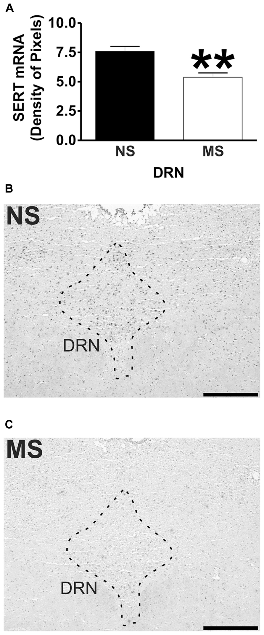
Densitometric analysis revealed that maternal separation (MS) increased 5-HT1A receptor mRNA expression in the amygdala, and reduced its expression in the dorsal raphé nucleus (DRN), but no changes were observed in the hippocampus in comparison to non-separated (NS) controls. Also, MS reduced SERT mRNA expression in the DRN when compared to NS rats. These results suggest that early-life stress induces persistent changes in 5-HT1A receptor and SERT mRNA expression in key brain regions involved in the development of stress-related psychiatric disorders.
All of this suggests that early stress causes an increase the serotonergic receptors in the amygdala, the fear processing center of the brain, and that the 5-HT system is instrumental in the fight or flight reaction to stress.

The same stress responses play a powerful role in mental illnesses as well. It's notable that one of the ACE questions involves emotional neglect. A subset of this group is the children of narcissists - Daniel Shaw has written the definitive book on that topic, Traumatic Narcissism: Relational Systems of Subjugation (2014).

Anyway, here is the article.

Full Citation: 
Bravo, JA, Dinan, TG, and Cryan, JF. (2014, Apr 10). Early-life stress induces persistent alterations in 5-HT1A receptor and serotonin transporter mRNA expression in the adult rat brain. Frontiers in Molecular Neuroscience; 7:24. doi: 10.3389/fnmol.2014.00024


Early-life stress induces persistent alterations in 5-HT1A receptor and serotonin transporter mRNA expression in the adult rat brain


1. Grupo de NeuroGastroBioquímica, Laboratorio de Química Biológica, Instituto de Química, Facultad de Ciencias, Pontificia Universidad Católica de Valparaíso, Valparaíso, Chile
2. Department of Psychiatry, University College Cork, Cork, Ireland
3. Laboratory of Neurogastroenterology, Alimentary Pharmabiotic Centre, University College Cork, Cork, Ireland
4. Department of Anatomy, University College Cork, Cork, Ireland
Early-life experience plays a major role in the stress response throughout life. Neonatal maternal separation (MS) is an animal model of depression with an altered serotonergic response. We hypothesize that this alteration may be caused by differences in 5-HT1A receptor and serotonin transporter (SERT) mRNA expression in brain areas involved in the control of emotions, memory, and fear as well as in regions controlling the central serotonergic tone. To test this, Sprague–Dawley rats were subjected to MS for 3 h daily during postnatal days 2–12. As control, age matched rats were non-separated (NS) from their dams. When animals reached adulthood (11–13 weeks) brain was extracted and mRNA expression of 5-HT1A receptor in amygdala, hippocampus and dorsal raphé nucleus (DRN) and SERT in the DRN was analyzed through in situ hybridisation. Densitometric analysis revealed that MS increased 5-HT1A receptor mRNA expression in the amygdala, and reduced its expression in the DRN, but no changes were observed in the hippocampus in comparison to NS controls. Also, MS reduced SERT mRNA expression in the DRN when compared to NS rats. These results suggest that early-life stress induces persistent changes in 5-HT1A receptor and SERT mRNA expression in key brain regions involved in the development of stress-related psychiatric disorders. The reduction in SERT mRNA indicates an alteration that is in line with clinical findings such as polymorphic variants in individuals with higher risk of depression. These data may help to understand how early-life stress contributes to the development of mood disorders in adulthood.


Introduction

In the early postnatal period of the rat, the brain is thought to be a developmental equivalent to the last trimester in utero and the perinatal period of human brain development (Romijn et al., 1991; Watson et al., 2006; Goodfellow et al., 2009), thus allowing for the use of postnatal rodent models in the investigation of the early programing of stress-related psychiatric disorders. It has been proposed that stress during developmental stages, can lead to developmental alterations that become evident in adult life (Barker, 1995), and moreover, during early-life the psychosocial milieu can substantially alter the nervous system, through mechanisms that permanently affect gene expression (Mathews and Janusek, 2011).

In the central nervous system (CNS), one of the key neurotransmitter systems involved in the response to stress and in the development of neuropsychiatric disorders is the 5-hydroxytriptamine (5-HT) system (Kirby et al., 1995; Graeff et al., 1996; Cryan et al., 2005; Savitz et al., 2009; O’Leary and Cryan, 2010). The majority of 5-HT neurons are located in the dorsal and median raphé nucleus in the brainstem (Graeff et al., 1996; Michelsen et al., 2007). Projections from these neurons innervate several structures of the limbic system, including the amygdala and hippocampus, and it has been described that through these projections the 5-HT system regulates the fight or flight reaction to stress (Graeff et al., 1996; Michelsen et al., 2007), by a region-specific release of 5-HT (Kreiss and Lucki, 1994; Kirby et al., 1995; Graeff et al., 1996).

There are 14 types of 5-HT receptors, divided into seven families, with different subtypes identified by letters (A–F in the case of 5-HT1 receptors; Hoyer et al., 1994; Barnes and Sharp, 1999; Bockaert et al., 2010). One of these receptors is 5-HT1A, a G protein-coupled receptor that has been described to play an important role in the development of psychiatric disorders (Bowen et al., 1989; López et al., 1998; Drevets et al., 1999; Gross et al., 2002; Savitz et al., 2009). The 5-HT1A receptor is predominantly a somatodendritic autoreceptor in the neurons of the raphé nucleus regulating the amount of 5-HT released and therefore serotonergic activity in the different projection areas (Blier and de Montigny, 1987; Hutson et al., 1989; Hjorth and Sharp, 1991; Kreiss and Lucki, 1994; Savitz et al., 2009). Also, 5-HT1A receptor expression has been described in forebrain areas (Chalmers and Watson, 1991; Pompeiano et al., 1992; Cryan et al., 2005; Savitz et al., 2009) including the hippocampus and amygdala, structures involved in learning, control of emotions, memory and fear related information (Vizi and Kiss, 1998; Nestler et al., 2002; LeDoux, 2007). Alterations in 5-HT1A receptor function have been related to mood disorders, as imaging analysis shows that depressive patients have reduced 5-HT1A receptor binding (Drevets et al., 1999; Sargent et al., 2000) as well as blunted responses to 5-HT1A receptor agonists (Lesch et al., 1990a,b).

Another component of the 5-HT system is the serotonin transporter (SERT), a presynaptic protein involved in the termination of the serotonergic signal through the reuptake of 5-HT from the synapse (Blakely et al., 2005). In the pharmacological treatment of depression, selective serotonin reuptake inhibitors (SSRIs) have been widely used (Frazer, 1997). SSRIs can readily inhibit SERT activity and elevate the serotonergic tone in the brain. However, full therapeutic effects become apparent only after chronic SSRI use, suggesting that alterations in this transporter are highly relevant to the development and treatment of psychiatric disorders (Frazer and Benmansour, 2002).

Neonatal maternal separation (MS) is a well validated animal model of depression and increases anxiety resulting in behavioral alterations (Lippmann et al., 2007) and functional changes in the hypothalamus-pituitary-adrenal (HPA) axis responsiveness in adulthood (Ladd et al., 1996; Schmidt et al., 2004; O’Mahony et al., 2009). In addition, MS animals have been reported to display alterations in their central corticotrophin releasing factor (CRF) system (Bravo et al., 2010; O’Malley et al., 2011), which is suggestive of an altered gene expression in key brain areas as result of early-life stress.

There is evidence suggesting an enhanced serotonergic response in animals subjected to MS, as there are differences in brain stem levels of 5-HT and its metabolite 5-hydroxyindole acetic acid (5-HIAA; O’Mahony et al., 2008), as well as increased responsiveness to the SSRI citalopram (Arborelius et al., 2004).Therefore, differences in central serotonergic modulation in adult rats subjected to early-life stress could arise as a result of altered 5-HT1A receptor and SERT expression in areas of the brain involved in the control of emotions, memory, and fear as well as in areas controlling the central serotonergic tone. To test this, in situ hybridization was used to study topographical differences in 5-HT1A receptor and SERT mRNA expression in the hippocampus, amygdala, and dorsal raphé nucleus (DRN) between MS and non-separated (NS) rats.

Materials and Methods

Animals

Adult male Sprague–Dawley (SD) rats that underwent a MS protocol were used (n = 6 MS from three different litters and n = 6 NS controls from three different litters). All animals were housed in standard conditions (room temperature of 21°C, with a 12 h light dark cycle) with access to regular chow and water ad libitum. Cages were cleaned once weekly to avoid excessive handling. Rats were of comparable weight (276–410 g) and age (11–13 weeks) at the moment of sacrifice All experimental procedures were carried out in accordance with the protocols approved by the Ethics Committee, at University College Cork, Cork, Ireland under a license issued from the Department of Health and Children (Cruelty to Animal Act 1876, Directive for the Protection of Vertebrate Animals used for Experimental and other Scientific Purposes [89/609/EEC]).

Maternal Separation

Early-life stress procedure (Hyland et al., 2009; O’Mahony et al., 2009, 2010) was adapted from a previously described protocol (Wigger and Neumann, 1999). Briefly, the litters that were randomly assigned to undergo MS, were removed from the home cage and placed into a smaller cage on heating pads set at 30–33°C for 3 h (9.00–12.00 h). After that time, pups were returned to the original home cage in the main colony room. This procedure was repeated from postnatal day 2 (P2) to P12. Control, NS litters remained undisturbed except for routine cage cleaning performed once a week. At P21, pups were weaned and group-housed (3–5 per cage), and left undisturbed until adulthood (11–13 weeks). We have previously shown that this MS protocol induces an array of behavioral and physiological changes that are indicative of increased anxiety and altered HPA axis function (O’Mahony et al., 2009).

Sacrifice and in Situ Hybridisation

Animals were lightly anesthetized with isoflurane, and killed by decapitation. The brain was immediately extracted and snap frozen in isopentane kept cold with dry ice. The brains were stored at -80°C before being processed for in situ hybridisation.

The in situ hybridisation was carried out with oligodeoxynucleotide (cDNA) probes complementary to 5-HT1A receptor mRNA (2107–2151 pb access number AF217200)and SERT mRNA (1719–1763 pb access number Y11024.1), labeled with a digoxigenin (DIG) oligonucleotide 3′-OH tailing kit (Roche, Molecular Biochemicals, Mannheim, Germany). The hybridisation was conducted as previously described (Bravo et al., 2009, 2010). Briefly, coronal brain sections of 10 μm thick were obtained from frozen brains and mounted on superfrost-plus glass slides (Menzel-Glaser, Menxel GmbH & Co., Germany). For hippocampus, four to five non-consecutive slices separated at least 100 μm from each other, approximately from bregma -2.56 mm to bregma -3.6 mm were analyzed bilaterally. For the amygdala: bilateral analysis of four to five slices of tissue, separated at least 100 μm from each other, approximately from bregma -1.80 mm to bregma -2.80 mm. In the case of the DRN at least three slices separated as a minimum as 100 μm from each other, approximately from bregma -7.64 mm to bregma -8.00 mm were obtained. These sections were post-fixed in 4% paraformaldehyde made in PBS for 30 min. Then the slides were permeabilized with proteinase K (0.5 mg/100 mL in TE buffer) and treated with acetic anhydride buffer. Next, the slides underwent dehydration through a series of ethanol dilutions (70, 95, and 100%) before being delipidated in chloroform for 5 min. The tissues were then rehydrated and placed in a humidity chamber with the hybridisation solution [formamide 50%, saline sodium citrate (SSC) buffer 4x, sheared salmon DNA 6.25 mg/mL, tRNA 125 μg/mL, and cDNA probe at fixed concentration of 100 pmol/mL for each probe] and incubated overnight at 37°C. After that, the sections were washed in ascending dilutions of SSC buffer (4, 2, 1, and 0.5x), and then equilibrated with maleic acid 0.1 M buffer before blocking for unspecific protein binding with Roche’s blocking reagent (Roche, Molecular Biochemicals, Mannheim, Germany). After 30 min of blocking, the DIG molecules attached to the hybridized probes were detected with an anti DIG antibody, conjugated with an alkaline phosphatase (Roche, Molecular Biochemicals, Mannheim, Germany). Finally, a substrate for the alkaline phosphatase (NBT/BCIP; Sigma, St. Louis, MO, USA) was added, and when a violet/blue precipitate was present on the tissues, the reaction was stopped. The slides were then left to air dry and cover-slips were mounted with DPX mounting media (Fisher Scientific, Loughborough, UK). Once the mounting media was dry, pictures of the areas of interest were taken with an Olympus DP71 digital camera attached to an Olympus BX51 microscope (Olympus Corporation, Tokyo, Japan). Specificity of the hybridisation was evaluated by the use of 100-fold excess of the unlabelled oligodeoxynucleotide. For semiquantitative analysis, densitometric measurements of each hippocampal, amygdala, and DRN were analyzed using FujiFilm’s Science Lab Multi Gauge v2.2 software (Fuji Photo Film Co., Ltd). All pictures were analyzed in gray scale and the value given by the software corresponds to the intensity of pixels (the darkest staining is the highest intensity; and the lightest staining the lowest intensity) in a given area (density of pixels). In the hippocampus, the hybridisation signal in the stratum radiatum was considered as background and was subtracted from the pixel density values obtained in the hippocampal cell layers. As for the amygdala, a small region between the analyzed areas was considered as background, and for the DRN a small region surrounding this structure was taken as background. For each animal the value represents the average from 4–5 non-consecutive brain sections (analyzed on both brain hemispheres for hippocampus and amygdala).

Statistical Analysis

All the values are expressed as the mean ± SEM. Data were analyzed with a two tailed Student’s t-test using GraphPad Prism 4 (GraphPad Software Inc., La Jolla, CA, USA). Statistical significance was accepted at the level p < 0.05.

Results

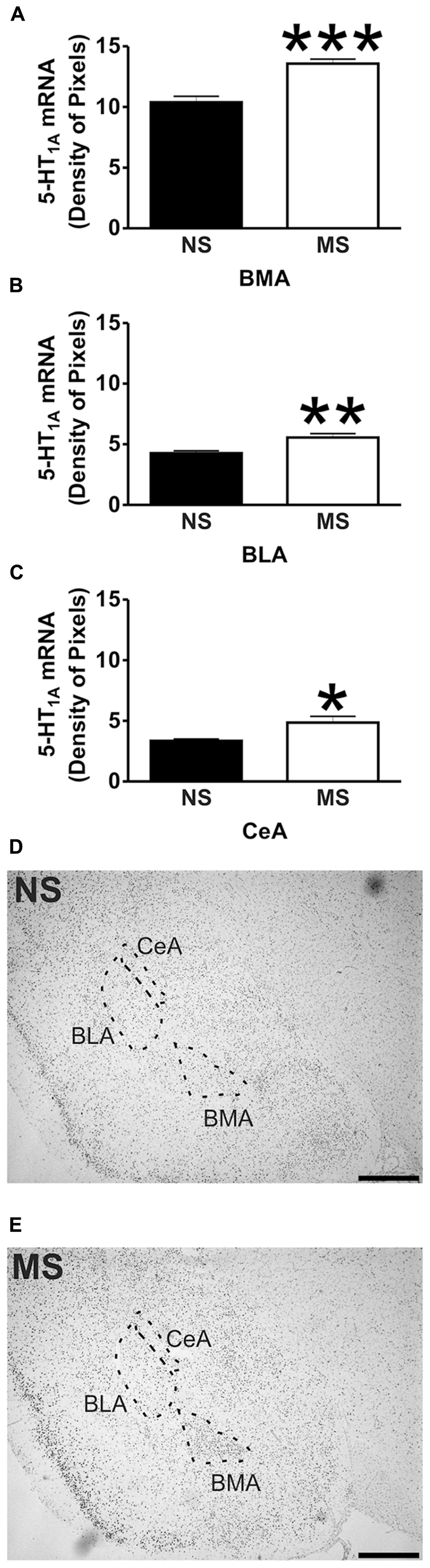
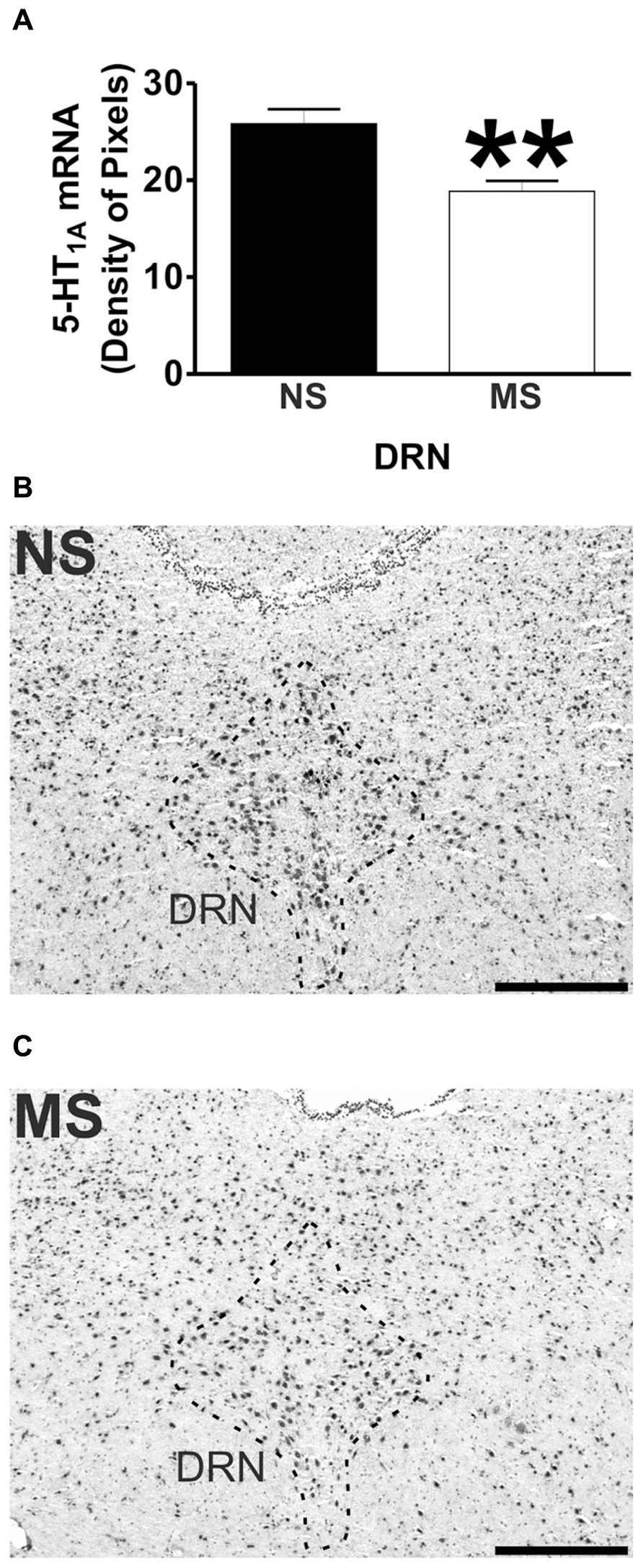
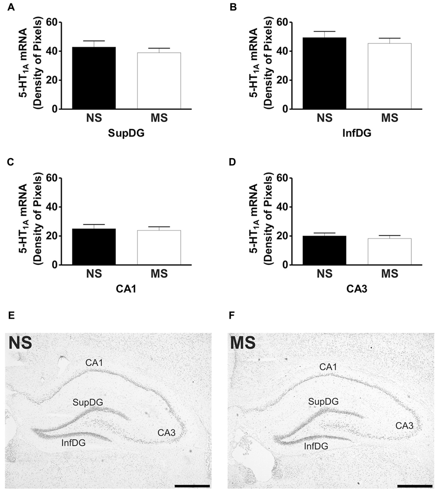
Signal for 5-HT1A receptor mRNA was detected in the amygdala (Figures 1D,E), DRN (Figures 2B,C), and hippocampus (Figures 3E,F), and for SERT mRNA in the DRN (Figures 4B,C). The level of staining in each case allowed densitometric analysis. Negative controls were performed using an excess of unlabelled cDNA probe during the hybridisation stage (not shown).
FIGURE 1
http://www.frontiersin.org/files/Articles/80981/fnmol-07-00024-HTML/image_m/fnmol-07-00024-g001.jpg FIGURE 1. 5-HT1A receptor mRNA expression in the amygdala. Maternal separation (MS) increases 5-HT1A receptor mRNA expression in three different areas of the amygdala in comparison to non-separated (NS) animals. Graphical representations of the densitometric analysis in the basomedial amygdala (BMA; ***p < 0.001; A), basolateral amygdala (BLA; **p < 0.01; B), and central amygdala (CeA; *p < 0.05; C). Representative microphotographs of 5-HT1A receptor mRNA expression in NS (D) and MS (E) animals (scale bar represents 1 mm; NS, n = 6 and MS, n = 6).
FIGURE 2
http://www.frontiersin.org/files/Articles/80981/fnmol-07-00024-HTML/image_m/fnmol-07-00024-g002.jpg FIGURE 2. 5-HT1A receptor mRNA expression in the DRN. Maternal separation (MS) reduces 5-HT1A receptor mRNA expression in the DRN in comparison to NS animals. Graphical representations of the densitometric analysis in the DRN (**p<0.01; A). Representative microphotographs of 5-HT1A receptor mRNA expression in NS (B) and MS (C) animals (scale bar represents 500 μm; NS, n = 6 and MS, n = 6).
FIGURE 3
http://www.frontiersin.org/files/Articles/80981/fnmol-07-00024-HTML/image_m/fnmol-07-00024-g003.jpg FIGURE 3. Hippocampal expression of 5-HT1A receptor mRNA. Maternal separation (MS) did not affect 5-HT1A receptor mRNA expression in the hippocampus when compared to NS animals. Graphical representations of the densitometric analysis in the suprapyramidal layer of the dentate gyrus (SupDG; A), infrapyramidal layer of the dentate gyrus (InfDG; B), cornus ammon field 1 (CA1; C) and cornus ammon field 3 (CA3; D). Representative microphotographs of 5-HT1A receptor mRNA expression in NS (E) and MS (F) animals (scale bar represents 1 mm; NS, n = 6 and MS, n = 6).

FIGURE 4
http://www.frontiersin.org/files/Articles/80981/fnmol-07-00024-HTML/image_m/fnmol-07-00024-g004.jpg FIGURE 4. SERT mRNA expression in the DRN. Maternal separation (MS) reduces SERT mRNA expression in the DRN in comparison to NS animals. Graphical representations of the densitometric analysis in the DRN (**p < 0.01; A). Representative microphotographs of SERT mRNA expression in NS (B) and MS (C) animals (scale bar represents 500 μm; NS, n = 6 and MS, n = 6).

5-HT1A Receptor mRNA Expression


Early-life stress significantly increased the levels of 5-HT1A receptor mRNA in the basomedial amygdala (BMA; Figure 1A; NS vs. MS: 10.39 ± 0.49 vs. 13.58 ± 0.36; t(10) = 5.226, p < 0.001), basolateral amygdala (BLA; Figure 1B; NS vs. MS: 4.27 ± 0.18 vs. 5.55 ± 0.33; t(10) = 3.373, p < 0.01) and central amygdala (CeA; Figure 1C; NS vs. MS: 3.35 ± 0.14 vs. 4.86 ± 0.51; t(10) = 2.838, p < 0.05), and decreased the expression of this transcript in the DRN (Figure 2A; NS vs. MS: 25.85 ± 1.49 vs. 18.87 ± 1.1; t(10) = 3.804, p < 0.01) when compared to NS controls. However, densitometric analysis of the 5-HT1A receptor transcript revealed no differences between MS and NS animals in any of the hippocampal layers (Figure 3).

SERT mRNA Expression


Maternal separation induced a significant reduction to the transcript for SERT in the DRN in comparison to NS rats (Figure 4A; NS vs. MS: 7.58 ± 0.43 vs. 5.38 ± 0.37; t(10) = 3.840, p < 0.01).

Discussion


The present data shows that early-life stress affects gene expression in adulthood, contributing to inadequate stress responses and could therefore lead to the manifestation of stress-related psychiatric disorders. Similar alterations have been described for another neurotransmitter system (Bravo et al., 2010), which further suggest that early-life stress does affects CNS function. Although MS did not affect 5-HT1A receptor mRNA expression in the hippocampus, a structure involved in memory and learning (Jacobson and Sapolsky, 1991; Vizi and Kiss, 1998), it increased its expression in three subregions of the amygdala, a structure related to the control of emotions and fear (LeDoux, 2007). In addition, MS reduced 5-HT1A receptor mRNA expression in the DRN, the major source of serotonergic input to the forebrain which is involved in the control of the central serotonergic tone (Graeff et al., 1996). Also, early-life stress reduced the expression of SERT mRNA in the DRN, which could have an impact on the bioavailability of 5-HT in projection areas of the DRN. These alterations suggest that the behavioral, physiological and molecular deficits described for this animal model (Ladd et al., 1996; Schmidt et al., 2004; Lippmann et al., 2007; O’Mahony et al., 2009) could arise as a consequence of changes in gene expression in key brain regions involved in the development of stress-related psychiatric disorders.


Early-life stress, such as that induced by MS, physical, sexual and emotional abuse and general neglect during childhood, has been associated with serious psychiatric impairments in adulthood (MacMillan et al., 2001; Lupien et al., 2009). During postnatal development the brain undergoes a variety of adaptive changes that depend mostly on the type of stimuli being received (Schmidt et al., 2004). In rats there is a period of reduced stress responsiveness during the first 2 weeks of life (Sapolsky and Meaney, 1986) which can be disinhibited by prolonged MS (Schmidt et al., 2004; O’Mahony et al., 2009). These long periods of MS immediately impact brain gene expression, including the 5-HT1A receptor. For example, Goodfellow et al. (2009) show that 5-HT1A electrophysiological activity in the prefrontal cortex is enhanced in the first 2 to 3 postnatal weeks after MS (3 h a day from P2 to P14). Moreover, mRNA expression of the 5-HT1A receptor in maternally separated animals is increased at postnatal day 9 (Goodfellow et al., 2009). However, when these animals reach adulthood (≥P40), the electrophysiological effects mediated by the 5-HT1A receptor are not different between maternally separated rats and their respective controls, and there is no difference in mRNA expression between rats subjected to early-life stress and control animals (Goodfellow et al., 2009). Nevertheless, exposure to social isolation stress reduces the 5-HT1A-elicited electrophysiological activity in the prefrontal cortex of animals that were exposed to early-life stress, in comparison to control rats (Goodfellow et al., 2009), thus suggesting that early-life stress increases the susceptibility toward stress-related psychiatric disorders in adulthood.


We have previously described alterations in serotonin metabolism in MS rats (O’Mahony et al., 2008), and additionally, alterations in the central serotonergic system have also been described as a result of different MS procedures (Arborelius and Eklund, 2007; Oreland et al., 2009). Oreland et al. (2009) have shown that brief exposures to MS (15 min from P1 to P13) reduces brain stem expression of 5-HT1A receptor mRNA (Oreland et al., 2009), a procedure that can also affect other neurotransmitter systems (Jaworski et al., 2005; Plotsky et al., 2005). This type of MS could be considered a more naturalistic stress as it mimics the natural rearing environment of rats, where the mother leaves her pups for short periods to forage (Arborelius et al., 2004; Arborelius and Eklund, 2007; Oreland et al., 2009). On the other hand, there is also evidence demonstrating that brief and long daily periods of MS do not affect 5-HT1A receptor and SERT mRNA expression (Arborelius et al., 2004), and furthermore, it has been shown that long periods of daily MS are more effective in producing changes in behavior and alterations in biomarkers associated to stress-related psychiatric disorders (Lippmann et al., 2007; O’Mahony et al., 2009; Bravo et al., 2010). Our previous studies demonstrated that a protocol consisting of 3 h of MS from P2 to P12 produces an increase in corticosterone levels (O’Mahony et al., 2009) and an increase in serotonin turnover (O’Mahony et al., 2008). However, quantitative real time PCR (qRT-PCR) revealed no differences in the expression of 5-HT1A receptor and SERT transcripts in complete brainstem homogenates of MS animals in comparison to NS rats (O’Mahony et al., 2008). Whilst qRT-PCR is a sensitive technique used to assess gene expression, is also a crude method which dilutes any localized changes in gene expression that might occur as a result of early-life stress. Therefore, the present findings corroborate that alterations in serotonin metabolism, induced by MS (O’Mahony et al., 2008), can be consequence of changes in gene expression within the DRN. However, it is important to note that the present observations only represent changes at the mRNA level and not protein, and they could be just a reflection of a more complex situation involving other neurotransmitter systems (Bravo et al., 2010) and a variety of intracellular cascades that can affect the expression of these transcripts in the different studied areas.


Maternal separation affected the expression of 5-HT1A receptor mRNA in the amygdala. The transcript for this receptor has been described in the rat BMA (Chalmers and Watson, 1991; Pompeiano et al., 1992), and binding of radio labeled 5-HT1A receptor antagonists has been shown in the BLA and CeA (Vicentic et al., 2006). In the present study, early-life stress increased the levels of the transcript for 5-HT1A receptor in the BMA, and also in the BLA and CeA, although in these areas the level of transcript was much lower than in the BMA. It has been shown that activation of 5-HT1A receptors within the amygdala using the agonist 8–Hydroxy-2-(dipropylamino)tetralin (8-OH-DPAT) reduces the levels of social interaction of male rats (Gonzalez et al., 1996), demonstrating that 5-HT1A activation in the amygdala of rats mediates anxiogenic effects. Moreover, and in line with the present findings, Vicentic et al. (2006) showed that non-handled rats (similar to our NS condition) have lower binding capacity of the 5-HT1A receptor antagonist 4-(2′-methoxyphenyl)-1-[2′-[N-(2′′-pyridinyl)-p-iodobenzamido] ethylpiperazine (pMPPI) in the BMA and BLA in comparison to MS animals. Therefore, the increase in 5-HT1A receptor mRNA expression within the amygdala of MS rats could account for some of the behavioral changes observed by Lippmann et al. (2007), where MS reduced locomotor activity, increased acoustic startle and also affects HPA axis responsiveness in adulthood (Ladd et al., 2005; Lippmann et al., 2007; O’Mahony et al., 2009).


In contrast to the increased expression of 5-HT1A receptor mRNA in the amygdala, there were lower levels of 5-HT1A receptor mRNA found in the DRN of MS rats in comparison to their controls. In this structure there is a high density of 5-HT1A receptors (Blier and de Montigny, 1987; Hutson et al., 1989; Hjorth and Sharp, 1991; Kreiss and Lucki, 1994; Cryan et al., 2005; Savitz et al., 2009), and activation of these presynaptically located receptors decreases the firing frequency, 5-HT synthesis and release from these neurons (Blier and de Montigny, 1987; Sprouse and Aghajanian, 1987; Hjorth and Magnusson, 1988; Hutson et al., 1989; Sharp et al., 1989; Kreiss and Lucki, 1994; Cryan et al., 2005). In addition, 5-HT1A receptor activation in the DRN has been shown to produce anxiolytic effects in different animal models (Andrews et al., 1994; Hogg and File, 1994; Jolas et al., 1995; Picazo et al., 1995; File et al., 1996; Remy et al., 1996; Romaniuk et al., 2001; Koprowska et al., 2002). The reduced levels of 5-HT1A receptor mRNA in the DRN of MS rats suggests an impaired regulation of the central serotonergic tone that could translate into inadequate behaviors toward stressful situations such as those observed by Lippmann et al. (2007). Moreover, we have previously shown that MS rats have altered 5-HT and 5-HIAA levels in the brain stem that clearly suggests an increased turnover of this neurotransmitter (O’Mahony et al., 2008). This could be a consequence of the lower 5-HT1A receptor expression in the DRN, as a lower level of this receptor could impact on the frequency of discharge and/or synthesis and release of 5-HT and therefore affect the neurotransmitter’s metabolism. In line with the previous suggestion, Ase et al. (2000) showed that 5-HT1A receptor knock-out mice have increased 5-HT turnover. However, these animals show no differences in basal levels of 5-HT in forebrain areas (Ase et al., 2000; He et al., 2001; Knobelman et al., 2001) and in the DRN (Ase et al., 2000; Bortolozzi et al., 2004) in comparison to wild-type controls. These observations argue against a role of presynaptic 5-HT1A receptors in the maintenance of the central serotonergic tone, and therefore reveal the complexity of 5-HT neurotransmission regulation.


Another level of regulation to the central 5-HT neurotransmission involves SERT. The levels of SERT mRNA were lower in the DRN of MS rats in comparison to NS rats, which suggest that the reuptake of the neurotransmitter could be affected. The importance of this finding is that changes in SERT expression have been related to psychiatric disorders. For instance, during treatment with SSRIs, the most widely prescribed antidepressants (Frazer, 1997), SERT gets downregulated, which seems to correlate with the efficacy of the treatment (Benmansour et al., 1999, 2002; Frazer and Benmansour, 2002; Gould et al., 2003; Thakker et al., 2004, 2005). However, SERT deficient mice display anxiety- and depression-like behaviors (Holmes et al., 2002; Lira et al., 2003), which suggest that the absence of this gene from early developmental stages affects the ability to cope with stressful situations throughout life. In addition, animals treated during early development with SSRIs also display altered behaviors in adulthood (Mirmiran et al., 1981; Vogel et al., 1990), as the antidepressant would down regulate SERT in early-life. Moreover, downregulation of SERT in adult animals resembles the effects of antidepressant treatment (Thakker et al., 2004, 2005), further highlighting an important role of SERT in the development of the serotonergic system during early-life. In the present study, the reduction in SERT mRNA expression in the DRN not only could affect local serotonin levels (and perhaps its turnover), but it could also impact the adequate development of the serotonergic system and therefore affect the ability to cope with stress. In addition, the reduction in SERT mRNA indicates an alteration that is in line with clinical findings. Individuals with a short allele for SERT, that reduce the efficiency of the gene’s transcription, showed more depressive symptoms in relation to stressful events than individuals with the long version of the allele (Caspi et al., 2003), and therefore are at a higher risk of developing psychiatric disorders such as depression.


In summary, the present findings, along with previous observations on other neuronal systems (Bravo et al., 2010) strongly suggest that early-life stress permanently affects gene expression in the CNS. These changes in 5-HT1A receptor and SERT mRNA reflect alterations in a neurotransmitter system that has been extensively related to the development of mood disorders. Moreover, these changes were observed in key brain areas related to the behavioral response to stress. Therefore, these data helps to understand how early-life stress contributes to the development of mood disorders later in life.

Author Contributions

Javier A. Bravo, Timothy G. Dinan and John F. Cryan designed research; Javier A. Bravo performed research and acquired data; Javier A. Bravo, Timothy G. Dinan and John F. Cryan interpreted and analyzed data; and Javier A. Bravo, Timothy G. Dinan and John F. Cryan drafted, revised and wrote the paper.


Conflict of Interest Statement

The authors declare that the research was conducted in the absence of any commercial or financial relationships that could be construed as a potential conflict of interest.
 

Acknowledgments

We would like to thank Drs. Gerard Clarke, Marcela Julio-Pieper, and Mr. Patrick Fitzgerald for their technical assistance. Javier A. Bravo is supported by a grant from the Vicerrectoría de Investigación y Estudios Avanzados from the Pontificia Universidad Católica de Valparaíso (Grant No. 037.499/2013). John F. Cryan and Timothy G. Dinan are supported in part by Science Foundation Ireland in the form of a Centre Grant (Alimentary Pharmabiotic Centre). The centre is also funded by GlaxoSmithKline.


References available at the Frontiers Site
* * * * *

Finding Your ACE Score


While you were growing up, during your first 18 years of life:

1. Did a parent or other adult in the household often or very often… Swear at you, insult you, put you down, or humiliate you?
or
Act in a way that made you afraid that you might be physically hurt?
Yes   No

2. Did a parent or other adult in the household often or very often… Push, grab, slap, or throw something at you?
or
Ever hit you so hard that you had marks or were injured?
Yes   No

3. Did an adult or person at least 5 years older than you ever… Touch or fondle you or have you touch their body in a sexual way?
or
Attempt or actually have oral, anal, or vaginal intercourse with you?
Yes   No

4. Did you often or very often feel that …No one in your family loved you or thought you were important or special?
or
Your family didn’t look out for each other, feel close to each other, or support each other?
Yes   No

5. Did you often or very often feel that …You didn’t have enough to eat, had to wear dirty clothes, and had no one to protect you?
or
Your parents were too drunk or high to take care of you or take you to the doctor if you needed
it?

Yes   No

6. Were your parents ever separated or divorced?
Yes   No

7. Was your parent or caretaker often or very often... pushed, grabbed, slapped, or had something thrown at them?
or
Sometimes, often, or very often kicked, bitten, hit with a fist, or hit with something hard?
or
Ever repeatedly hit at least a few minutes or threatened with a gun or knife?
Yes   No

8. Did you live with anyone who was a problem drinker or alcoholic or who used street drugs?
Yes   No

9. Was a household member depressed or mentally ill, or did a household member attempt suicide?
Yes   No

10. Did a household member go to prison?
Yes   No

Now add up your “Yes” answers: _______ This is your ACE Score

Here are some statistics:


63% of the people who participated in the study had experienced at least one category of childhood trauma. Over 20% experienced 3 or more categories of trauma which we call Adverse Childhood Experiences (ACEs).
• 11% experienced emotional abuse.
• 28% experienced physical abuse.
• 21% experienced sexual abuse.
• 15% experienced emotional neglect.
• 10% experienced physical neglect.
• 13% witnessed their mothers being treated violently.
• 27% grew up with someone in the household using alcohol and/or drugs.
• 19% grew up with a mentally-ill person in the household.
• 23% lost a parent due to separation or divorce.
• 5% grew up with a household member in jail or prison.

Number of Adverse Childhood Experiences (ACE Score) Women Men Total
0 34.5 38.0 36.1
1 24.5 27.9 26.0
2 15.5 16.4 15.9
3 10.3 8.6 9.5
4 or more 15.2 9.2 12.5

Here is a chart of the increased risk for various illnesses depending on the ACE score - notice how dramatically the risk goes up with each increasing number of ACEs:


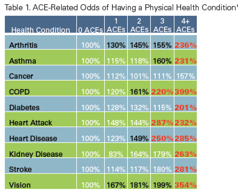
The more categories of trauma experienced in childhood, the greater the likelihood of experiencing:
• alcoholism and alcohol abuse
• chronic obstructive pulmonary disease (COPD)
• depression
• fetal death
• poor health-related quality of life
• illicit drug use 
• ischemic heart disease (IHD)
• liver disease
• risk for intimate partner violence
• multiple sexual partners
• sexually transmitted diseases (STDs)
• smoking
• obesity
• suicide attempts
• unintended pregnancies