Showing posts with label medial prefrontal cortex. Show all posts

Friday, August 22, 2014

What Can the Organization of the Brain’s Default Mode Network Tell Us About Self-Knowledge?


Here is the article referenced in the Nautilus article about silence (posted earlier this morning). It seems like a good piece of writing with some valuable insights.

Full Citation: 
Moran, JM, Kelley, WM, and Heatherton, TF. (2013, Jul 17). What can the organization of the brain’s default mode network tell us about self-knowledge? Frontiers in Human Neuroscience; 7:391. doi: 10.3389/fnhum.2013.00391

What can the organization of the brain’s default mode network tell us about self-knowledge?

Joseph M. Moran [1,2], William M. Kelley [3] and Todd F. Heatherton [3]
1. U.S. Army Natick Soldier Research, Development, and Engineering Center, Natick, MA, USA
2. Center for Brain Science, Harvard University, Cambridge, MA, USA
3. Psychological and Brain Sciences, Dartmouth College, Hanover, NH, USA
Abstract

Understanding ourselves has been a fundamental topic for psychologists and philosophers alike. In this paper we review the evidence linking specific brain structures to self-reflection. The brain regions most associated with self-reflection are the posterior cingulate and medial prefrontal (mPFC) cortices, together known as the cortical midline structures (CMSs). We review evidence arguing that self-reflection is special in memory, while noting that these brain regions are often engaged when we think about others in our social worlds. Based on the CMSs’ patterns of connectivity and activity, we speculate about three possible interpretations of their role in supporting self-reflection that are somewhat overlapping, and not intended to be mutually exclusive. First, self may be a powerful, but ordinary case for a cognitive system specialized for thinking about people. Second, mPFC may serve as a processing “hub,” binding together information from all sensory modalities with internally generated information. Third, mPFC may serve as a cortical director of thought, helping to guide moment-by-moment conscious processing. Suggestions are made for future research avenues aimed at testing such possibilities.

How do we know what we are like? How do we determine the boundaries between ourselves and the world around us? How do we know what is ours, and what is not? Questions like these have engaged philosophers for millennia, and psychological scientists throughout psychology’s relatively brief history. Great progress has been made by practitioners of these fields, who have recently been joined by neuroscientists bearing the promise of going beyond introspection, self-report, and behavior to the source of our sense of self, the brain. This work is theoretically useful in at least two ways. First, it enables characterization of how the brain implements the psychological process(es) of self-reflection, allowing for links between the neural and psychological levels of analysis. Second, it may suggest new ways to interpret and modify accounts of self-reflection at the psychological-level, allowing neural-level data to influence psychological-level theorizing. Wielding two major empirical breakthroughs, cognitive neuroscientists have made significant headway in understanding how the brain gives rise to a sense of self, revealing surprising knowledge about the organization of the neuronal networks responsible for self-reflection.

The Default Mode Network and Self-Reflection

In brief, these breakthroughs consisted first of the discovery of what has come to be known as the default mode network (Shulman et al., 1997; Raichle et al., 2001), and second of the independent identification that a subset of these brain regions are enlisted when we engage in self-reflection (Gusnard et al., 2001; Johnson et al., 2002; Kelley et al., 2002). To be clear, this network’s involvement is observed most closely during the psychological task of reflecting on one’s personalities and characteristics (self-reflection), rather than during self-recognition, thinking of the self-concept, or thinking about self-esteem, for example. As such, this paper will focus on self at the level of self-reflection and the neural networks responsible for this task. The set of regions contributing to self-reflection consists primarily of the medial prefrontal cortex (mPFC), encompassing the medial surface of the medial frontal gyrus [Brodmann’s Areas (BAs) 8 and 10], and the medial parietal cortex, roughly encompassing the retrosplenial and posterior aspects of the cingulate cortex, the area bounded at the anterior by the paracentral lobule, and at the posterior by the parieto-occipital sulcus (BAs 23, 31, 7). For ease of reference, we will refer to this medial parietal cluster together as posterior cingulate cortex (pCC). These regions have come to be known together as the cortical midline structures (CMSs) (Northoff and Bermpohl, 2004), and are the regions most closely associated with self-reflection in meta-analyses (e.g., Northoff et al., 2006; Qin and Northoff, 2011).

The default mode network concept arose to explain the puzzling observation that when subjects rest quietly with eyes closed, CMS activity is elevated (as measured by positron emission tomography), along with that of anterior temporal lobes and lateral parietal cortices (Shulman et al., 1997). This set of regions is more active when people rest than when they are engaged in goal-directed tasks, and display functional connectivity: these regions’ activity rises and falls together during the normal course of cognitive engagement and disengagement from the external world (Greicius et al., 2003; Fox et al., 2005). This led Raichle and colleagues to propose that this set of regions formed a default mode network; a network that may serve to generate internal mental stimuli and pay attention to our stream of consciousness, but whose activity is attenuated when we turn our attention to the outside world (as in goal-directed tasks) (Gusnard et al., 2001; Raichle et al., 2001). Following these observations, several labs demonstrated direct overlap between the brain regions engaged during rest and during self-reflection (Wicker et al., 2003; D’Argembeau et al., 2005; Schneider et al., 2008; Whitfield-Gabrieli et al., 2011). This relation is further supported by a meta-analysis (Qin and Northoff, 2011), which reported that the same finding occurred across many studies.

Medial Prefrontal Cortex

Several aspects of these regions’ neuroanatomy may support these well-characterized roles. mPFC is larger than any other prefrontal region in humans (Ongur et al., 2003). By proportion, it covers more of the cortex in humans and has more space available for connections with other supramodal areas than in other primates (Semendeferi et al., 2001). It has a greater density of dendritic spines (69% more on average than primary sensory cortex) and smaller density of cell bodies on the average than other cortical regions, suggesting more complex associative processing (Jacobs et al., 2001). Finally, mPFC is almost exclusively interconnected with other heteromodal processing regions in the prefrontal cortex (Barbas and Pandya, 1989; Petrides and Pandya, 1999), anterior temporal cortex (Amaral and Price, 1984; MorĂ¡n et al., 1987), and the cingulate gyrus (Morecraft and Van Hoesen, 1993; Arikuni et al., 1994). Most of these connections are reciprocal in nature (Passingham et al., 2002).

These regions are considered to be part of the “social brain”: a network implicated by neuroimaging and lesion work in representing the people that populate our social worlds (Adolphs, 2001; Heatherton, 2011; Lewis et al., 2011). mPFC’s enlargement in humans, preponderance of interconnections rather than cell bodies, and connections with other “social brain” nodes are all features that point toward a role in social abstraction, a skill for which humans are evidently selected (Dunbar, 2009). Indeed, humans form much larger social networks than do other animals (Dunbar, 1998). Lewis et al. (2011) showed further that the size of particular mPFC regions is correlated both with the degree to which we are able to represent multiple others’ viewpoints and the size of our social networks. Underscoring the role of mPFC in social processing in general, and self-processing in specific, a recent meta-analysis further subdivides mPFC into ventral and dorsal aspects (Denny et al., 2012; Wagner et al., 2012), showing that ventral mPFC responds more to self, and dorsal mPFC responds more to others.

Medial Parietal Cortex

Posterior cingulate cortex shares many reciprocal connections with mPFC. In addition, the subregions of pCC are reciprocally connected with one another in a bilateral manner (Cavanna and Trimble, 2006). Along with mPFC, pCC is disproportionately large in humans relative to non-human primates (Goldman-Rakic, 1987). pCC shares many connections with subcortical and cortical regions and serves as “association cortex,” allowing the brain to “integrate both external and self-generated information and to produce much of the mental activity that characterizes Homo sapiens” (Cavanna and Trimble, 2006, p. 568). This set of neuroanatomical features suggests that these regions would be good candidates for those able to perform the inward-focusing and self-generation of stimuli that constitute mental activity when we are not focused on the external world (Mason et al., 2007; Smallwood et al., 2008). That these regions are disproportionately developed in humans, and that humans congregate in the largest social networks, suggests that much of this mental activity at rest might be about ourselves and others.

If we were to plan to design a system that would be able to retain information about itself, to determine what is and is not self, and to update that store of information in a flexible and goal-dependent manner, we could do worse than to outfit it with the array of connections and features that are possessed by the CMS. While the neuroanatomical evidence is certainly suggestive of a set of regions that are specialized for self-reflection, stronger evidence has emerged in cognitive neuroscience. Work that we and others have done has repeatedly demonstrated that reflecting on the self engages the CMS relative to reflecting about (certain) other people, or non-social classes of stimuli (Craik et al., 1999; Johnson et al., 2002; Kelley et al., 2002; Heatherton et al., 2006; Moran et al., 2011; Whitfield-Gabrieli et al., 2011). This work has supported the idea that the self is a special cognitive structure, providing a superordinate means by which information can be encoded into memory (Fossati et al., 2004; Macrae et al., 2004). This position is further supported by neuropsychological work from Klein et al. (1999) that revealed a post-lesion dissociation in patients’ abilities to form memories about the self versus about general semantic categories. The theoretical position that self-is-special is in direct contrast to the notion that the self is a “powerful, but ordinary” structure in memory; a view which suggests that our improved memory for information encoded in reference to the self is simply a result of the greater familiarity of the self-concept (Greenwald and Banaji, 1989), but that the semantic structures of self are no different from the semantic structures of sailboats and silver jewelry. Even though the cognitive neuroscience evidence strongly supports the self-is-special view, Denny et al.’s (2012) meta-analytical finding of a dorsal-ventral axis along which mPFC appears to be differentiated for other- and self-representation appears contradictory. Why is it the case that, on the one hand, our neural representations of self and other are so closely allied, but on the other hand these representations occur in regions of the cortex distinct from (and largely anatomically disconnected from) those networks that are engaged when we reflect about non-social sources of information?

Why Does Self-Reflection Engage the Cortical Midline Structures?

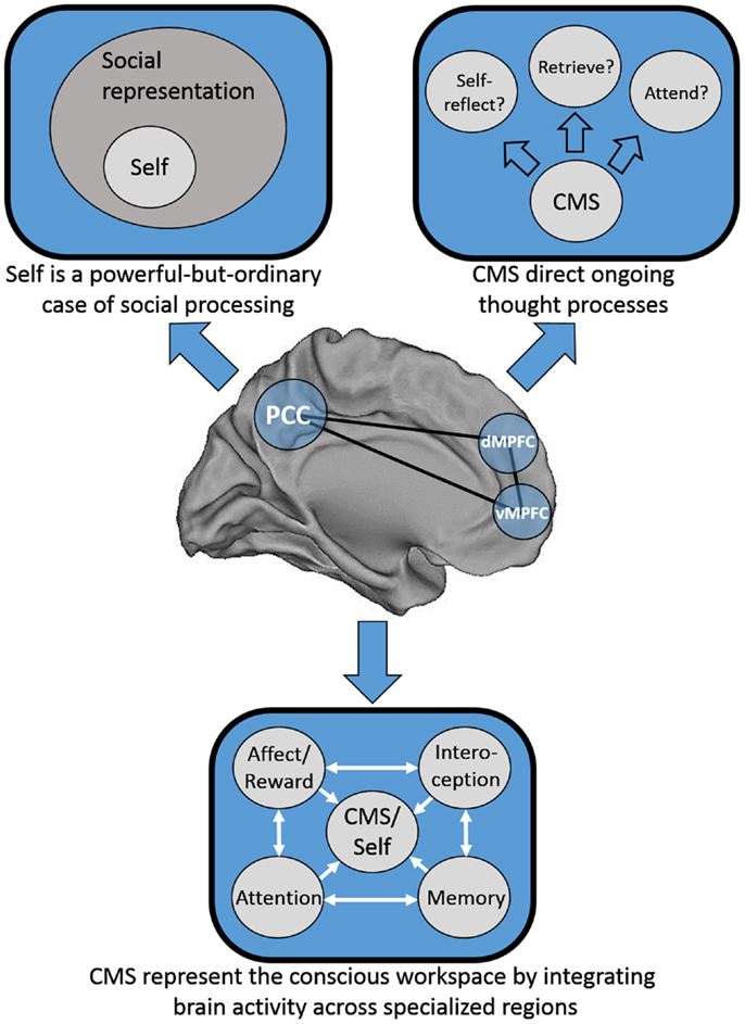
We consider three possible explanations for this pattern of results that are speculative, not intended to be mutually exclusive, and are at least partially overlapping. First, one possibility is that Greenwald and Banaji (1989) may have been half-right: it may be that social information is special, and that the self is a powerful-but-ordinary social knowledge structure. Second, Heatherton (2011) has proposed that mPFC serves as a “hub,” binding together heavily processed information from secondary sensory areas from each of the senses with internally generated information to represent the conscious “workspace.” Third, mPFC may act in a meta-cognitive fashion by guiding our moment-to-moment thought processes; in essence, in deciding what to think about next. See Figure 1 for a schematic representation of each of these models.
FIGURE 1
http://www.frontiersin.org/files/Articles/55238/fnhum-07-00391-HTML/image_m/fnhum-07-00391-g001.jpg

Figure 1. Schematic representation of three possible distinct, but not mutually exclusive models of cortical midline structure (CMS) function. Top left: the CMS are specialized for representing social information, of which the self is a powerful-but-ordinary subset. Top right: the CMS serve as a set of regions responsible for the direction of our thought processes on a moment-to-moment basis. Bottom: the CMS serve as a hub integrating information from disparate neural processing systems into a “conscious workspace.”

Is the Self a Powerful-But-Ordinary Social Construct?

On the self is a powerful-but-ordinary social construct view, the CMS could be seen as representing social information per se, and their seeming selectivity for self-relevant information might simply represent an extreme case of social information processing about a social target (the self) that by definition is more familiar than all other social targets. The overarching view of simulation theory (Gordon, 1986) is that in order to understand others we run a mental simulation of how we might act in given social situations. Conversely, the emerging discipline of neural hermeneutics (Gallotti and Frith, 2013) suggests that in order to understand ourselves, we pay close attention to the social behavior of others. Both of these viewpoints converge on the idea that the self might be a powerful, but ordinary social target.

One obvious prediction of this idea is that the CMS might be differentially engaged by the representation of (and processing about) social targets that are differentially familiar to us. Familiarity contains the concepts of both closeness and similarity: close individuals are those we feel close to (including family and friends), whereas similar individuals are those who share characteristics with us (like members of our race, political affiliation, or age group). Indeed, in Qin and Northoff’s (2011) meta-analysis, they observe that stimulus familiarity drives activation in a similar ventral mPFC region just as much as does self-reflection. In addition, Denny et al.’s (2012) meta-analysis shows that ventral aspects of mPFC are preferentially engaged by reflecting on the self versus others. If this region is sensitive to the familiarity (or “selfness”) of social information, then it should respond more to information that is more self-relevant than not. Several studies have found such a pattern of results (e.g., Phan et al., 2004; Moran et al., 2006). Indeed, Mitchell et al. (2006) observed that social targets manipulated to be similar to the self engaged this ventral mPFC region, whereas social targets manipulated to be dissimilar to the self engaged dorsal mPFC. Krienen et al. (2010) clarified Mitchell et al.’s findings by demonstrating that the driver of activation in mPFC was closeness rather than similarity per se, suggesting that the familiarity of repeated exposure to individuals drives their self-relevance.

Converging on this idea, a series of studies investigating self-reflection in different cultures have provided support for the notion that in individuals whose cultures are more interdependent, the same ventral mPFC region does not differentiate thinking about self from thinking about close family members (like participants’ own mothers) (Zhang et al., 2006; Zhu et al., 2007; Chen et al., 2013), but that this does not necessarily hold true in Western, more independent cultures (Kelley et al., 2002; Kjaer et al., 2002; Heatherton et al., 2006; Vanderwal et al., 2008). These cross-cultural findings are best interpreted in the context of recent criticisms suggesting that standard delineations between Western and Eastern cultures are not as clear-cut as has been suggested (Martinez Mateo et al., 2013). In this context, Moran et al. (2011) provide data that clarify the distinction between independent and interdependent cultures. In their paper, consideration of one’s mother’s personality traits, but not her physical characteristics produced activation levels midway between those of thinking about one’s own traits versus those of former US President, George W. Bush. To the degree that we represent the traits of a close other as being like our own (rather than their physical characteristics), this suggests again that “selfness” may be driving this difference in ventral mPFC). Considered as a unit, these lines of research reveal a quantitative dimension along which social targets of greater familiarity activate ventral mPFC to a greater degree, with the self sitting at the top as the most familiar social target of all.

A further prediction of the notion that the CMS are specialized for social processing (and that the self is a powerful subset of such processing) is that we might be able to differentiate their relative contributions along lines in which thinking about ourselves and thinking about others naturally cleave. To the degree that our representations of ourselves are first-person, and our representations of others are third-person, one would imagine that neural systems implicated in social processing that preferentially receive visual information would be more responsive to third-person representations. Based on the patterns of connectivity that we introduced at the beginning of this paper, it should be clear that the regions of pCC implicated in the default mode (and in self-reflection) are strongly linked to regions that create complex visual representations. Indeed, Raichle et al. (2001) advocate for a domain-general role for the pCC regions in providing complex visual representations to consciousness. Other work in cognitive neuroscience supports and extends this view, showing via meta-analysis that pCC regions participate in a network engaged in autobiographical memory, prospective future thinking, and navigation (Spreng et al., 2009). All such tasks require complex visual representation, and it is interesting that mPFC did not emerge in this meta-analysis. More direct evidence in support of the idea that pCC supports the third- rather than first-person representations more common in thinking about others rather than the self comes again from the meta-analysis of Denny et al. (2012). In their paper, they found across 107 studies that the precuneus was more active when participants thought about others than when they thought about themselves. Single-study evidence of the idea that visual rather than conceptual representations of people engage pCC comes from Moran et al. (2011), who showed that thinking about social targets’ appearance (e.g., Does George W. Bush have a beard?) versus thinking about their character traits (e.g., Is George W. Bush kind?) produces more activation in pCC. This relationship also held true when the social target was the self. Direct investigations of adopting third- versus first-person perspectives have also shown greater pCC involvement during third-person perspective taking (Ruby and Decety, 2001).

Is Medial Prefrontal Cortex a Hub for Integrating Internal and External Information?

Our second possibility is that the ventral mPFC region identified by Heatherton (2011) serves as a hub that integrates internal and external information into a conscious workspace. On this view, self-reflection would be the canonical task for such a region because it so strongly requires the flexible and ongoing integration of our own knowledge about ourselves with our ever-changing knowledge gained from our sense organs about how we are interacting with the environment, and about how social actors in our environment think about us. Thinking about those social actors independent from ourselves (theory of mind) would drive this machinery to a lesser degree (but still more than thinking about non-social aspects of the world) because rapid and complex integration of sensory, external, and non-sensory conceptual knowledge is required to understand others’ goals, intentions, and beliefs, whereas such dynamic processing is much less necessary for thinking about tools or cars or jewelry. This sort of integration into a conscious workspace is also a hallmark of the cognitive processes engaged during “rest,” and engendered by the default mode of brain functioning.

Is Medial Prefrontal Cortex Specialized for Directing Conscious Thought Processes?

Finally, our third possibility is that the ventral mPFC region identified in self-reflection tasks is specialized for helping to decide in which direction our thought processes should proceed. The convergence of heavily processed external sensory inputs with internally generated inputs would also support this view, which of course is not mutually exclusive with the view that mPFC serves as a hub for integration of information from disparate neural processing units. To the degree that deciding where our thoughts should go is a representational process, and that reflection on those thoughts (and our enduring personality traits) is a meta-representational version of the same process, one would imagine that a system with such functional-anatomic properties would be well-placed to perform both conscious direction of thoughts and self-reflection. That rest and self-reflection so consistently overlap (Qin and Northoff, 2011; Whitfield-Gabrieli et al., 2011) suggests that being free to direct our own thoughts (i.e., not responding directly to the environment or an experimenter-provided task) is a state that mimics the natural process observed when we are asked to reflect directly on our own selves. A prediction of this viewpoint is that decision-making might be tied to activity in the CMS, and indeed research shows that CMS activity predicts freely made decisions up to 7 s before participants indicate becoming aware of the decision having been made (Soon et al., 2008). This third possibility thus may account for the still-puzzling observation that the mPFC is perhaps the most important actor in the brain’s default mode network, which itself perhaps serves as a proxy for our ongoing conscious awareness of both our internal and external words. This conjecture awaits empirical investigation however, not least because sampling the ongoing representational processes of the default mode requires disrupting such processes.

Conclusion

In summary, we have speculated about several different explanations for the observation that the CMS are observed so consistently to participate in self-reflection. Neuroanatomical connectivity suggests that these regions are heteromodal association areas that derive much of their inputs from upstream regions associated with social information processing, and that pCC in particular gains its inputs from regions of the brain responsible for complex visual representations. Because these regions are associated with social processing, are developed strongly in humans relative to other animals, and humans travel in much larger social networks than do other animals, we speculate that they may form the basis of a special neurocognitive system evolved for social processing. More fundamental characterizations of this system suggest that the anterior midline structure, mPFC, is in fact a domain-general region dedicated as a hub of information processing about the internal and external worlds, and relatedly, that the purpose of such a confluence of representations is to direct our conscious awareness from one moment to the next, switching flexibly between representations of our internal mental life and of the world around us. On this view, mPFC’s seeming specialization for social information processing merely reflects its response to stimuli (self and others) that drive the integration of internal and external information sources more strongly than non-social stimuli.

Much research remains to be done to gain greater understanding of how and why the self, other social targets, and the default mode of thought are related to one another, and why they so reliably involve the CMS. Initial support for the idea that mPFC regions might be necessary for self-reflection comes from a study with patients with ventral mPFC damage at the site implicated by Kelley et al. (2002) as being maximally involved in self-reflection (Philippi et al., 2012). These patients did not show the self-reference effect in memory, suggesting that mPFC is necessary for encoding information in relation to oneself. Emerging advances in TMS may allow researchers to target more closely these regions for temporary, reversible lesions, or for theta-burst stimulation for temporary increases in excitability of these regions (Vernet et al., 2013). Such studies could provide more controlled evidence to determine whether these regions are necessary for reflection about self and other. In parallel, advances in real-time fMRI techniques (deCharms et al., 2004; Hinds et al., 2011) allow for the exquisite control of presentation parameters, such that we can manipulate when participants are asked to reflect on self and others to moments when activation in either mPFC or pCC are high or low, and determine with a great degree of accuracy what effects natural fluctuations in the default mode at any given moment might have on our abilities to accurately represent ourselves.

Conflict of Interest Statement

The authors declare that the research was conducted in the absence of any commercial or financial relationships that could be construed as a potential conflict of interest.

Acknowledgments

This work was supported by a grant from the National Institutes of Health (R01MH059282) to Todd F. Heatherton.

References available at the Frontiers site.
Thursday, May 29, 2014

Transcriptomic Evidence for Immaturity of the Prefrontal Cortex in Patients with Schizophrenia


This new research from Molecular Brain shows, for the first time, that the gene expression patterns in the schizophrenic prefrontal cortex (PFC) resemble those in the juvenile PFC. This can possibly open new avenues for therapeutic interventions (not with drugs), including some of the Mindsight techniques developed by Dan Siegel that target the medial prefrontal cortex.

Full Citation:
Hagihara, H, Ohira, K, Takao, K and Miyakawa, T. (2014, May 29). Transcriptomic evidence for immaturity of the prefrontal cortex in patients with schizophrenia. Molecular Brain; 7:41. doi:10.1186/1756-6606-7-41

Transcriptomic evidence for immaturity of the prefrontal cortex in patients with schizophrenia

Hideo Hagihara, Koji Ohira, Keizo Takao and Tsuyoshi Miyakawa
Author Affiliations

Abstract (provisional)


Background

Schizophrenia, a severe psychiatric disorder, has a lifetime prevalence of 1%. The exact mechanisms underlying this disorder remain unknown, though theories abound. Recent studies suggest that particular cell types and biological processes in the schizophrenic cortex have a pseudo-immature status in which the molecular properties partially resemble those in the normal immature brain. However, genome-wide gene expression patterns in the brains of patients with schizophrenia and those of normal infants have not been directly compared. Here, we show that the gene expression patterns in the schizophrenic prefrontal cortex (PFC) resemble those in the juvenile PFC.

Results

We conducted a gene expression meta-analysis in which, using microarray data derived from different studies, altered expression patterns in the dorsolateral PFC (DLFC) of patients with schizophrenia with those in the DLFC of developing normal human brains, revealing a striking similarity. The results were replicated in a second DLFC data set and a medial PFC (MFC) data set. We also found that about half of the genes representing the transcriptomic immaturity of the schizophrenic PFC were developmentally regulated in fast-spiking interneurons, astrocytes, and oligodendrocytes. Furthermore, to test whether medications, which often confound the results of postmortem analyses, affect on the juvenile-like gene expressions in the schizophrenic PFC, we compared the gene expression patterns showing transcriptomic immaturity in the schizophrenic PFC with those in the PFC of rodents treated with antipsychotic drugs. The results showed no apparent similarities between the two conditions, suggesting that the juvenile-like gene expression patterns observed in the schizophrenic PFC could not be accounted for by medication effects. Moreover, the developing human PFC showed a gene expression pattern similar to that of the PFC of naive Schnurri-2 knockout mice, an animal model of schizophrenia with good face and construct validity. This result also supports the idea that the transcriptomic immaturity of the schizophrenic PFC is not due to medication effects.

Conclusions

Collectively, our results provide evidence that pseudo-immaturity of the PFC resembling juvenile PFC may be an endophenotype of schizophrenia.

The complete article is available as a provisional PDF. The fully formatted PDF and HTML versions are in production.

Thursday, January 02, 2014

Art Reaches Within: Aesthetic Experience, the Self, and the Default Mode Network


From the open access Frontiers in Neuroscience, this is an intriguing study that asked subjects to rate images of artworks while in an fMRI scanner. The researchers found that regions in the medial prefrontal cortex that are known to be part of the default mode network (DMN) were positively activated on the highest-rated trials (presumably, these same regions were not activated during the lowest-rated trials).

Typically, at least in our current understanding, the DMN is somewhat off-line when we are engaged in externally oriented activity; conversely, it is more active when we are not engaged in a mental activity, for example, when we are daydreaming. However, the DMN is activated when the activities involve self-referential tasks or if tasks involve self-relevant information.

It seems that rating an artwork as a 4 (1-4 scale) required subjects to be somewhat self-referential, perhaps requiring them to determine why they felt moved by that particular work of art. If this is true, then the study adds to the data around the DMN being active in self-referential tasks. The authors reach the same conclusion:
This account is consistent with the modern notion that individuals' taste in art is linked with their sense of identity, and suggests that DMN activity may serve to signal “self-relevance” in a broader sense than has been thought so far.
It's an interesting article - give it a read.

Full Citation: 
Vessel EA, Starr GG, and Rubin N. (2013, Dec 30). Art reaches within: aesthetic experience, the self and the default mode network. Frontiers in Neuroscience; 7:258. doi: 10.3389/fnins.2013.00258

Art reaches within: aesthetic experience, the self and the default mode network
 
Edward A. Vessel [1], G. Gabrielle Starr [2], and Nava Rubin [3,4]
1. Center for Brain Imaging, New York University, New York, NY, USA
2. Department of English, New York University, New York, NY, USA
3. Center for Neural Science, New York University, New York, NY, USA
4. ICREA and DTIC, Universitat Pompeu Fabra, Barcelona, Spain

Abstract


In a task of rating images of artworks in an fMRI scanner, regions in the medial prefrontal cortex that are known to be part of the default mode network (DMN) were positively activated on the highest-rated trials. This is surprising given the DMN's original characterization as the set of brain regions that show greater fMRI activity during rest periods than during performance of tasks requiring focus on external stimuli. But further research showed that DMN regions could be positively activated also in structured tasks, if those tasks involved self-referential thought or self-relevant information. How may our findings be understood in this context? Although our task had no explicit self-referential aspect and the stimuli had no a priori self-relevance to the observers, the experimental design we employed emphasized the personal aspects of aesthetic experience. Observers were told that we were interested in their individual tastes, and asked to base their ratings on how much each artwork “moved” them. Moreover, we used little-known artworks that covered a wide range of styles, which led to high individual variability: each artwork was rated highly by some observers and poorly by others. This means that rating-specific neural responses cannot be attributed to the features of any particular artworks, but rather to the aesthetic experience itself. The DMN activity therefore suggests that certain artworks, albeit unfamiliar, may be so well-matched to an individual's unique makeup that they obtain access to the neural substrates concerned with the self—access which other external stimuli normally do not get. This mediates a sense of being “moved,” or “touched from within.” This account is consistent with the modern notion that individuals' taste in art is linked with their sense of identity, and suggests that DMN activity may serve to signal “self-relevance” in a broader sense than has been thought so far.


Introduction


The burgeoning field of neuroaesthetics attempts to address the mysteries of the human preoccupation with art by studying the underlying brain mechanisms. And, while understanding the artistic creative process itself is certainly a formidable challenge, many of the open questions concern the response to works of art by their viewers, listeners, and readers. What makes us so drawn to certain artistic creations, so influenced and moved by them? In recent years, we have learned a considerable amount from brain imaging studies about the neural correlates of aesthetic experience and how they relate to sensory, reward, and emotion neural processes (for reviews see Di Dio and Gallese, 2009; Brown et al., 2011; Chatterjee, 2011; Nadal and Pearce, 2011). One aspect that has so far received little investigation is that of individual differences: although it is widely recognized that individuals can differ markedly in their aesthetic response, previous research in neuroaesthetics tended to utilize art pieces that were manipulated in a manner intended to have a consistent effect on observers' preferences or that were generally highly regarded and often, widely known (e.g., the Mona Lisa). It seems reasonable to expect that studying widely admired artwork can help uncover the universal aspects of aesthetic experience. But studying artworks that generate a diversity of responses can also be valuable. Brain imaging can, in principle, be used to probe the neural correlates of an experience in a manner dissociable from the external stimuli that gave rise to this experience. In particular, it is possible to capitalize on the differences in individual's responses to artworks to search for commonalities in brain activity associated with the aesthetic experience itself, irrespective of the stimulus properties of specific works of art that gave rise to it. We have used this strategy in a recent study (Vessel et al., 2012) and the results underscore its power and promise, by confirming known results while at the same time revealing new and hitherto unsuspected findings.

KEY CONCEPT 1. Neuroaesthetics
A multi-disciplinary field aimed at understanding the neural basis of aesthetic experience and behavior. This includes interactions with art-objects as well as aesthetic modes of interaction with non-art objects, such as faces, natural objects, and scenes.

KEY CONCEPT 2. Aesthetic experience
Aesthetics is a discipline concerned with the perception, appreciation, and production of art. Aesthetic experiences, such as looking at paintings, listening to music or reading poems, are linked to the perception of external objects, but not to any apparent functional use the objects might have. Aesthetic experience involves more than preference, encompassing a variety of emotional responses ranging from beauty to awe, sublimity, and a variety of other (often knowledge-based) emotions.

Highly Individualized Responses to Visual Art


As in much previous work in neuroaesthetics, we wished to compare fMRI brain activity during observation of visual art that elicited a high level of aesthetic appreciation with responses to unappreciated artworks. But there was an important difference: a primary goal of our study was to move away from the scenario whereby different observers tend to respond similarly to the art presented to them. (The rationale for this goal is explained below, section Neural Correlates of Aesthetic Appreciation: Two Distinct Activity Patterns). To achieve this, we collated a set of images of two-dimensional visual artwork spanning a wide variety of periods, regions, styles and genres (fifteenth to twentieth century, Western and Eastern works, including a range of representational and abstract genres). Importantly, although the images were taken from museum collections, the artworks were not commonly reproduced and were therefore novel to our observers. Moreover, the instructions to the participants emphasized that we were interested in their own, individual response (rather than in what may be the “normative” assessment of each artwork), and that aesthetic experiences may come in a variety of forms: “The paintings may cover the entire range from ‘beautiful’ to ‘strange’ or even ‘ugly.’ Respond on the basis of how much this image moves you.” Each observer (N = 16) was shown the same series of 109 color artworks (in randomized order) while being scanned using fMRI, and was asked to rate each artwork on a 4-point scale according to these instructions. For a list of artworks and other experimental details, see Vessel et al. (2012), Materials and Methods and List of Artworks.

Analysis of the behavioral responses revealed that responses were indeed highly individual: there was little agreement between observers regarding how moving each painting was (0.13 average correlation between the ratings of pairs of observers, computed over the entire set of images; SD = 0.17). This means that, on average, each image was rated as highly moving by one subset of observers and rated poorly by another subset of observers (Figure 1). These results stand in contrast with the rather high agreement obtained when observers make preference judgments for real-world scenes [e.g., 0.46 between-observer correlation in Vessel and Rubin (2010)] or attractiveness judgments for faces [0.41 correlation between pairs of strangers in Bronstad and Russell (2007); 0.40 in Honeköpp (2006)]. As we shall see below, the low agreement between individuals in terms of their aesthetic response is what allowed us to disentangle the external attributes of specific stimuli from the internal (neural) states to which they gave rise.

FIGURE 1

http://c431376.r76.cf2.rackcdn.com/52124/fnins-07-00258-HTML/image_m/fnins-07-00258-g001.jpg

Figure 1. Aesthetic appreciation of visual art is highly individual. (A) Two sample images from the set observers were shown. Images were reproductions of museum artworks that are not commonly reproduced (see Acknowledgments for image credits). Observers rated each image for how much the artwork “moved” them on a scale of 1 (lowest) to 4 (highest). (B) Ratings of all 16 observers for the two images in (A). As was typical for the artworks used in the experiment, observers differed widely in their response to the pair of images. In particular, some observers rated the top image (blue bars) to be highly moving, while others rated the bottom image (red bars) to be highly moving. (For this bar plot, observers were first sorted by their rating to the top image, then by their rating to the bottom image).
Another finding from the behavioral data that will play a role in interpreting the brain imaging results is that, on average, observers used the highest (“4”) rating significantly less than 25% of the time (mean: 16.7%; SD = 11.6%; 4 of 16 observers gave more than 25% “4” responses). This is interesting given that there was no special mention of the highest rating in our instructions, and that in rating sensory/perceptual attributes (e.g., perceived brightness) observers tend to distribute their responses across all available options. That the observers in our experiments behaved differently, and did not calibrate their responses so as to give a rating of “4” to roughly a quarter of the stimuli, suggests that they reserved this response for images which met a certain internal (and generally high) criterion.
 

Neural Correlates of Aesthetic Appreciation: Two Distinct Activity Patterns


The fMRI data were analyzed to compare responses during trials in which the artworks were highly-rated with trials of low-rated artwork. Contrasting brain activity between conditions that differ by the observers' own responses, or performance, has been used successfully in many domains of cognitive neuroscience (e.g., studying neural correlates of memory encoding by contrasting activity in subsequently-remembered and forgotten trials; Brewer et al., 1998; Wagner et al., 1998). But in the context of neuroaesthetics, extra care must be taken to dissociate neural correlates of the aesthetic experience itself from other aspects of brain activity elicited by the stimuli. As a simple example, suppose observers are presented with a set of paintings comprised mainly of portraits and landscapes, and suppose further that most of them happen to appreciate portraiture more than landscapes. Face-selective brain regions would then likely show up in a contrast between highly-rated and low-rated trials, but is it warranted to interpret their activity as pertaining to aesthetic experience? In this case, the (conjured) agreement in aesthetic preference is simple enough, and our knowledge of face-selectivity in the brain sound enough, to easily discern that the activity can be explained by other aspects of the stimuli (the types of objects depicted). But in fact, such potential confounds are present whenever there is high agreement between observers about the art: the highly-rated and low-rated trials in such cases correspond to different sets of (artwork) stimuli, which may well result in some differential activation unrelated to the aesthetic experience they produce. Conversely, high variability between different observers' aesthetic judgments alleviates the potential confound: in the limit of completely uncorrelated ratings, the highly-rated trials and the low-rated trials contain identical sets of stimuli (each contributed by a different observer to each set). This was therefore our motivation in creating a stimulus set that generated highly individualized responses: rating-specific neural responses would then not be attributable to the features of any particular artworks, thus allowing us to isolate neural correlates of the aesthetic experience itself.

We performed several different analyses, using both statistical activation maps and regions of interest (ROIs) generated from the same data set or from separate “localizer” runs. We first created whole-brain activation maps by contrasting the group-level brain response to the most moving trials (rated as “4”) with the responses to the least moving trials (rated as “1”). This “4-vs.-1” analysis revealed a network of regions distributed across posterior, anterior, and subcortical structures (Figure 2A; note that, in addition, extensive portions of visual sensory cortex were strongly activated by all stimuli, but the magnitude of response did not differ by rating; Figure 2B). This is consistent with conclusions from previous research using a variety of stimuli that multiple brain regions are engaged during aesthetic appreciation (Aharon et al., 2001; Blood and Zatorre, 2001; Cela-Conde et al., 2004; Kawabata and Zeki, 2004; Vartanian and Goel, 2004; Jacobsen et al., 2006; Koelsch et al., 2006; Di Dio et al., 2007; Kim et al., 2007; Yue et al., 2007; Calvo-Merino et al., 2008; Fairhall and Ishai, 2008; Cupchik et al., 2009; Ishizu and Zeki, 2011; Lacey et al., 2011; Salimpoor et al., 2011; Jacobs et al., 2012; Kuhn and Gallinat, 2012). Note that the large inter-observer variability in behavioral responses to our stimulus set means that the common (group-level) activation in the 4-vs.-1 contrast must reflect effects of the aesthetic experience itself, i.e., it could not be due to any attributes of particular art stimuli that gave rise to this experience. This is because, at the group level, the set of highly rated trials consisted mostly of the same images as the poorly rated trials (recall that for every image rated as high by one observer there was, on average, another observer that rated it as low). This also means, however, that our approach is more restrictive than that in some other studies, which could give rise to differences in the activations observed. We will not go here into details of comparing and contrasting the loci of activation with those previously reported in the literature (see Vessel et al., 2012). Instead, we focus below on those aspects most relevant for a novel and intriguing finding: the activation by highly moving stimuli of the default mode network (DMN).

FIGURE 2

http://c431376.r76.cf2.rackcdn.com/52124/fnins-07-00258-HTML/image_m/fnins-07-00258-g002.jpg

Figure 2. Distinct patterns of response to artworks as a function of their ratings in a distributed network of brain regions. (A) Center panel: a whole-brain analysis contrasting trials on which observers rated artworks as highly moving (4) vs. trials where artworks were given the lowest rating (1), showing a lateral (top) and ventral (middle) view of an inflated left hemisphere, and a coronal section (bottom) through the striatum (data thresholded at a False Discovery Rate of q < 0.05 in volumetric space and projected on a hemisphere of a single observer for visualization). Right-side panels: a linear increase with rating was observed for the activation loci in occipitotemporal cortex and some subcortical loci (shown here: left inferior temporal sulcus, lITS; left parahippocampal cortex, lPHC; left striatum, lSTR; see (Vessel et al., 2012) for additional ROIs and further detail). Left-side panels: a nonlinear, “step”-like response pattern was observed in the anterior activation loci; responses did not differ for images rated 1, 2, or 3, but were significantly elevated for images rated 4 (shown here: left inferior frontal gyrus par triangularis, lIFGt; left lateral orbitofrontal cortex, lLOFC). (B) Extensive portions of early visual cortex were strongly activated by all paintings, but the magnitude of fMRI response did not differ by rating.
The bar graphs surrounding the activation map in Figure 2A show fMRI response magnitude as a function of observers' ratings for select ROIs, revealing that different ROIs exhibited distinct response patterns. Moreover, ROIs could be grouped in two main categories: for one set of ROIs, response magnitudes varied linearly with rating (right-side panels: lITS, lPHC, and lSTR). The linear response pattern was observed in different variations in terms of its relation to the baseline (“rest”) level: in occipitotemporal cortex, higher ratings were accompanied by linearly changing BOLD signals that either increased well above a resting baseline (lITS, and lPHC) or, in one case, decreased well below it (rSTG, not shown). In subcortical regions, fMRI activity was suppressed below its resting level for low-rated stimuli and rose progressively to above-rest for highly rated stimuli [lSTR, bottom right panel; PRF, not shown; see Vessel et al. (2012) for ROIs not shown here and further details]. Since the 4-vs.-1 contrast selects for regions that responded differently to trials rated “4” compared with trials rated “1,” the pattern of response for the intermediate ratings of 2 or 3 in these regions is a priori unknown. It is therefore noteworthy that responses in these ROIs followed a linear trend so closely. Moreover, regions whose response patterns were significantly non-linear all showed the same distinct pattern, as follows.

A second category of regions revealed by the 4-vs.-1 contrast were characterized by a distinct “step” pattern: fMRI responses in those regions did not differ significantly for images rated 1, 2, or 3; only for the highest (4) rating was there a significant difference in response magnitude, and it was marked and dramatic (Figure 2A, left-side panels; see Vessel et al., 2012 for other examples; see also below, Figure 3). We performed several additional analyses in order to examine more closely the nature and spatial distribution of these nonlinear “step” responses. A whole-brain analysis contrasting the highest-rated trials with an average of all other trials (4-vs.-321; Vessel et al., 2012) gave us more power to detect regions that may not have reached the significance threshold in the 4-vs.-1 contrast due to the lower number of trials. A conjunction was subsequently computed to specifically capture the regions that, while showing a differential response to the highest-rated stimuli (“4”), showed no significant differences in responses within the lower ratings (1, 2, and 3). The resulting statistical map contained large swaths of highly significant differences in several regions known to be part of the DMN, and further examination indicated that the pattern of responses in those regions consisted of a strong deactivation in trials rated 1, 2, or 3 (with no significant differences in magnitude), which was greatly alleviated or even eliminated in the highest-rated trials [“4”; see Vessel et al. (2012), Figure 6]. To better underscore the commonalities and differences from what is currently known about the DMN, below we represent our results in a different format than before, which is modeled after that used in the DMN literature.

FIGURE 3

http://c431376.r76.cf2.rackcdn.com/52124/fnins-07-00258-HTML/image_m/fnins-07-00258-g003.jpg

Figure 3. The default mode network (DMN) deactivation during task performance is alleviated when viewing highly moving artworks. (A) Lateral (left) and medial (right) views of an inflated cortical surface are overlaid with statistical maps comparing fMRI responses during task (viewing and rating of artworks) vs. “rest” periods. Maps were computed separately for trials from each of the four possible ratings, 1 (top) to 4 (bottom). The warm colors indicate greater fMRI response during task; the cool colors indicate greater response during rest (“deactivation”; data were thresholded at a False Discovery Rate of q < 0.05 before projection onto one observer's inflated cortex). In trials rated 1, 2, or 3 (top three panels) there were deactivations in medial prefrontal cortex (MPFC), posterior cingulate cortex (PCC), lateral temporal cortex (LTC), temporoparietal junction (TPJ), and superior frontal gyrus (SFG). The suppression was greatly reduced for the highest-rated trials (4; bottom panel). (B) The spatial pattern of deactivation during the lower-rated trials (1–3) closely resembles that of the default mode network [DMN; image adapted with permission from Fox et al. (2005) Copyright 2005 National Academy of Sciences, U.S.A.]. (C) Average fMRI response in the MPFC region of interest (ROI) was markedly and uniformly below rest for trials rated 1, 2, or 3, but was not different from rest for the highest-rated trials (4). (D) fMRI signal timecourse in the MPFC for the lower-rated trials (cyan) and the highest-rated trials (magenta). Note that activity initially fell below its level during rest also for the highest rated trials, yet it rapidly recovered and then proceeded to increase above rest level. The fMRI response used for both C and D was estimated from an ROI defined via a contrast of the response on “4” trials vs. the other trials (4-vs.-321), conjoined with a map of regions showing no difference in the low-rated trials. The timecourses for each rating level were extracted by modeling the average timecourse from this ROI as a set of four finite impulse response functions (Dale and Buckner, 1997).
Figure 3A shows statistical activation maps contrasting the task-induced fMRI responses with “Rest”—intervals interspersed between the trials when only a blank screen was shown—overlaid on the inflated surface of the left hemisphere. The maps were generated separately for each of the four sets of trials corresponding to the four possible ratings (from top to bottom: 1-vs.-Rest to 4-vs.-Rest). Large regions in occipital cortex, as well as portions of parietal and frontal cortex, showed activation above rest for all four rating levels (warm colors, red-yellow). The cool colors (blue–green) denote regions that showed a reduced fMRI signal during viewing and rating of the artworks, compared to during rest. For the sets of trials rated 1, 2, or 3 (top three panels), extensive regions of reduced activity can be seen; their anatomical loci and spatial distribution closely resembles that observed in studies that contrasted activity during a wide range of cognitive and perceptual tasks with periods of rest (Shulman et al., 1997; Simpson et al., 2001), shown in Figure 3B (adapted from Fox et al., 2005). Specifically, reduced activity was observed in the medial prefrontal cortex (MPFC), posterior cingulate cortex (PCC), precuneus (PCu), temporo-parietal junction (TPJ), lateral temporal cortex (LTC) and superior frontal gyrus (SFG). Studies of blood flow and oxygen utilization indicate that the baseline level of these regions—that measured during rest—corresponds not to a lack of activity, but rather to activity associated with an ongoing, organized “default mode” of brain processing, which is suspended during performance of tasks that require externally directed attention (Gusnard and Raichle, 2001; Raichle et al., 2001). The reduced fMRI response in regions of this default mode network (DMN) during task performance is therefore widely referred to as deactivation (although the mechanisms giving rise to it are not fully understood).

KEY CONCEPT 3. Default mode network
A network of brain regions typically found to be suppressed when observers engage in externally oriented tasks, which includes the medial prefrontal cortex (MPFC), posterior cingulate cortex (PCC), temporo-parietal junction (TPJ), lateral temporal cortex (LTC), superior frontal gyrus (SFG) and the hippocampus. Patterns of spatial correlation measured in the absence of directed tasks (resting state fMRI) support this network structure and suggest that the DMN is composed of midline hub regions (MPFC, PCC) and two subsystems.
In contrast with the pattern observed for trials rated 1, 2, and 3, DMN regions showed markedly less deactivation during the highest-rated trials (“4”; bottom panel in Figure 3A). Indeed, in some portions of the DMN—most notably, in the MPFC—the deactivation seems all but gone. ROI analysis confirmed that the MPFC was strongly and uniformly deactivated for lower-rated trials (1–3), but not at all during those trials when the artworks were given the highest rating (4), resulting in a step-like response pattern [Figure 3C; for plots of several other DMN components, see Vessel et al. (2012)].
 

The Default Mode Network and Self-Referential Mental Processing


A defining characteristic of the DMN—indeed, how it was discovered—is that it is suppressed when observers are engaged in demanding tasks that require them to focus on external stimuli, compared with its level of activity during passive viewing or periods of rest between the tasks (Shulman et al., 1997; Buckner et al., 2008). The ubiquity of DMN deactivation during many different cognitive tasks with a variety of stimuli and response demands, along with studies of functional connectivity during rest, have led to the view that the DMN represents a “task-negative” network of brain regions that normally functions in an anti-correlated manner from “task-positive” networks such as sensory-semantic pathways and the dorsal attention network (Ingvar, 1979; Corbetta and Shulman, 2002; Fox et al., 2005; Buckner and Carroll, 2007). The finding that, in our own task, the cortical regions that overlap with previously identified components of the DMN (MPFC, PCC, TPJ, LTC) showed significant deactivation below their baseline (rest) level during a majority of the trials, those rated 1–3 (Figure 3A, top three panels) is therefore consistent with what is known about the DMN. From this same perspective, the dramatic reduction of deactivation in the trials rated “4” (Figure 3A, bottom panel) and its complete absence in the MPFC (Figure 3C) therefore seems puzzling. But consideration of additional findings about the DMN offers a potential explanation.

Following its initial identification, further research showed that the DMN regions can maintain their baseline activity not only during periods of (waking) rest, but that they can escape deactivation, or even become activated above baseline, also during the performance of structured tasks. Ventral portions of the MPFC are involved in affective decision making processes, including (but not restricted to) encoding the subjective value of future rewards and assessing the emotional salience of stimuli (Bechara et al., 1999; Knutson et al., 2005; Kringelbach, 2005; Kable and Glimcher, 2007; Schmitz and Johnson, 2007; Levy and Glimcher, 2011). The anterior and dorsal portions of MPFC are active in tasks involving self-knowledge such as making judgments about oneself as well as about close others (family and friends), self-relevant moral decision-making (Reniers et al., 2012) and in “theory of mind” tasks that require gauging others' perspectives (Zysset et al., 2002; Ochsner et al., 2004, 2005; Amodio and Frith, 2006; Mitchell et al., 2006; Enzi et al., 2009; Andrews-Hanna et al., 2010; Whitfield-Gabrieli et al., 2011). The PCC and medial temporal lobe regions are active during tasks that involve retrieving autobiographical memories as well as planning or simulating the future (Buckner and Carroll, 2007; Buckner et al., 2008; Andrews-Hanna et al., 2010).

The DMN is thus emerging as a highly interconnected network of brain regions that support self-referential mental processing (Northoff et al., 2006). Such processing is, of course, ubiquitous in everyday life and is undoubtedly important for normal functioning. In experimental settings it can occur spontaneously (e.g., as “mind wandering” during periods of rest) but it can also be triggered in structured tasks, by external stimuli that cause observers to draw on self-referential information (intentionally or automatically), or to engage in inwardly focused attention. Could this have been the case with the images that our observers rated as “highly moving”? We propose that the answer is yes, as detailed in the account provided below.

KEY CONCEPT 4. DMN and self-referential mental processing
Structured tasks can activate the DMN if they require some self-referential processing (e.g., introspection, autobiographical memory recall). Similarly, it is presumed that the DMN is metabolically active during baseline non-task periods (e.g., fixation or “rest” conditions) because observers engage in such processes spontaneously.

Intense Aesthetic Experience: A (Non-Personal) External Stimulus Reaches the Self


Taste in art is highly individual and can be hard to predict by even the most well-informed bystander (e.g., Bell and Koren, 2007), yet it is strongly felt. Indeed, many individuals consider their artistic taste to be an important part of their identity, their sense of who they are. This is not limited to connoisseurs of “high art”: from teenagers whose tumultuous struggles for self-determination are conducted to the soundtrack of meticulously compiled music collections, to adults of all ages who repeatedly turn to their favorite genres of fiction or film to escape the tedium of their daily lives, our taste in art is intertwined with the choices we make about how to spend our time and with whom to spend it, and as such it is part of who we are. How does this come about? What gives certain artworks their mysterious “pull”? Our data say nothing about this in terms of the attributes of the artwork itself. (Whether this will remain a mystery forever or may yield to future research is an interesting question that will not be discussed here). But our results suggest that the strong effect of certain artworks can be understood in terms of the physiological state they generate and how this state is experienced, or interpreted, by the observer.

We propose that certain artworks can “resonate” with an individual's sense of self in a manner that has well-defined physiological correlates and consequences: the neural representations of those external stimuli obtain access to the neural substrates and processes concerned with the self—namely to regions of the DMN. This access, which other external stimuli normally do not obtain, allows the representation of the artwork to interact with the neural processes related to the self, affect them, and possibly even be incorporated into them (i.e., into the future, evolving representation of self). This hypothesis gains considerable support from the way that the fMRI responses evolved over time in the MPFC, the region most associated with evaluations of self-relevance. As can be seen from the time course plots in Figure 3D, immediately following stimulus presentation the fMRI signal in the MPFC fell below baseline for all images, i.e., also for those images that were (later) rated by the observer as highly moving (4). Thus, the initial predisposition of this DMN region was, for all external stimuli, to deactivate. But in contrast with the MPFC response to the artworks rated 1, 2, or 3, which was suppressed during image presentation and remained below baseline throughout the subsequent recovery (Figure 3D, cyan line), in the 4-rated trials activity started recovering soon after stimulus presentation and then continued to rise above baseline (magenta line). This is reminiscent of the MPFC recovery from deactivation observed when a highly self-relevant stimulus such as one's own name is presented in a stream of self-irrelevant stimulation, as in the “cocktail party effect” (e.g., Cherry, 1953; Bargh, 1982; Wood and Cowan, 1995; Perrin et al., 2005). But why should a hitherto unseen artwork, that has no a priori personal relevance for the observer, have this effect of engaging the DMN system? Again, we cannot say what attributes make specific artworks so exquisitely attuned to an individual's unique makeup. And yet this hypothesis provides a coherent explanation of our data in that it is consistent not only with what we know about the DMN, but also with what we know about art.

Great art is, almost by definition, universal: the wide appeal it commands comes from a connection with fundamental aspects of human nature and human cognition (Kant, 1790/1987). Yet, at its best, art in any of its forms—visual art, music, literature, etc.—can feel strikingly personal. Intense aesthetic experience often carries with it a sense of intimacy, “belonging,” and closeness with the artwork. It may be hard to imagine that the experiences of our observers, lying in an MRI scanner watching images of little-known artworks selected by an experimenter who knew nothing about them, reached the profound levels that give art its intense power. And yet the data are compellingly in line with the phenomenology of aesthetic experience: in the small subset of the trials that observers rated as “highly moving,” DMN regions and in particular the MPFC were released from deactivation and even activated above baseline, a hallmark of self-relevant neural processing. Perhaps the key to this was in our experimental design, which relied on a stimulus set that maximized individual differences in behavioral response. As already mentioned, the original motivation for this design was to measure neural correlates of aesthetic experience in the absence of potential confounds with effects of stimulus attributes. But the emphasis on a diversity of artistic styles and topics may have, serendipitously, also increased the chances that a few of the artworks resonate with each observer in a particularly powerful way.

Note that the “resonance” between certain artworks and observers' sense of self that, we propose, occurs during intense aesthetic experience, is different from explicitly self-referential emotions such as pride, shame, guilt and embarrassment, as these involve an appraisal of self-responsibility for an event (Silvia, 2012). It is also interesting to note in this context that intense aesthetic experience can sometimes be thrillingly bidirectional: not only does the perceiver feel as if they understand the artwork, but there is a sense that the artwork “understands” the perceiver, expressing one's own innermost thoughts, feelings, or values. The latter sense points to the possibility that it is the artist, not the artwork, who has understood something deep about the perceiver's experience; hence the intensely personal connection felt by many people toward favorite artists who are, after all, strangers to them. In some cases, this bidirectionality is accompanied by a perceived or real congruence with the intentions of the artist (Jucker and Barrett, 2011; Tinio, 2013). Thus, unlike in self-referential emotions, in aesthetic experience the relation to others is not focused on appraisal but on a sense of understanding, gained insight and meaning. The extraction of meaning has been suggested previously as a primary factor of aesthetic experience (Martindale, 1984; Leder et al., 2004). But, while those authors suggest that an appeal to self-related information is but one way in which viewers extract meaning from artwork, the release of the DMN from suppression on only the trials rated “4” suggests that, in fact, self-relevance is an integral aspect of intensely moving aesthetic experience.

What internal signal did the observers use to provide their responses? It is tempting to think that they were able to detect the unusual release from deactivation in the DMN when viewing artworks which they (later) rated “highly moving,” and that they based their responses on this internal signal. Indeed, the MPFC and PCC respond to self-relevant information even when there is no explicit requirement to evaluate self-relevance, and such information is in fact task-irrelevant (Moran et al., 2009; Reniers et al., 2012). Perhaps observers conferred the highest rating on those artworks that invoked in them a sense of self-relevance, even though they were not instructed to do so, and may well be unable to explicitly state this as their strategy. Yet given the poor temporal information provided by fMRI, it is too early to rule out the possibility that responses on the “4” trials arose from posterior regions whose activity grew linearly with rating or from other frontal regions that showed positive activation for only the “4” trials, and that the release from suppression in the DMN for highly moving artworks occurred subsequent to the evaluation. A recent MEG study of aesthetic appreciation reported coherence between frontal midline, posterior and temporal regions that was detectable 1 s after onset of images deemed “beautiful” (1000–1500 ms analysis window) but not in an earlier epoch (250–750 ms; Cela-Conde et al., 2013). This finding is consistent with our proposal that the release of the DMN from suppression for intensely moving artworks occurs subsequent to an initial perceptual and semantic analysis, and early enough to be a potential basis for response selection; however, it leaves open the question of how, in time, explicit evaluation relates to these dynamics.

A coactivation of the DMN and stimulus-driven sensory system as we have observed for strongly moving aesthetic experiences has so far not been reported in other contexts. Yet, if our self identity is to be influenced by the world we inhabit, it may be that similar moments should occur with greater frequency than would be expected based on the current conceptualization of the DMN as a network that is invariably suppressed during mental activity which is directed at the external world. It may be that our findings are just the “tip of the iceberg”—i.e., that instances of resonance between external stimuli and internal, self-related processing are more commonplace in daily life than what has so far been captured in fMRI experiments in the laboratory. By that view, much of our existence may be well-served by switching between periods of dominance of externally-directed (“task-positive”) brain networks over the DMN and vice versa, but those periods are punctuated by significant moments when our brains detect a certain “harmony” between the external world and our internal representation of the self—allowing the two systems to co-activate, interact, influence and reshape each other.


Conflict of Interest Statement


The authors declare that the research was conducted in the absence of any commercial or financial relationships that could be construed as a potential conflict of interest.
 

Acknowledgments


This project was supported by an ADVANCE Research Challenge Grant funded by the NSF ADVANCE-PAID award # HRD-0820202 and by the Andrew W. Mellon Foundation (as a New Directions Fellowship). Cloud Study, c. 1822. John Constable. Oil on paper, 29.21 × 48.26 cm. The Frick Collection, Bequest of Henrietta E.S. Lockwood in memory of her father and mother, Ellery Sedgwick and Mabel Cabot Sedgewick, 2001.3.134. An Ecclesiastic, c. 1874. Mariano JosĂ© Maria Bernardo Fortuny y Carbo. Oil on panel, 19 × 13 cm. The Walters Art Museum, Bequest of William T. Walters. 37.150.

References available at the Frontiers site.


Sunday, December 15, 2013

Bessel van der Kolk - The Body Keeps the Score (Part Two)


The title of this talk is the nearly identical to that of a new book from Bessel van der Kolk due out in June, 2014 The Body Keeps the Score: Brain, Mind, and Body in the Healing of Trauma (pre-order at Amazon). I will be excited to see this new work - his research in the recent years has focused on yoga, tapping (Emotional Freedom Technique), chi gong, and neurofeedback, among other body-centered modalities for healing trauma.

What follows are my notes, as best as I can make them sensible from yesterday's 3 hour talk. This is part two - part one is here. This second installment is more than half of the talk and it gets into the neuroscience a lot more.


The Body Keeps the Score, Part II

Mental illness is now conceived of as a dysfunction in brain wiring or function. However, 80-90% of our brain function is outside of our conscious control (fast thinking, or Type I), and only 10-20% of our brain function is consciously controlled (slow thinking, or Type II). [I am including the references to Daniel Kahneman's work, BvdK didn' t make these references].


Our brain stem does the basic housekeeping in the brain - controlling arousal, sleep, breathing, food/elimination, and chemical balance, among other things. In working with trauma, these core regulation functions must be stabilized BEFORE we can do any kind of deeper work. All of these functions, however, are outside of our verbal influence - we cannot talk our way to better sleep or out of hyper-arousal. Traditional talk therapy is helpless to reset these physiological regulatory functions.

Development


The limbic system comes online at birth and develops extensively through about age six, after which the primate brain is more the developmental focus. The limbic system controls right brain function (see Allen Schore), including affect regulation, interpersonal skills, and the core map of our self in relation to the world. [When Schore writes about affect dys/regulation and the development of the self, he is basically outlining the ways trauma impacts this core self map.]

Survivors of incest or molestation, and/or extreme neglect often talk about how they are evil or damaged or worthless. When we tell them that is not true, it can make them feel even worse, more alone and misunderstood - despite our good intentions, we have just told them again how wrong they are, even about their own reality. They need for us, as their therapists, to get how ugly they feel about themselves, how ugly their core self map really is.

We need to help them go inside themselves with an adult ego and notice what happened to them without dissociating or avoiding. There is no need to relive the memories, only to witness them as an observer (the reliving of a memory is known entering the memory field). If they bring adult awareness to wounded child-part of themselves, it becomes easier to regulate the core brain stem functions. [This is the foundation of self compassion training.]

Brain Anatomy


According to Antonio Damasio, fear is held in the cerebellum and brain stem (including the amygdala), but these systems are not accessible by the cerebral cortex or the prefrontal cortex. In addition, the insula (which plays a major role in sense of self, acting as an integration point between body systems and higher order functions), is nearly always damaged in trauma survivors.

Because of this, the core experiential self (Damasio's proto-self) gets hijacked by the trauma - yet this experiential self is essential in healing the trauma. The only way to heal this self through verbal approaches is to describe it in very precise sensory detail (smells, sounds, tastes, pressure on the skin, and so on). Again, this is a challenge because the left anterior prefrontal cortex (including Broca's Area) goes offline when the trauma system is activated, which limits the ability to talk about it.


When the trauma system is activated there is a shift to right brain function, including the amygdala, the insula, and the anterior temporal lobe. As this occurs, the dorsal lateral prefrontal cortex (site of working memory, integrating past, present, and future) goes offline, which is why we get stuck in the trauma as if we are always in that horrific moment/experience. Negative cognition's are often a form of verbal flashback to thoughts we had while in the neurochemical soup of the trauma experience.

The thalamus integrates sensory and temporal data into a story explaining who we are, where we are, and what we are doing. This process is seriously compromised in trauma so any sense data similar to the original sensory data triggers a flashback experience.

People who shut down or dissociate during the trauma experience can often remain in that state even while retelling their story - unless we can get them to focus on their interiority (interception) as experienced in sensory data during the traumatic event. In these survivors, brain activity throughout the whole brain is two standard deviations lower than the norm.

Emotional Freedom Technique


BvdK uses "tapping" to get dissociated people back into their bodies. EFT, which is based on pressure points, causes a decrease in limbic system activity, making it a solid grounding technology even where the verbal system fails. He is currently researching EFT, qi gong, chanting, and "om-ing," which seem to offer similar benefits.

Amygdala


The following information is based on a graph based on the work of Joseph LeDoux.

There are two pathways for threats to follow when they activate the limbic/amygdala (LA):
1. The threat can move from the LA to the basal ganglia, associated with movement, which leads to active coping (planning, action)
2. The threat can move from the LA to the central nucleus of the amygdala, which leads to passive coping (freeze, despondency). [BvdK did not mention this directly, but this what we often see in those with a number of adverse childhood experiences.]
Van den Kolk believes an amygdala stuck in these patterns can be rewired. Action resets the amygdala. Activities like boxing, tai chi, akido, and other martial arts are treatments, not simply physical activity. We need a visceral impact of something that felt bad (being connected to and in our bodies) now feeling good in order to rewire the amygdala. He is doing research on exactly this idea.

More information from Joseph LeDoux that supports BvdK's model:

As mentioned above, the dorsal lateral prefrontal cortex is where our working memory resides, as well as being the location of planning. It has no direct access to the amygdala and the limbic system, information out simply feeds back in.

However, the medial prefrontal cortex (and to a lesser extent, the posterior cingulate), which is where we process inner experience or interoception has a direct link to the amygdala and limbic system. This is the only system through which we can access and change our emotional self. This is the power of mindfulness practice, it's centered in the MPC. Dan Siegel is the current expert in this realm.

The moment of trauma often feels like forever because the dorsal lateral prefrontal cortex is offline during the initial trauma experience. The fact that Broca's Area also is offline during the experience means we have no words or language associated with the experience. We have images and other sense data, but not language.

The Body in Trauma


Trauma survivors often can't tell us where they feel things in their bodies. The body is too scary of a place to go into for them. We need to be persistent to get them to go inside, to activate the MPC. However, the earlier the trauma the harder it is to get them to go inside because they have no experience of interception that is not terrifying.

Part of the healing process involves helping them to feel safe in their own interior world, possibly for the first time. However, when clients go into the images, sounds, scents - into the wounding - the arousal system is activated, so we must monitor their reactions to keep them in the experience and not retreating into the story.

Trauma Repetition


BvdK has a theory that part of trauma repetition might be due to the release of endogenous opioid chemicals (about 8 mg worth) during the original trauma experience. Replaying the trauma activates all of them same brain chemicals as the original trauma, but in the absence of pain, the opioid drugs alter consciousness and can also generate nausea (many clients describe feeling sick after a replay of the original trauma.

Part of trauma repetition may be self-medicating with our own brain chemicals.

Internal Family Systems


BvdK has done considerable work with Richard Schwartz on his Internal Family Systems Therapy model, including appearances at the IFS conference. In this final piece of the talk, he brings in IFS as a way to work with emotions "exiled" in the body.

Allowing ourselves to feel the grief or fear or terror of the trauma and then use our adult self to comfort that wounded part of us brings the medial prefrontal cortex into connection with the trauma. It's somatic, experiential, and nonverbal.

Incest survivors almost universally hate and/despise the child part that was the victim of the molestation, which is likely true in survivors of repeated physical abuse or neglect.

These hated and despised parts of ourselves are known as exiles in the IFS model. It is the exile that holds the trauma memories and sensory data.

[ME: The psyche, through manager parts (pleaser, perfectionist, inner critic, for example) try to keep the exiled part locked a psychological closet, preferably forever. Should the managers fail, there are parts called firefighters whose job it is to jump into action and prevent those pesky exiles from breaking through into consciousness, usually through addictive behaviors (and even the addictive behaviors will one day fail.)