Pages

Saturday, November 01, 2014

Buddhist Geeks 336: How to HEAL the Brain’s Negativity Bias (w/ Rick Hanson)

A while ago I posted part one of this Buddhist Geeks Conference keynote address by Dr. Rick Hanson, and now the second part is available.

Rick Hanson, Ph.D., is a neuropsychologist and New York Times best-selling author of Hardwiring Happiness: The New Brain Science of Contentment, Calm, and Confidence (2013), Buddha's Brain: The Practical Neuroscience of Happiness, Love, and Wisdom (2009), Just One Thing: Developing a Buddha Brain One Simple Practice at a Time (2011), and Mother Nurture: A Mother's Guide to Health in Body, Mind, and Intimate Relationships (2002). Hansom is also Founder of the Wellspring Institute for Neuroscience and Contemplative Wisdom.

BG 336: How to HEAL the Brain’s Negativity Bias


by


Episode Description:


Rick Hanson, Ph.D., is a neuropsychologist, Senior Fellow of the Greater Good Science Center at UC Berkeley, and New York Times best-selling author. He’s been an invited speaker at Oxford, Stanford, and Harvard, and taught in meditation centers worldwide.

In the conclusion to his 2013 Buddhist Geeks Conference keynote address, Rick answers questions from the audience and leads them through the HEAL exercise, a process which trains the brain to reprogram its natural negativity bias towards the positive.
This is part two of a two part series.

Listen to part one BG 335: Practicing with the Brain in Mind.

Episode Links:

Rick Hanson
 
Rick Hanson, Ph.D., is a neuropsychologist and New York Times best-selling author. His books include Hardwiring Happiness, Buddha's Brain, Just One Thing, and Mother Nurture. Founder of the Wellspring Institute for Neuroscience and Contemplative Wisdom, and on the Advisory Board of the Greater Good Science Center at UC Berkeley, he's been an invited speaker at Oxford, Stanford, and Harvard, and taught in meditation centers worldwide. He has several audio programs and his free Just One Thing newsletter has over 100,000 subscribers.

Website: RickHanson.net

Why Do Zombies Lumber?

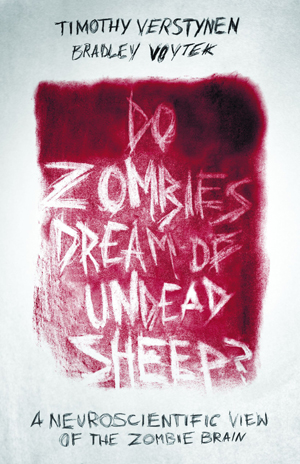
In the spirit of the Halloween weekend, here is a cool article from Slate on why zombies lumber when they walk. The article is an excerpt from the new book Timothy Verstynen and Bradley Voytek, Do Zombies Dream of Undead Sheep?: A Neuroscientific View of the Zombie Brain (2014). Not only is it a good explainer for zombie walking patterns, but it also introduces some basic neuroscience.

Why Do Zombies Lumber?

Two neuroscientists explain why zombies have so much trouble walking.


By Timothy Verstynen and Bradley Voytek
October 30, 2014 

Drawing by Anne Karetnikov

Excerpted from Do Zombies Dream of Undead Sheep?: A Neuroscientific View of the Zombie Brain by Timothy Verstynen and Bradley Voytek. Out now from Princeton University Press.

In the movie Dawn of the Dead (1978) there is a scene when anarchist outlaws break into the mall that the movie’s heroes had secured and lived in for weeks. This invasion subsequently allows the horde of zombies—which had been aggregating outside—free range of the place. The humans are zipping around playing games while the zombies lumber along slowly and clumsily. The humans easily dispatch the threat of individual zombies because the undead are just too darn slow; there’s no real threat until the humans are outnumbered.

The slow and uncoordinated movements of zombies are perhaps the most identifiable feature of their behavior (next to the whole biting and flesh-eating thing of course). Ask anyone to impersonate a zombie and the first thing she’ll do is hold her arms out, widen her stance, stiffen her legs, and utter a low, guttural moan. That’s because, in the movies, as soon as zombies rise from the dead, they begin walking. Well not walking ... more like lumbering. Each step is slow and arduous. Their stance is wide and stiff. This presents us with a very important clue about what’s happened to their brains.

So what does it take to turn the normally smooth, fast, coordinated movements of a healthy person into the traditional zombie lumber? First, let’s consider the pathways in the brain that give rise to our movements.

While “higher” cognitive functions (aka thinking) tend to get all the glory in neuroscience, before the brain did a lot of deep thinking it did a lot of moving. In fact, some scientists have argued that the entire reason we have a brain is to get us moving around in the environment.

The logic for this argument arises from observations of a little ocean creature called the sea squirt. Seriously, that’s its name. The sea squirt is a small and evolutionarily old animal of the phylum Chordata (when scientists say “evolutionarily old,” we mean that the life form has been in a relatively unchanged state for millions and millions of years). In its young life, the sea squirt is a little larval creature that has a very primitive brain and sensory organs. Its goal during its larval stage of development is to swim around and find a rock to perch on. Once it has found a suitable home, like, say, a nice secure rock with plenty of organic food just flowing by, the sea squirt will attach itself with its head facing out. Then it basically just sits there catching food as it floats by. As it matures into a full-grown adult creature the sea squirt does something quite strange: It digests its own brain.

Yeah, you read that right. Let that sink in. It digests its own brain.

Biologists and neuroscientists have argued that this is evolutionarily advantageous. See, the brain is really expensive, from a metabolic standpoint. Meaning that it takes a lot of energy to keep the brain going, and energy (food) is pretty hard to come by when you’re little more than a stick on a rock with a mouth attached. So when you no longer need a metabolically expensive organ like the brain, it is better to just get rid of it. Thus, no longer needing to navigate around its environment, the sea squirt simply has lost the need for its brain and does away with it. But waste not, want not, in nature. So “doing away with it” means “eating it.” And thus the sea squirt digests its own brain.

Now luckily for us, we humans are more than mouth-sticks attached to rocks. We need to keep moving. We can’t just sit around and digest away our own brain, because food doesn’t just come to us. No, we still have to go out and get our food, even if it is just by driving to the local fast-food chain down the block. This means we get to keep our brains because, for the brain, movement is life.

Unfortunately, the same is true for zombies. Because humans rarely run to zombies, the walking dead have to go to their food source. Which means the zombies also still need their brains. Well, at least part of their brains.

If we presume that the primary function of the brain is to get us moving around in the world, then it’s not surprising that a lot of neural real estate is devoted to the planning and execution of actions. In fact, the computations required to simply move around our environment are distributed across vast swaths of both cortical and subcortical areas. So let’s take a walk through the multitude of brain systems that move us around, shall we?

Most of our voluntary movements start in the neocortex, in two of the four major lobes: the frontal and parietal lobes. Neurons in the parietal lobe that primarily maintain spatial awareness, and those in the frontal lobe that control decision making, are constantly negotiating with one another as to what action we should do next. We might imagine the dialogue going something like this:
  • PARIETAL LOBE: “Hey, there’s a tasty piece of broccoli 30 degrees to the left.”
  • FRONTAL LOBE: “Broccoli??? No way! I want something more awesome!”
  • PARIETAL LOBE: [sigh] “OK, how about that doughnut 10 degrees to the right?”
  • FRONTAL LOBE: “Now you’re talking. Hey! Right arm! Attention, right arm! Prepare the triceps, deltoids, and hand muscles for action. We’re going to make a reach.”
  • MOTOR CORTEX: “Jawohl, Lord Frontal Cortex!”
In our silly little sketch here, the parietal lobe tells us where to attend to things that are in the environment while the frontal cortex in the front of the head decides what to do. Then the motor areas, in the back part of the frontal cortex, make the movement happen.

Contrary to what you may have heard, there’s not just a single motor cortex. In fact, there are several “motor” areas that are spread out across the frontal lobe and provide the groundwork for planning your movements. You can think of these as the middle management of motor planning. They take the decisions handed down from frontal areas and turn them into plans that the heavy lifters in the arms, legs, and other muscles know what to do with. Which isn’t as easy as it sounds.

Let’s consider the following scenario: You’re a zombie sitting very patiently on the examination table, hand resting on your desiccated, disgusting lap. The nerdy scientists in their awkward lab coats then place a tasty chunk of human flesh right in front of you. What remains of your undead frontal lobes will immediately say “GO GET THAT!” because, hey, free thigh.


Before you can actually grab that tasty piece of meat, however, the motor planning areas in your undead brain, called premotor regions, have to figure out how to get your hand from your lap to the yummy flesh. Now remember, while you can see the tasty morsel, the process of getting your hand off of your lap and to the chunk of meat is pretty complicated. Somehow your brain has to convert a map of the world that’s being projected from the back of your eyeballs to a plan of muscle contractions that uses your bones as levers, much as a puppet master has to coordinate the strings of a marionette doll to make it dance—except here the puppet master is your own brain.

Let’s return our attention to that horde of walking dead outside. While zombie movements are slow, stiff, and uncoordinated, zombies do seem to be able to plan movements in the right direction. That is, when a zombie wants to lunge toward you, it mostly gets the direction right. Once it gets its hands on you, it has no problem grasping and holding on. Therefore it would appear that the cortical motor systems are all intact. So what could be wrong? The only real neural culprits left as plausible candidates for the motor dysfunction seen in zombies are the basal ganglia and the cerebellum.

Given this restriction, let’s consider what happens when the basal ganglia are malfunctioning and compare that with when something goes wrong with the cerebellum. In both cases, people have trouble walking and coordinating their movements, but in dramatically different ways. For example, in Parkinson’s disease, people develop a slouched posture and walk by taking short, shuffling steps. They also have difficulty generating actions without a very obvious goal (they tend to freeze up). In contrast, people with spinocerebellar ataxia develop a stiff, wide-legged stance and take big, lumbering steps. And unlike those afflicted with Parkinson’s disease, these patients have no problem initiating movements.

How can we use this information to diagnose a zombie’s brain? We know that the walking dead are shown in movies as having a stiff, wide-legged stance and a big, lumbering walk. They tend to move slowly (most of the time) and lack smooth, coordinated actions. Yet they don’t seem to have trouble initiating movements. In fact, zombies are almost constantly on the move, they never have problems starting a movement (say reaching for a new victim), and they don’t stall in the middle of movements. They also don’t shuffle or have curvature in their posture.

For these reasons we argue that the cluster of symptoms seen in zombies, the wide stance, lumbering walk, lack of freezing, ease in general planning and execution of actions, reflects a pattern of cerebellar degeneration. That is, cerebellar dysfunction would lead to many of the motor symptoms of the zombie infection. However, cortical motor areas and basal ganglia pathways should be relatively intact.

At about this point, the really astute zombie movie fan will ask, “What about fast zombies?” For those who haven’t seen movies like World War Z, 28 Days Later, or the 2004 remake of Dawn of the Dead, “fast zombies” don’t appear to have any motor dysfunctions. They can move quickly and don’t appear to have any coordination problems. Given the terrifyingly coordinated movements that “fast zombies” exhibit, it is our belief that their cerebellums are likely intact. Any difficulty fast zombies may have moving are likely more to do with the fact that their arms and legs are rotting than any sort of neural damage.

In fact, this difference in presentation may allow us to develop neurological classifications of different subtypes of the disorder that may give important clues to the etiology of the zombie epidemic.
  • Subtype I (slow-moving subtype): First observed variant of the disease. 

  • Subtype II (fast-moving subtype): Distinguished from Subtype I variant by healthy motor coordination.
Hey folks ... sometimes diseases mutate. Why wouldn’t zombism?

Truth be told, when we had the opportunity to ask George Romero why he made his ghouls walk the way they did in the Living Dead movies, he responded, “They’re supposed to be dead. They’re stiff. That’s how you’d walk if you were dead.” Not quite the answer that appeals to our neuroscience instincts, but a good alternative hypotheses to test in the next zombie apocalypse.
____

Excerpted from Do Zombies Dream of Undead Sheep?: A Neuroscientific View of the Zombie Brain by Timothy Verstynen and Bradley Voytek © 2014 by Princeton University Press. Reprinted by permission.

Timothy Verstynen is an assistant professor in the department of psychology and at the Center for the Neural Basis of Cognition at Carnegie Mellon University.

Bradley Voytek is an assistant professor of computational cognitive science and neuroscience at the University of California–San Diego.

Friday, October 31, 2014

Michael White - Why DNA Is One of Humanity’s Greatest Inventions

From Pacific Standard, this is an interesting article on how humans are using DNA -- ours and other species' -- to change the world. One wonders if that is such a good thing considering how little we still know about how DNA functions.

Why DNA Is One of Humanity’s Greatest Inventions


By Michael White • October 24, 2014 


dna-bulb
(Photo: Renzo.luo/Shutterstock)


How we’ve co-opted our genetic material to change our world.


Humans and their ancestors have been using tools for millions of years. We owe our prolific capacity for making tools to our DNA, and now we’ve reached the point were we’ve made DNA itself into a tool. We use DNA as a crucial research tool in the lab, we use it to engineer food crops and biofuel-producing microbes, we use it as a forensic tool and a medical diagnostic, and we use it to access our ancient history. We’ve even put DNA to uses that have nothing to do with biology whatsoever, as a nanomaterial with amazing chemical properties. Our genetic material is turning out to be one of our most useful high-tech tools.

It didn’t have to be this way. Before the 20th century, scientists didn’t expect the physical make-up of our genes to be something as simple, elegant, and useful as DNA. Darwin, for example, proposed that our genetic material was basically a chemical soup, made of different kinds of molecules that were secreted by each body part and gathered in the reproductive cells. Like the diffuse nebular gases that self-organize into solar systems, our genetic material was often thought to be a complex mix of ingredients that spontaneously organize themselves to direct the development of a new organism.

If this theory were true, the science of genetics—and our society—would now be very different. It would be much more difficult, if not impossible, to do many of the things we do with DNA, such as engineer microbes to make drugs or biofuels, or discover our relationship with Neanderthals. Paternity tests and viruses wouldn’t exist. Perhaps life wouldn’t exist either; in the 1930s and ’40s, physicists convincingly argued that a genetic chemical soup was thermodynamically impossible.

By that time, biologists were already well on their way to discovering what genes actually are: segments of giant molecules of DNA. Our genetic material turns out to be surprisingly similar to a text, with genes spelled out in a linear string of chemical letters. The analogy to a text isn’t perfect, but it’s remarkably good and partially explains why DNA is such a useful tool. The ability to represent genes as a text on our computers is a boon to biologists, who now analyze genomes with the same text-parsing algorithms that go into spam filters. One of the most basic tools of a molecular biology lab are enzymes that cut, copy, and paste DNA text; without these, much of the last 40 years of biological research would have been impossible. So would commercial genetic engineering, which, aside from its role in agriculture, is increasingly important for producing drugs and chemicals using processes that are more efficient and less environmentally damaging.

Because of DNA’s useful properties, “molecular biology” is not only a scientific discipline, but also a set of essential tools used by biologists of all sorts. By manipulating DNA, scientists tackle questions that have little to do with DNA’s biological role. They modify DNA to test hypotheses or to insert chemical sensors into an experimental organism, and they use DNA as a convenient way to read the outcome of an experiment. In my own work, I use DNA “barcodes,” short, easy-to-read sequences of DNA that tag the process I’m interested in. In paternity testing and forensics DNA is merely an identifier; its molecular function is irrelevant. That DNA is a tool with many different biological applications explains the enormous impact of the recent, dramatic improvements in technologies to read and synthesize DNA—DNA science dominates biology more than ever before.

We’ve now extended the uses of DNA beyond biology. DNA is not only life’s hackable source code; it’s a nanomaterial with very useful chemical properties. In particular, DNA is a programmable polymer that reliably folds into very small and fantastically intricate shapes. Engineering shapes from folded DNA is called, appropriately enough, DNA origami. Two key properties of DNA make this molecular origami possible: With its 4-letter chemical alphabet, the number of possible designs is huge. There are over a trillion variations of even a very short, 20-unit long DNA segment. And second, strands of DNA like to stick to each other in predictable ways, typically forming the iconic double helix.

Using DNA origami, engineers have built all sorts of miniature DNA widgets: motors, sensors, rulers, stencils, boxes, smiley faces, and “dolphin-shaped structures with flexible tails.”

These aren’t just demonstration pieces; DNA origami is key element of nanotechnology. In a paper published this month, researchers from MIT and Harvard’s Wyss Institute for Biologically Inspired Engineering describe how they built DNA “molds” to cast precisely shaped gold and silver nanoparticles. These nanoparticles are used in a variety of technologies, and they are typically etched using beams of electrons. But this process is slow and limited in its resolution. By building small molds out of strands of DNA and filling them with gold or silver particles, the researchers were able to cast nano-spheres, triangles, and cubes. They argue that their strategy “points to a new kind of manufacture framework: DNA-directed, digitally programmable fabrication of inorganic nanostructures and devices”—essentially 3-D printing on a nanometer level.

As a tool-using species, we’ve long been making tools out of whatever materials we can get our hands on. We’re lucky that we’ve got the DNA to build tools. We’re also lucky that DNA itself makes such a great tool.

Michael White
Michael White is a systems biologist at the Department of Genetics and the Center for Genome Sciences and Systems Biology at the Washington University School of Medicine in St. Louis, where he studies how DNA encodes information for gene regulation. He co-founded the online science pub The Finch and Pea. Follow him on Twitter @genologos.

More From Michael White

C. Nathan DeWall - Magic May Lurk Inside Us All

From the New York Times, here is a nice essay in the spirit of the season. DeWall looks at the persistence of magical thinking in adults, all of whom would likely deny their thinking is magical.

We typically associate this type of thinking with children, for example, believing that if their parents are fighting it must be because they are bad kids. Another example, from my own childhood, is "step on a crack [in the sidewalk] and break your mother's back." I was really young when I learned that this did not work as advertised.

However, as DeWall illustrates, magical thinking often persists into adulthood. Think of The Secret, as a recent adult example of believing ones thoughts can alter reality. Might as well place your intent on making it rain or generating world peace. Ain't gonna happen.

Magic may Lurk Inside Us All

C. Nathan DeWall | Oct. 27, 2014


How many words does it take to know you’re talking to an adult? In “Peter Pan,” J. M. Barrie needed just five: “Do you believe in fairies?”

Such belief requires magical thinking. Children suspend disbelief. They trust that events happen with no physical explanation, and they equate an image of something with its existence. Magical thinking was Peter Pan’s key to eternal youth.

The ghouls and goblins that will haunt All Hallows’ Eve on Friday also require people to take a leap of faith. Zombies wreak terror because children believe that the once-dead can reappear. At haunted houses, children dip their hands in buckets of cold noodles and spaghetti sauce. Even if you tell them what they touched, they know they felt guts. And children surmise that with the right Halloween makeup, costume and demeanor, they can frighten even the most skeptical adult.

We do grow up. We get jobs. We have children of our own. Along the way, we lose our tendencies toward magical thinking. Or at least we think we do. Several streams of research in psychology, neuroscience and philosophy are converging on an uncomfortable truth: We’re more susceptible to magical thinking than we’d like to admit. Consider the quandary facing college students in a clever demonstration of magical thinking. An experimenter hands you several darts and instructs you to throw them at different pictures. Some depict likable objects (for example, a baby), others are neutral (for example, a face-shaped circle). Would your performance differ if you lobbed darts at a baby?

It would. Performance plummeted when people threw the darts at the baby. Laura A. King, the psychologist at the University of Missouri who led this investigation, notes that research participants have a “baseless concern that a picture of an object shares an essential relationship with the object itself.”

Paul Rozin, a psychology professor at the University of Pennsylvania, argues that these studies demonstrate the magical law of similarity. Our minds subconsciously associate an image with an object. When something happens to the image, we experience a gut-level intuition that the object has changed as well.

Put yourself in the place of those poor college students. What would it feel like to take aim at the baby, seeking to impale it through its bright blue eye? We can skewer a picture of a baby face. We can stab a voodoo doll. Even as our conscious minds know we caused no harm, our primitive reaction thinks we tempted fate.

How can well-educated people — those who ought to know better — struggle to throw a dart at a piece of paper? Some philosophers argue that magical thinking is, in some ways, adaptive. Tamar Gendler, a philosopher at Yale University, has coined the term “aliefs” to refer to innate and habitual reactions that may be at odds with our conscious beliefs — as when pictures of vipers, snarling dogs or crashing airplanes make our hearts race.

Aliefs motivate us to take or withhold action. You might enjoy sweets, but would you eat a chocolate bar shaped like feces? Dr. Rozin and his colleagues showed that college students would not, though they knew it would not harm them. Our conscious beliefs tell us to shape up, use our wits and act rationally. But our subconscious aliefs set off deeply ingrained reactions that protect us from disease. The alief often wins.

We may have evolved to be this way — and that is not always a bad thing. We enter the world with innate knowledge that helped our evolutionary ancestors survive and reproduce. Babies know mother from stranger, scalding heat from soothing warmth. When we grow up, our minds cling to that knowledge and, without our awareness, use it to try to make sense of the world.

Can magical beliefs offer a window into the aggressive mind? My colleagues and I examined this idea in recent research published in the journal Aggressive Behavior. In one illustrative study, 529 married Americans were shown a picture of a doll and were told that it represented their spouse. They could insert as many pins into the doll as they wished, from zero to 51. Participants also reported how often they had perpetrated intimate partner violence, which included psychological aggression and physical assault.

Voodoo dolls can measure whether your romantic partner is “hangry”— that dangerous combination of hunger and anger. If we let our blood sugar drop, it becomes harder to put the brakes on our aggressive urges. In a study published in Proceedings of the National Academy of Sciences, we showed that on days when their blood sugar dropped, married people stabbed the voodoo doll with more pins.

Do people take the voodoo doll seriously? If they don’t, their responses should not relate to actual violent behavior. But they do. The more pins people used to stab the voodoo doll, the more psychological and physical aggression they perpetrated.

Stabbing a voodoo doll can also satisfy the desire for vengeance, another study found. When German students imagined an upsetting situation, they began to see the world through blood-colored glasses, increasing their tendency to ruminate on aggression-related thoughts. Stabbing a voodoo doll that represented the provocateur returned their glasses to their normal hue. By quenching their aggressive appetite, magical beliefs enabled provoked students to satisfy their aggressive goal without harming anyone.

Yes, children believe in magic because they don’t know any better. Peter Pan never grew up because he embraced magical beliefs. But such beliefs make for more than happy Halloweeners and children’s books. They give a glimpse into how the mind makes sense of the world.

We can’t overcome magical thinking. It is part of our evolved psychology. Our minds may fool us into thinking we are immune to magical thoughts. But we are only fooling ourselves. That’s the neatest trick of all.

~ C. Nathan DeWall is a professor of psychology at the University of Kentucky. With David G. Myers, he is co-author of the forthcoming book, Psychology (11th Edition).

Thursday, October 30, 2014

Box - Where Does Reality End and Digital Space Begin?

http://cdn-imgs-mag.aeon.co/images/2014/10/BOX-FINAL-960x540.jpg

Via Aeon . . .

Box


Where does reality end and digital space begin? Projection mapping shifts the bounds of visual expression and perception.

Bot & Dolly

5 MINUTES

Daniel Levitin: "The Organized Mind: Thinking Straight in an Age of Information Overload"

https://gallery.mailchimp.com/f64149d518f47ed9cd8b89911/images/96a73506-1881-4d2f-9240-eafadd5b85d9.jpg

Daniel Levitin is the author of The Organized Mind: Thinking Straight in the Age of Information Overload (2014), as you may have guessed by the title of this post. From the Amazon page for the book (linked to in previous sentence), here is the ad copy:
New York Times bestselling author and neuroscientist Daniel J. Levitin shifts his keen insights from your brain on music to your brain in a sea of details.

The information age is drowning us with an unprecedented deluge of data. At the same time, we’re expected to make more—and faster—decisions about our lives than ever before. No wonder, then, that the average American reports frequently losing car keys or reading glasses, missing appointments, and feeling worn out by the effort required just to keep up.

But somehow some people become quite accomplished at managing information flow. In The Organized Mind, Daniel J. Levitin, PhD, uses the latest brain science to demonstrate how those people excel—and how readers can use their methods to regain a sense of mastery over the way they organize their homes, workplaces, and time.

With lively, entertaining chapters on everything from the kitchen junk drawer to health care to executive office workflow, Levitin reveals how new research into the cognitive neuroscience of attention and memory can be applied to the challenges of our daily lives. This Is Your Brain on Music showed how to better play and appreciate music through an understanding of how the brain works. The Organized Mind shows how to navigate the churning flood of information in the twenty-first century with the same neuroscientific perspective.
Levitin stopped by Google recently to talk about his new book.

Daniel Levitin: "The Organized Mind: Thinking Straight in an Age of Information Overload"

Published on Oct 28, 2014


The information age is drowning us with an unprecedented deluge of data. At the same time, we’re expected to make more—and faster—decisions about our lives than ever before. No wonder, then, that the average American reports frequently losing car keys or reading glasses, missing appointments, and feeling worn out by the effort required just to keep up.

But somehow some people become quite accomplished at managing information flow. In The Organized Mind, Daniel J. Levitin, PhD, uses the latest brain science to demonstrate how those people excel—and how readers can use their methods to regain a sense of mastery over the way they organize their homes, workplaces, and time.

This Is Your Brain on Psychedelic Drugs (via Discover)

Dr. David Nutt and a team of researchers have published a study on the psychoactive substance in mushrooms, psilocybin, and how it impacts the brain circuits. As you can see in the picture below, the effect of psilocybin is a much more interconnected brain (which suggests some other circuits that limit activity are inactive under the influence of psilocybin).

Interesting stuff.

The summary below is from Discover, then the whole article, which is open access, is also included below.

This Is Your Brain on Psychedelic Drugs


By Ben Thomas | October 29, 2014 4:16 pm


Left, the stable brain activity in a normal brain. Right, under the influence of psilocybin, diverse brain regions not normally in communication become strongly linked.

Psychedelic substances can change a user’s mindset in profound ways — a fact that’s relevant even to those who’ve never touched the stuff, because such altered states of consciousness give scientists a window into how our brains give rise to our normal mental states. But neuroscientists are only beginning to understand how and why those mental changes occur.

Now some mathematicians have jumped into the fray, using a new mathematical technique to analyze the brains of people on magic mushrooms.

Psychedelic Puzzles

Scientists have known for decades that many of psychedelic drugs’ most famous effects — visual hallucinations, heightened sensory and emotional sensitivity, etc. — are linked to elevated levels of the neurotransmitter serotonin.

But increasingly neuroscience researchers are interested not just in single chemicals but also in overall brain activity, because the most complicated brain functions arise from lots of different regions working together. Over the last several years, a branch of mathematics known as network theory has been applied to study this phenomenon.

Paul Expert, a complexity researcher at the Imperial College London, and his team took this approach to analyzing fMRI data from people who’d taken psilocybin, the psychedelic chemical in magic mushrooms. The team had recently been working on a new technique for network modeling — one designed to highlight small but unusual patterns in network connectivity.

Brains on Drugs

The team used fMRI data from a previous study, in which 15 healthy people rested inside an fMRI scanner for 12 minutes on two separate occasions. The volunteers received a placebo in one of those sessions, and a mild dose of psilocybin during the other, but they weren’t told which was which.

The investigators crunched the data, specifically studying the brain’s functional connectivity — the amount of active communication among different brain areas.

They found two main effects of the psilocybin. First, most brain connections were fleeting. New connectivity patterns tended to disperse more quickly under the influence of psilocybin than under placebo. But, intriguingly, the second effect was in the opposite direction: a few select connectivity patterns were surprisingly stable, and very different from the normal brain’s stable connections.

This indicates “that the brain does not simply become a random system after psilocybin injection, but instead retains some organizational features, albeit different from the normal state,” the authors write in their paper in the Journal of the Royal Society Interface.

Far Out
The findings seem to explain some of the psychological experiences of a psilocybin trip. Linear thinking and planning become extremely difficult, but nonlinear “out of the box” thinking explodes in all directions. By the same token, it can become difficult to tell fantasy apart from reality during a psilocybin trip; but focusing on a certain thought or image — real or imagined — often greatly amplifies that thought’s intensity and vividness.

The authors suggest that effects like these may be rooted in the two connectivity traits they spotted, since the connectivity patterns that rapidly disperse may reflect unorganized thinking, while the stable inter-regional connections may reflect information from one sensory domain “bleeding” into other areas of sensory experience. In fact, the researchers also suggest that synesthesia — the sensory blurring that causes users of psychedelics to experience sounds as colors, for example — may be a result of these connectivity changes too.

The researchers hope that the patterns they’ve found will provide neuroscientists with new approaches for studying the brain on psychedelic drugs, and therefore better understand the strange psychological effects their users report.
* * * * *

Full Citation:
Petri, G, Expert, P,  Turkheimer, F, Carhart-Harris, R, Nutt, D, Hellyer, PJ, and Vaccarino, F. (2014, Oct 29). Homological scaffolds of brain functional networks. J. R. Soc. Interface; 11(101). doi: 10.1098/​rsif.2014.0873 [Print: 6 December 2014] 

Homological scaffolds of brain functional networks


G. Petri [1], P. Expert [2], F. Turkheimer [2], R. Carhart-Harris [3], D. Nutt [3], P. J. Hellyer [4] and
F. Vaccarino [1,5]
1. ISI Foundation, Via Alassio 11/c, 10126 Torino, Italy
2. Centre for Neuroimaging Sciences, Institute of Psychiatry, Kings College London, De Crespigny Park, London SE5 8AF, UK
3. Centre for Neuropsychopharmacology, Imperial College London, London W12 0NN, UK
4. Computational, Cognitive and Clinical Neuroimaging Laboratory, Division of Brain Sciences, Imperial College London, London W12 0NN, UK
5. Dipartimento di Scienze Matematiche, Politecnico di Torino, C.so Duca degli Abruzzi no 24, Torino 10129, Italy
Abstract

Networks, as efficient representations of complex systems, have appealed to scientists for a long time and now permeate many areas of science, including neuroimaging (Bullmore and Sporns 2009 Nat. Rev. Neurosci. 10, 186–198. (doi:10.1038/nrn2618)). Traditionally, the structure of complex networks has been studied through their statistical properties and metrics concerned with node and link properties, e.g. degree-distribution, node centrality and modularity. Here, we study the characteristics of functional brain networks at the mesoscopic level from a novel perspective that highlights the role of inhomogeneities in the fabric of functional connections. This can be done by focusing on the features of a set of topological objects—homological cycles—associated with the weighted functional network. We leverage the detected topological information to define the homological scaffolds, a new set of objects designed to represent compactly the homological features of the correlation network and simultaneously make their homological properties amenable to networks theoretical methods. As a proof of principle, we apply these tools to compare resting-state functional brain activity in 15 healthy volunteers after intravenous infusion of placebo and psilocybin—the main psychoactive component of magic mushrooms. The results show that the homological structure of the brain's functional patterns undergoes a dramatic change post-psilocybin, characterized by the appearance of many transient structures of low stability and of a small number of persistent ones that are not observed in the case of placebo.

1. Motivation

The understanding of global brain organization and its large-scale integration remains a challenge for modern neurosciences. Network theory is an elegant framework to approach these questions, thanks to its simplicity and versatility [1]. Indeed, in recent years, networks have become a prominent tool to analyse and understand neuroimaging data coming from very diverse sources, such as functional magnetic resonance imaging (fMRI), electroencephalography and magnetoencephalography [2,3], also showing potential for clinical applications [4,5]. 

A natural way of approaching these datasets is to devise a measure of dynamical similarity between the microscopic constituents and interpret it as the strength of the link between those elements. In the case of brain functional activity, this often implies the use of similarity measures such as (partial) correlations or coherence [68], which generally yield fully connected, weighted and possibly signed adjacency matrices. Despite the fact that most network metrics can be extended to the weighted case [913], the combined effect of complete connectedness and edge weights makes the interpretation of functional networks significantly harder and motivates the widespread use of ad hoc thresholding methods [7,1418]. However, neglecting weak links incurs the dangers of a trade-off between information completeness and clarity. In fact, it risks overlooking the role that weak links might have, as shown for example in the cases of resting-state dynamics [19,20], cognitive control [21] and correlated network states [22]. 

In order to overcome these limits, Rubinov & Sporn [13,23,24] recently introduced a set of generalized network and community metrics for functional networks that among others were used to uncover the contrasting dynamics underlying recollection [25] and the physiology of functional hubs [26]. 

In this paper, we present an alternative route to the analysis of brain functional networks. We focus on the combined structure of connections and weights as captured by the homology of the network. A summary of all the keywords and concepts introduced in this paper can be found in table 1.
View this table:
Table 1. List of notations.
2. From networks to topological spaces and homology

Homology is a topological invariant that characterizes a topological space X by counting its holes and their dimensions. By hole, we mean a hollow region bounded by the parts of that space. The dimension of a hole is directly related to the dimension of its boundary. The boundary of a two-dimensional hole is a one-dimensional loop; the three-dimensional inner part of a doughnut, where the filling goes, is bounded by two-dimensional surface; for dimensions higher than 2, it becomes difficult to have a mental representation of a hole, but k-dimensional holes are still bounded by (k − 1) dimensional faces. In our work, we start with a network and from it construct a topological space. We now use figure 1 to show how we proceed and make rigorous what we mean by boundaries and holes. 

Figure 1.
Figure 1. Panels (a,b) display an unweighted network and its clique complex, obtained by promoting cliques to simplices. Simplices can be intuitively thought as higher-dimensional interactions between vertices, e.g. as a simplex the clique (b,c,i) corresponds to a filled triangle and not just its sides. The same principle applies to cliques—thus simplices—of higher order. (Online version in colour.) 
In a network like that of figure 1a, we want the ring of nodes (a,b,c,d) to be a good candidate for a one-dimensional boundary, whereas the other rings of three nodes should not constitute interesting holes. The reason for this choice comes from the formalization of the notion of hole. One way to formalize this is by opposition that is we define what we mean by a dense subnetwork in order to highlight regions of reduced connectivity, i.e. holes. The most natural and conservative definition we can adopt for a dense subnetwork is that of clique, a completely connected subgraph [27]. Moreover, cliques have the crucial property, which will be important later, of being nested, i.e. a clique of dimension k (k-clique) contains all the m-cliques defined by its nodes with m < k. Using this definition and filling in all the maximal cliques, the network in figure 1a can be represented as in figure 1b: 3-cliques are filled in, becoming tiles, and the only interesting structure left is the square (a,b,c,d). It is important at this point to note that a k-clique can be seen as a k − 1 simplex, i.e. as the convex hull of k-points. Our representation of a network can thus be seen as a topological space formed by a finite set of simplices that by construction satisfy the condition that defines the type of topological spaces called abstract simplicial complexes [28]: each element of the space is a simplex, and each of its faces (or subset in the case of cliques) is also a simplex. 

This condition is satisfied, because each clique is a simplex, and subsets of cliques are cliques themselves, and the intersection of two cliques is still a clique. 

The situation with weighted networks becomes more complicated. In the context of a weighted network, the holes can be thought of as representing regions of reduced connectivity with respect to the surrounding structure. 

Consider, for example, the case depicted in figure 2a: the network is almost the same as figure 1 with the two exceptions that it now has weighted edges and has an additional very weak edge between nodes a and c. The edges in the cycle [a,b,c,d] are all much stronger than the link (a,c) that closes the hole by making (a,b,d) and (b,c,d) cliques and therefore fills them. The loop (e,f,g,h,i) has a similar situation, but the difference in edge weights between the links along the cycle and those crossing, is not as large as in the previous case. It would be therefore useful to be able to generalize the approach exposed earlier for binary networks to the case of weighted networks in such a way as to be able to measure the difference between the two cases (a,b,c,d) and (e,f,g,h,i). As shown by figure 2b, this problem can be intuitively thought of as a stratigraphy in the link-weight fabric of the network, where the aim is to detect the holes, measure their depth and when they appear as we scan across the weights' range. 

Figure 2.
Figure 2. Panels (a­–c) display a weighted network (a), its intuitive representation in terms of a stratigraphy in the weight structure according the weight filtration described in the main text (b) and the persistence diagram for H1 associated with the network shown (c). By promoting cliques to simplices, we identify network connectivity with relations between the vertices defining the simplicial complex. By producing a sequence of networks through the filtration, we can study the emergence and relative significance of specific features along the filtration. In this example, the hole defined by (a,b,c,d) has a longer persistence (vertical solid green bars) implying that the boundary of the cycle are much heavier than the internal links that eventually close it. The other hole instead has a much shorter persistence, surviving only for one step and is therefore considered less important in the description of the network homological properties. Note that the births and deaths are defined along the sequence of descending edge weights in the network, not in time. (Online version in colour.) 
From figure 2b, it becomes clear that the added value of this method over conventional network techniques lies in its capability to describe mesoscopic patterns that coexist over different intensity scales, and hence to complement the information about the community structure of brain functional networks. A way to quantify the relevance of holes is given by persistent homology. We describe it and its application to the case of weighted networks in full detail in §3.
3. A persistent homology of weighted networks

The method that we adopt was introduced in references [29,30] and relies on an extension of the metrical persistent homology theory originally introduced by references [31,32]. Technical details about the theory of persistent homology and how the computation is performed can be found in the works of Carlsson, Zomorodian and Edelsbrunner [28,3135]. Persistent homology is a recent technique in computational topology developed for shape recognition and the analysis of high dimensional datasets [36,37]. It has been used in very diverse fields, ranging from biology [38,39] and sensor network coverage [40] to cosmology [41]. Similar approaches to brain data [42,43], collaboration data [44] and network structure [45] also exist. The central idea is the construction of a sequence of successive approximations of the original dataset seen as a topological space X. This sequence of topological spaces X0, X1, … , XN = X is such that Graphic whenever i < j and is called the filtration. Choosing how to construct a filtration from the data is equivalent to choosing the type of goggles one wears to analyse the data. 

In our case, we sort the edge weights in descending order and use the ranks as indices for the subspaces. More specifically, denote by Graphic the functional network with vertices V, edges E and weights Graphic. We then consider the family of binary graphs Gω = (V, Eω), where an edge e ∈ E is also included in Gω if its weight ωe is larger than ω (Graphic). 

To each of the Gω, we associate its clique, or flag complex Kω, that is the simplicial complex that contains the k-simplex [n0, n1, n2, … nk − 1] whenever the nodes n0, n1, n2, … nk −1 define a clique in Gω [27]. As subsets of cliques and intersections of cliques are cliques themselves, as we pointed out in §2, our clique complex is thus a particular case of a simplicial complex. 

The family of complexes {Kω} defines a filtration, because we have Graphic for ω > ω′. At each step, the simplices in Kω inherit their configuration from the underlying network structure and, because the filtration swipes across all weight scales in descending order, the holes among these units constitute mesoscopic regions of reduced functional connectivity.

Moreover, this approach also highlights how network properties evolve along the filtration, providing insights about where and when lower connectivity regions emerge. This information is available, because it is possible to keep track of each k-dimensional cycle in the homology group Hk. A generator uniquely identifies a hole by its constituting elements at each step of the filtration process. The importance of a hole is encoded in the form of ‘time-stamps' recording its birth βg and death δg along the filtration {Kω} [31]. These two time-stamps can be combined to define the persistence πg = δgβg of a hole, which gives a notion of its importance in terms of it lifespan. Continuing the analogy with stratigraphy, βg and δg correspond, respectively, to the top and the bottom of a hole and πg would be its depth. As we said above, a generator Graphic, or hole, of the kth homology group Hk is identified by its birth and death along the filtration. Therefore, Graphic is described by the point Graphic. A standard way to summarize the information about the whole kth persistent homology group is then to consider the diagram obtained plotting the points corresponding to the set of generators. The (multi)set {(βg,δg} is called the persistence diagram of Hk. In figure 2c, we show the persistence diagram for the network shown in figure 2a for H1. Axes are labelled by weights in decreasing order. It is easy to check that the coordinates correspond exactly to the appearance and disappearance of generators. The green vertical bars highlight the persistence of a generator along the filtration. The further a point is from the diagonal (vertically), the more persistent the generator is. In §4, we introduce two objects, the persistence and the frequency homological scaffolds, designed to summarize the topological information about the system. 

4. Homological scaffolds

Once one has calculated the generators Graphic of the kth persistent homology group Hk, the corresponding persistence diagram contains a wealth of information that can be used, for example, to highlight differences between two datasets. It would be instructive to obtain a synthetic description of the uncovered topological features in order to interpret the observed differences in terms of the microscopic components, at least for low dimensions k. Here, we present a scheme to obtain such a description by using the information associated with the generators during the filtration process. As each generator, Graphic is associated with a whole equivalence class, rather than to a single chain of simplices, we need to choose a representative for each class, we use the representative that is returned by the javaplex implementation [46] of the persistent homology algorithm [47]. For the sake of simplicity in the following, we use the same symbol Graphic to refer to a generator and its representative cycle. 

We exploit this to define two new objects, the persistence and the frequency homological scaffolds Graphic and Graphic of a graph G. The persistence homological scaffold is the network composed of all the cycle paths corresponding to generators weighted by their persistence. If an edge e belongs to multiple cycles g0,g1, … ,gs, its weight is defined as the sum of the generators' persistence:

Formula 4.1

Similarly, we define the frequency homological scaffold Graphic as the network composed of all the cycle paths corresponding to generators, where this time, an edge e is weighted by the number of different cycles it belongs to

Formula 4.2

where Graphic is the indicator function for the set of edges composing gi. By definition, the two scaffolds have the same edge set, although differently weighted. 

The construction of these two scaffolds therefore highlights the role of links which are part of many and/or long persistence cycles, isolating the different roles of edges within the functional connectivity network. The persistence scaffolds encodes the overall persistence of a link through the filtration process: the weight in the persistence scaffold of a link belonging to a certain set of generators is equal to the sum of the persistence of those cycles. The frequency scaffold instead highlights the number of cycles to which a link belongs, thus giving another measure of the importance of that edge during the filtration. The combined information given by the two scaffolds then enables us to decipher the nature of the role different links have regarding the homological properties of the system. A large total persistence for a link in the persistence scaffold implies that the local structure around that link is very weak when compared with the weight of the link, highlighting the link as a locally strong bridge. We remark that the definition of scaffolds we gave depends on the choice of a specific basis of the homology group, and the choice of a consistent basis is an open problem in itself, therefore the scaffolds are not topological invariants. Moreover, it is possible for an edge to be added to a cycle shortly after the cycle's birth in such a way that it creates a triangle with the two edges composing the cycle. In this way, the new edge would be part of the shortest cycle, but the scaffold persistence value would be misattributed to the two other edges. This can be checked, for example, by monitoring the clustering coefficient of the cycle's subgraph as edges are added to it. We checked for this effect and found that in over 80% of the cases the edges do not create triangles that would imply the error, but instead new cycles are created, whose contribution to the scaffold is then accounted for by the new cycle. Finally, we note also that, when a new triangle inside the cycle is created, the two choices of generator differ for a path through a third strongly connected node, owing to the properties of boundary operators. Despite this ambiguity, we show in the following that they can be useful to gain an understanding of what the topological differences detected by the persistent homology actually mean in terms of the system under study.
5. Results from fMRI networks

We start from the processed fMRI time series (see Methods for details). The linear correlations between regional time series were calculated after covarying out the variance owing to all other regions and the residual motion variance represented by the 24 rigid motion parameters obtained from the pre-processing, yielding a partial-correlation matrix χα for each subject. The matrices χα were then analysed with the algorithm described in the previous sections. We calculated the generators Graphic of the first homological group H1 along the filtration. As mentioned before, each of these generators identifies a lack of mesoscopic connectivity in the form of a one-dimensional cycle and can be represented in a persistence diagram. We aggregate together the persistence diagrams of subjects belonging to each group and compute an associated persistence probability density (figure 3). These probability density functions constitute the statistical signature of the groups' H1 features. 

Figure 3.
Figure 3. Probability densities for the H1 generators. Panel (a) reports the (log-)probability density for the placebo group, whereas panel (b) refers to the psilocybin group. The placebo displays a uniform broad distribution of values for the births–deaths of H1 generators, whereas the plot for the psilocybin condition is very peaked at small values with a fatter tail. These heterogeneities are evident also in the persistence distribution and find explanation in the different functional integration schemes in placebo and drugged brains. (Online version in colour.) 
We find that, although the number of cycles in the groups are comparable, the two probability densities strongly differ (Kolmogorov–Smirnov statistics: 0.22, p-value less than 10−10). 

The placebo group displays generators appearing and persisting over a limited interval of the filtration. On the contrary, most of the generators for the psilocybin group are situated in a well-defined peak at small birth indices, indicating a shorter average cycle persistence. However, the psilocybin distribution is also endowed with a longer tail implying the existence of a few cycles that are longer-lived compared with the placebo condition and that influences the weight distribution of the psilocybin persistence scaffold. The difference in behaviour of the two groups is made explicit when looking at the probability distribution functions for the persistence and the birth of generators (figure 4), which are both found to be significantly different (Kolmogorov–Smirnov statistics: 0.13, p-value < 10−30 for persistence and Kolmogorov–Smirnov statistics: 0.14, p-value < 10−35 for births). In order to better interpret and understand the differences between the two groups, we use the two secondary networks described in §4, Graphic and Graphic for the placebo group and Graphic and Graphic for the psilocybin group. The weight of the edges in these secondary networks is proportional to the total number of cycles an edge is part of, and the total persistence of those cycles, respectively. They complement the information given by the persistence density distribution, where the focus is on the entire cycle's behaviour, with information on single links. In fact, individual edges belonging to many and long persistence cycles represent functionally stable ‘hub’ links. As with the persistence density distribution, the scaffolds are obtained at a group level by aggregating the information about all subjects in each group. These networks are slightly sparser than the original complete χα networks

Formula 5.1 

and Formula 5.2 

and have comparable densities. A first difference between the two groups becomes evident when we look at the distributions for the edge weights (figure 5a). In particular, the weights of Graphic display a cut-off for large weights, whereas the weights of Graphic have a broader tail (Kolmogorov–Smirnov statistics: 0.06, p-value < 10−20; figure 5a). Interestingly, the frequency scaffold weights probability density functions cannot be distinguished from each other figure 5a (inset) (Kolmogorov–Smirnov statistics: 0.008, p-value = 0.72). Taken together, these two results imply that while edges statistically belong to the same number of cycles, in the psilocybin scaffold, there exist very strong, persistent links. 

Figure 4.
Figure 4. Comparison of persistence π and birth β distributions. Panel (a) reports the H1 generators' persistence distributions for the placebo group (blue line) and psilocybin group (red line). Panel (b) reports the distributions of births with the same colour scheme. It is very easy to see that the generators in the psilocybin condition have persistence peaked at shorter values and a wider range of birth times when compared with the placebo condition. (Online version in colour.) 

Figure 5.
Figure 5. Statistical features of group homological scaffolds. Panel (a) reports the (log-binned) probability distributions for the edge weights in the persistence homological scaffolds (main plot) and the frequency homological scaffolds (inset). While the weights in the frequency scaffold are not significantly different, the weight distributions for the persistence scaffold display clearly a broader tail. Panel (b) shows instead the scatter plot of the edge frequency versus total persistence. In both cases, there is a clear linear relationship between the two, with a large slope in the psilocybin case. Moreover, the psilocybin scaffold has a larger spread in the frequency and total persistence of individual edges, hinting to a different local functional structure within the functional network of the drugged brains. (Online version in colour.) 
The difference between the two sets of homological scaffolds for the two groups becomes even more evident when one compares the weights between the frequency and persistence scaffolds of the same group. Figure 5b is a scatter plot of between the weights of edges from both scaffolds for the two groups. The placebo group has a linear relationship between the two quantities meaning that edges that are persistent also belongs to many cycles (R2 = 0.95, slope = 0.23). Although the linear relationship is still a reasonable fit for the psilocybin group (R2 = 0.9, slope = 0.3), the data in this case display a larger dispersion. In particular, it shows that edges in Graphic can be much more persistent/longer-lived than in Graphic but still appear in the same number of cycles, i.e. the frequency of a link is not predictive of its persistence or simply put: some connections are much more persistent in the psychedelic state. Moreover, the slopes of linear fits of the two clouds are statistically different (p-value < 1020, npla = 13 200 and npsi = 13 275 [48]) pointing to a starkly different local functional structure in the two conditions. 

The results from the persistent homology analysis and the insights provided by the homological scaffolds imply that although the mesoscopic structures, i.e. cycles, in the psilocybin condition are less stable than in the placebo group, their constituent edges are more stable.
6. Discussion

In this paper, we first described a variation of persistent homology that allows us to deal with weighted and signed networks. We then introduced two new objects, the homological scaffolds, to go beyond the picture given by persistent homology to represent and summarize information about individual links. The homological scaffolds represent a new measure of topological importance of edges in the original system in terms of how frequently they are part of the generators of the persistent homology groups and how persistent are the generators to which they belong to. We applied this method to an fMRI dataset comprising a group of subjects injected with a placebo and another injected with psilocybin. 

By focusing on the second homology group H1, we found that the stability of mesoscopic association cycles is reduced by the action of psilocybin, as shown by the difference in the probability density function of the generators of H1 (figure 3). 

It is here that the importance of the insight given by the homological scaffolds in the persistent homology procedure becomes apparent. A simple reading of this result would be that the effect of psilocybin is to relax the constraints on brain function, ascribing cognition a more flexible quality, but when looking at the edge level, the picture becomes more complex. The analysis of the homological scaffolds reveals the existence of a set of edges that are predominant in terms of their persistence although they are statistically part of the same number of cycles in the two conditions (figure 5). In other words, these functional connections support cycles that are especially stable and are only present in the psychedelic state. This further implies that the brain does not simply become a random system after psilocybin injection, but instead retains some organizational features, albeit different from the normal state, as suggested by the first part of the analysis. Further work is required to identify the exact functional significance of these edges. Nonetheless, it is interesting to look at the community structure of the persistence homological scaffolds in figure 6. The two pictures are simplified cartoons of the placebo (figure 6a) and psilocybin (figure 6b) scaffolds. In figure 6a,b, the nodes are organized and coloured according to their community membership in the placebo scaffold (obtained with the Louvain algorithm for maximal modularity and resolution 1 [50]). This is done in order to highlight the striking difference in connectivity structure in the two cases. When considering the edges in the tail of the distribution, weight greater than or equal to 80, in figure 5a, only 29 of the 374 edges present in the truncated psilocybin scaffold are shared with the truncated placebo scaffold (165 edges). Of these 374 edges, 217 are between placebo communities and are observed to mostly connect cortical regions. This supports our idea that psilocybin disrupts the normal organization of the brain with the emergence of strong, topologically long-range functional connections that are not present in a normal state. 


Figure 6. Simplified visualization of the persistence homological scaffolds. The persistence homological scaffolds (a) and (b) are shown for comparison. For ease of visualization, only the links heavier than 80 (the weight at which the distributions in figure 5a bifurcate) are shown. This value is slightly smaller than the bifurcation point of the weights distributions in figure 5a. In both networks, colours represent communities obtained by modularity [49] optimization on the placebo persistence scaffold using the Louvain method [50] and are used to show the departure of the psilocybin connectivity structure from the placebo baseline. The width of the links is proportional to their weight and the size of the nodes is proportional to their strength. Note that the proportion of heavy links between communities is much higher (and very different) in the psilocybin group, suggesting greater integration. A labelled version of the two scaffolds is available as GEXF graph files as the electronic supplementary material. (Online version in colour.)
The two key results of the analysis of the homological scaffolds can therefore be summarized as follows (i) there is an increased integration between cortical regions in the psilocybin state and (ii) this integration is supported by a persistent scaffold of a set of edges that support cross modular connectivity probably as a result of the stimulation of the 5HT2A receptors in the cortex [51]. 

We can speculate on the implications of such an organization. One possible by-product of this greater communication across the whole brain is the phenomenon of synaesthesia which is often reported in conjunction with the psychedelic state. Synaesthesia is described as an inducer-concurrent pairing, where the inducer could be a grapheme or a visual stimulus that generates a secondary sensory output—like a colour for example. Drug-induced synaesthesia often leads to chain of associations, pointing to dynamic causes rather than fixed structural ones as may be the case for acquired synaesthesia [52]. Broadly consistent with this, it has been reported that subjects under the influence of psilocybin have objectively worse colour perception performance despite subjectively intensified colour experience [53]. 

To summarize, we presented a new method to analyse fully connected, weighted and signed networks and applied it to a unique fMRI dataset of subjects under the influence of mushrooms. We find that the psychedelic state is associated with a less constrained and more intercommunicative mode of brain function, which is consistent with descriptions of the nature of consciousness in the psychedelic state.
7. Methods

7.1. Dataset


A pharmacological MRI dataset of 15 healthy controls was used for a proof-of-principle test of the methodology [54]. Each subject was scanned on two separate occasions, 14 days apart. Each scan consisted of a structural MRI image (T1-weighted), followed by a 12 min eyes-close resting-state blood oxygen-level-dependent (BOLD) fMRI scan which lasted for 12 min. Placebo (10 ml saline, intravenous injection) was given on one occasion and psilocybin (2 mg dissolved in 10 ml saline) on the other. Injections were given manually by a study doctor situated within the scanning suite. Injections began exactly 6 min after the start of the 12-min scans, and continued for 60 s. 

7.1.1. Scanning parameters

The BOLD fMRI data were acquired using standard gradient-echo EPI sequences, reported in detail in reference [54]. The volume repetition time was 3000 ms, resulting in a total of 240 volumes acquired during each 12 min resting-state scan (120 pre- and 120 post-injection of placebo/psilocybin). 

7.1.2. Image pre-processing

fMRI images were corrected for subject motion within individual resting-state acquisitions, by registering all volumes of the functional data to the middle volume of the acquisition using the FMRIB linear registration motion correction tool, generating a six-dimension parameter time course [55]. Recent work demonstrates that the six parameter motion model is insufficient to correct for motion-induced artefact within functional data, instead a Volterra expansion of these parameters to form a 24 parameter model is favoured as a trade-off between artefact correction and lost degrees of freedom as a result of regressing motion away from functional time courses [56]. fMRI data were pre-processed according to standard protocols using a high-pass filter with a cut-off of 300 s.
Structural MRI images were segmented into n = 194 cortical and subcortical regions, including white matter cerebrospinal fluid (CSF) compartments, using Freesurfer (http://surfer.nmr.mgh.harvard.edu/), according to the Destrieux anatomical atlas [57]. In order to extract mean-functional time courses from the BOLD fMRI, segmented T1 images were registered to the middle volume of the motion-corrected fMRI data, using boundary-based registration [58], once in functional space mean time-courses were extracted for each of the n = 194 regions in native fMRI space. 

7.1.3. Functional connectivity

For each of the 194 regions, alongside the 24 parameter motion model time courses, partial correlations were calculated between all couples of time courses (i,j), non-neural time courses (CSF, white matter and motion) were discarded from the resulting functional connectivity matrices, resulting in a 169 region cortical/subcortical functional connectivity corrected for motion and additional non-neural signals (white matter/CSF). 

7.2. Persistent homology computation


For each subject in the two groups, we have a set of persistence diagrams relative to the persistent homology groups Hn. In this paper, we use the H1 persistence diagrams of each group to construct the corresponding persistence probability densities for H1 cycles. 

Filtrations were obtained from the raw partial-correlation matrices through the Python package Holes and fed to javaplex [46] via a Jython subroutine in order to extract the persistence intervals and the representative cycles. The details of the implementation can be found in reference [30], and the software is available at Holes [59].
Funding statement

G.P. and F.V. are supported by the TOPDRIM project supported by the Future and Emerging Technologies programme of the European Commission under Contract IST-318121. I.D. P.E. and F.T. are supported by a PET methodology programme grant from the Medical Research Council UK (ref no. G1100809/1). The authors acknowledge support of Amanda Feilding and the Beckley Foundation and the anonymous referees for their critical and constructive contribution to this paper.

© 2014 The Authors. Published by the Royal Society under the terms of the Creative Commons Attribution License http://creativecommons.org/licenses/by/4.0/, which permits unrestricted use, provided the original author and source are credited.
References at the Journal of the Royal Society Interface site